. 2025 Mar;24(3):e14422.
doi: 10.1111/acel.14422. Epub 2024 Dec 19.

A subset of human dermal fibroblasts overexpressing Cockayne syndrome group B protein resist UVB radiation-mediated premature senescence

Affiliations

A subset of human dermal fibroblasts overexpressing Cockayne syndrome group B protein resist UVB radiation-mediated premature senescence

Asimina Fotopoulou et al. Aging Cell. 2025 Mar.

Abstract

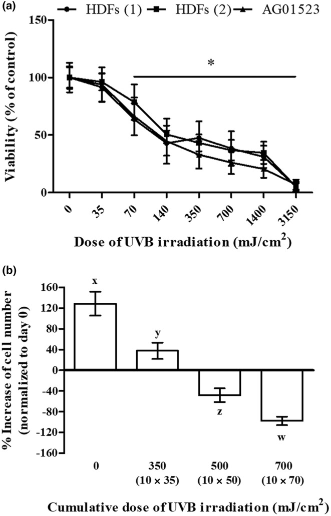
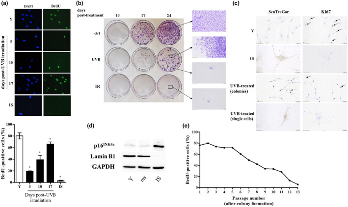
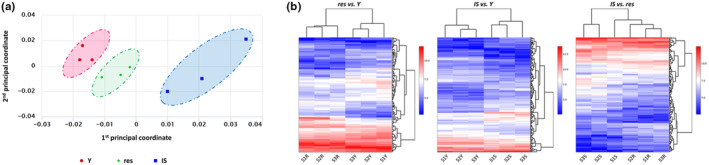
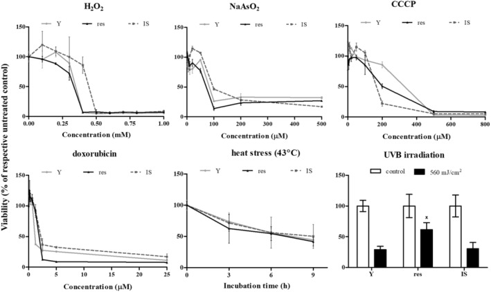
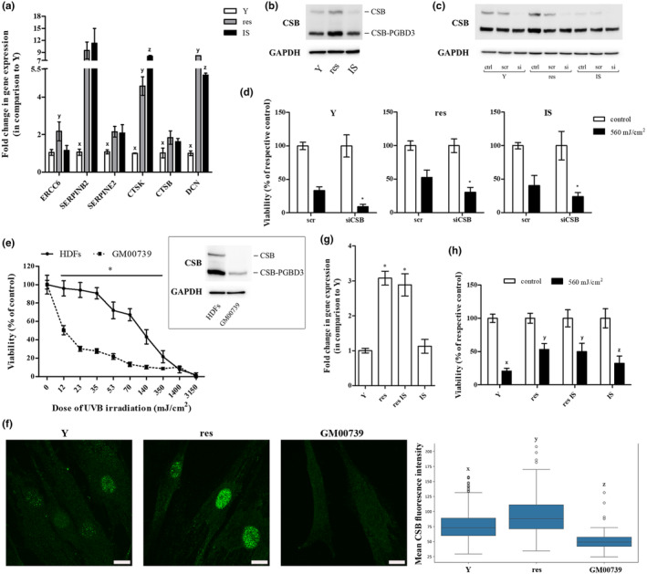
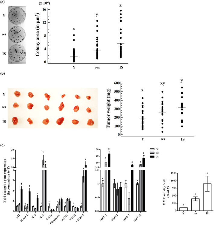
Ultraviolet B (UVB) radiation is a major contributor to skin photoaging. Although mainly absorbed by the epidermis, UVB photons managing to penetrate the upper dermis affect human dermal fibroblasts (HDFs), leading, among others, to the accumulation of senescent cells. In vitro studies have shown that repeated exposures to subcytotoxic UVB radiation doses provoke HDFs' premature senescence shortly after the end of the treatment period. Here, we found that repetitive exposures to non-cytotoxic UVB radiation doses after several days lead to mixed cultures, containing both senescent cells and fibroblasts resisting senescence. "Resistant" fibroblasts were more resilient to a novel intense UVB radiation stimulus. RNA-seq analysis revealed that ERCC6, encoding Cockayne syndrome group B (CSB) protein, is up-regulated in resistant HDFs compared to young and senescent cells. CSB was found to be a key molecule conferring protection toward UVB-induced cytotoxicity and senescence, as siRNA-mediated CSB loss-of-expression rendered HDFs significantly more susceptible to a high UVB radiation dose, while cells from a CSB-deficient patient were found to be more sensitive to UVB-mediated toxicity, as well as senescence. UVB-resistant HDFs remained normal (able to undergo replicative senescence) and non-tumorigenic. Even though they formed a distinct population in-between young and senescent cells, resistant HDFs retained numerous tissue-impairing characteristics of the senescence-associated secretory phenotype, including increased matrix metalloprotease activity and promotion of epidermoid tumor xenografts in immunodeficient mice. Collectively, here we describe a novel subpopulation of HDFs showing increased resistance to UVB-mediated premature senescence while retaining undesirable traits that may negatively affect skin homeostasis.

Keywords: CSB; RNA‐seq; UVB; human dermal fibroblasts; photoaging; resistance; senescence.


Conflict of interest statement

None declared.

Figures

FIGURE 1
FIGURE 1
UVB irradiation is cytotoxic for human dermal fibroblasts (HDFs) in a dose‐dependent manner. (a) Primary HDFs from two different adult donors, as well as the commercially available cell line of foreskin fibroblasts AG01523, were exposed to UVB radiation doses ranging from 35 to 3150 mJ/cm2. After incubation at 37°C for 72 h, cells were detached by trypsinization, stained with neutral red, and measured using a hemocytometer. Unirradiated cells served as the control sample. Numerical values are the means ± standard deviations of two independent experiments conducted in duplicates. Asterisks denote statistically significant differences in comparison to unirradiated cells (Student's t test, p < 0.05). (b) Cells were exposed to various doses of UVB radiation (either 35, 50, or 70 mJ/cm2) twice a day for 5 consecutive days (cumulative UVB radiation dose of 350, 500, and 700 mJ/cm2, respectively). Unirradiated cells served as the untreated control. At the end of the ten doses' scheme (day 5), cells were detached by trypsinization, the number of viable cells was counted after staining with neutral red using a hemocytometer, and the obtained values were normalized to that of unirradiated cells at day 0. Statistically significant differences between any pair of means at the given UVB radiation dose are depicted by diverse letters (ANOVA, Tukey's test, p < 0.05).
FIGURE 2
FIGURE 2
A subset of UVB‐treated human dermal fibroblasts (HDFs) regain their proliferative potential after an initial growth arrest with no signs of transformation. (a) HDFs were exposed to a 10 × 35 mJ/cm2 UVB irradiation scheme and they were allowed to attach onto glass coverslips for 3, 10, and 17 days post‐treatment before their labeling with 50 μM BrdU. After fixation, samples were stained with an anti‐BrdU‐FITC antibody, counterstained with DAPI, and observed under a fluorescence microscope. Unirradiated early‐passage (young, Y) cells served as the untreated control, while cells exposed to ionizing radiation served as the positive control for cellular senescence (ionizing radiation‐induced senescent cells, IS). BrdU incorporation is expressed as a % ratio of the number of BrdU‐positive nuclei to the number of DAPI‐positive nuclei. Microscopic pictures and means ± standard deviations in the graph are from a representative experiment out of three similar ones. Asterisks denote statistically significant differences in comparison to Y (Student's t test, p < 0.05). (b) Untreated HDFs (ctrl), HDFs exposed to a 10 × 35 mJ/cm2 UVB irradiation scheme (UVB) and HDFs exposed to ionizing irradiation (IR) were very sparsely plated and left to grow onto the surface of petri dishes for 10, 17, and 24 days. After fixation, colonies formed were stained with crystal violet. (c) Unirradiated early‐passage (Y), ionizing radiation‐induced senescent (IS) and exposed to a 10 × 35 mJ/cm2 UVB irradiation scheme (UVB‐treated) HDFs were allowed to attach onto glass coverslips for 10 days before their fixation and the staining of lipofuscin‐containing cells with the SenTraGor reagent and of proliferating cells with an anti‐Ki67 antibody. Representative microscopic pictures from one experiment out of three similar ones are depicted here. (d) Protein extracts from Y, resistant (res) and ionizing radiation‐induced senescent (IS) HDFs were subjected to western blot analysis with primary antibodies raised against p16INK4a and lamin B1. Western blot analysis using an anti‐GAPDH antibody was also performed to validate equal loading among samples. Representative blots from three independent experiments are presented here. (e) Resistant UVB‐treated HDFs were serially subcultured, and nuclear BrdU incorporation was estimated at every passage post‐colony formation as described in (a). A representative experiment of three similar ones is shown here.
FIGURE 3
FIGURE 3
Resistant HDFs constitute a separate population from both young and ionizing radiation‐induced senescent cells based on their transcriptional profile. (a) Total RNA was extracted from young (Y), resistant (res), and ionizing radiation‐induced senescent (IS) HDFs and was then used for RNA‐seq analysis. Levels of similarities/dissimilarities and clustering of samples are visualized by a multidimensional scaling (MDS) plot of RNA‐sequencing datasets corresponding to Y, res, and IS HDFs. (b) Differentially expressed genes (DEGs) heatmaps depicting clustering of samples according to their expression values after normalization and statistical testing and possible clusters of co‐expressed genes.
FIGURE 4
FIGURE 4
Tolerance of young (Y), resistant (res), and ionizing radiation‐induced senescent (IS) HDFs toward various types of stresses. Cells were treated with 0–1000 μΜ H2O2, 0–500 μΜ NaAsO2, 0–800 μΜ CCCP, 0–25 μΜ doxorubicin and exposed to 43°C for 3, 6, and 9 h or to a single dose of UVB radiation (560 mJ/cm2). Cell viability after staining of the cells with the vital dye neutral red was estimated 72 h post‐treatment. Untreated cells served as the control sample. Numerical values are the means ± standard deviations of at least two independent experiments. Not sharing a common letter denotes statistically significant differences between any pair of means at a particular concentration, time‐point, or dose (ANOVA, Tukey's test, p < 0.05).
FIGURE 5
FIGURE 5
Cockayne syndrome group B (CSB) plays a crucial role in the protection of HDFs toward UVB‐mediated cytotoxicity. (a) Total RNA was extracted from young (Y), resistant (res), and ionizing radiation‐induced senescent (IS) HDFs and was then used for RT‐qPCR analysis. Glyceraldehyde‐3‐phosphate dehydrogenase (GAPDH) served as the reference gene. Numerical values are the means ± standard deviations of at least two independent experiments conducted in duplicates. Samples not sharing a common letter per gene tested are significantly different (ANOVA, Tukey's test, p < 0.05). (b) Total protein extracts from young (Y), resistant (res), and ionizing radiation‐induced senescent (IS) HDFs were subjected to western blot analysis with a primary antibody raised against the N‐terminus of CSB protein, able to hybridize with both the functional CSB and the CSB‐piggyBac fusion protein (CSB‐PGBD3). Western blot analysis for GAPDH served as the loading control. A representative blot from three independent experiments is shown here. (c) Young (Y), resistant (res), and ionizing radiation‐induced senescent (IS) HDFs were transfected with 50 nM of either a predesigned scramble (scr) or the CSB siRNA (si) before total protein extraction 48 h post‐transfection and western blot analysis for CSB protein. GAPDH served as the loading control. A representative blot from at least three independent experiments is depicted. (d) Transfected with CSB siRNA (siCSB) young (Y), resistant (res), and ionizing radiation‐induced senescent (IS) HDFs were exposed to a high dose of UVB radiation (560 mJ/cm2), stained with neutral red 72 h post‐treatment and measured. Cells transfected with scramble (scr) served as the control sample. Values in the graph are the means ± standard deviations of at least two independent experiments conducted in quadruplicates. Asterisks denote statistically significant differences in comparison to the respective scramble (Student's t test, p < 0.05). (e) Primary HDFs and dermal fibroblasts from a Cockayne syndrome patient carrying a mutation in the ERCC6 gene (GM00739) were exposed to UVB radiation doses ranging from 12 to 3150 mJ/cm2. After incubation at 37°C for 72 h, cells were detached, stained with neutral red, and counted. Unirradiated cells served as the control sample. Numerical values are the means ± standard deviations of two independent experiments conducted in duplicates. Asterisk denotes statistically significant differences in comparison to normal HDFs at the given UVB radiation doses (Student's t test, p < 0.05). In the insert, the expression profile of CSB and CSB‐PGBD3 of HDFs and GM00739 dermal fibroblasts is presented. (f) Young (Y) untreated and resistant (res) HDFs were allowed to attach onto glass coverslips, fixed, stained with an anti‐CSB antibody, counterstained with DAPI, and observed under a confocal laser scanning microscope. Representative images of the CSB channel maximum intensity projection (MIP) from three independent experiments are shown here (Scale bar: 15 μm). GM00739 cells not expressing a functional CSB protein served as the negative control. The mean CSB fluorescence intensity of Y, res and GM00739 HDFs is presented in the box plot. Statistically significant differences between any pair of samples are marked by dissimilar letters (ANOVA, Tukey's test, p < 0.05). (g) RNA was extracted from young (Y), resistant (res), resistant‐rendered senescent after exposure to ionizing radiation (res IS), and ionizing radiation‐induced senescent (IS) HDFs before RT‐qPCR analysis for ERCC6 gene expression. GAPDH served as the reference gene. Values in the graph are the means ± standard deviations of two experiments conducted in duplicates. Samples sharing a common symbol are not significantly different (ANOVA, Tukey's test, p < 0.05). (h) Young (Y), resistant (res), resistant rendered senescent after exposure to ionizing radiation (res IS) and ionizing radiation‐induced senescent (IS) HDFs were exposed to 560 mJ/cm2 of UVB radiation and measured 72 h post‐irradiation after their staining with neutral red. Unirradiated cells served as the control sample. Values in the graph are the means ± standard deviations of four independent experiments in quadruplicates. Dissimilar letters denote statistically significant differences between any pair of means (ANOVA, Tukey's test, p < 0.05).
FIGURE 6
FIGURE 6
Resistant HDFs promote cancer cells' growth in vitro and in vivo. (a) Confluent cultures of young (Y), resistant (res), and ionizing radiation‐induced senescent (IS) HDFs were overlaid with A431 cells and further incubated for 12 days. At the end of the 12‐day incubation period, co‐cultures were stained with rhodanile blue, observed under the microscope, and colonies' area in μm2 was calculated. Dissimilar letters denote statistically significant differences among compared samples (ANOVA, Tukey's test, p < 0.05). (b) A431 cells were injected in three different spots in the back of severe combined immunodeficiency (SCID) mice along with an equal number of young (Y), resistant (res), or ionizing radiation‐induced senescent (IS) HDFs. Animals were sacrificed 2 weeks post‐injections, tumors were removed, and weighed. Photographs of tumors excised from 6 representative animals are presented here. Groups not sharing a common letter are considered significantly different (ANOVA, Tukey's test, p < 0.05). (c) Total RNA was extracted from young (Y), resistant (res), and ionizing radiation‐induced senescent (IS) HDFs and was then used for RT‐qPCR analysis using specific primers for the designated genes. Glyceraldehyde‐3‐phosphate dehydrogenase (GAPDH) was used as the reference gene. Numerical values are the means ± standard deviations of at least two independent experiments conducted in duplicates. Dissimilar letters denote statistically significant differences between any pair of means for every gene tested (ANOVA, Tukey's test, p < 0.05). Conditioned media of young (Y), resistant (res) and ionizing radiation‐induced senescent (IS) HDFs were collected and concentrated 10‐fold, before the determination of MMP activity using the fluorogenic substrate Dabcyl‐Gaba‐Pro‐Gln‐Gly‐Leu‐Glu‐(EDANS)‐Ala‐Lys‐NH2. After incubation at 37°C for 48 h in the dark, fluorescence was measured at 480 nm after excitation at 340 nm. Enzymatic activity per cell was expressed as a % ratio of that of Y cells. Numerical values are the means ± standard deviations of three independent experiments conducted in duplicates. Dissimilar letters denote statistically significant differences in the enzymatic activity among the three compared samples (ANOVA, Tukey's test, p < 0.05).

Similar articles

Cited by

References

    1. Ananthaswamy, H. N. , & Pierceall, W. E. (1990). Molecular mechanisms of ultraviolet radiation carcinogenesis. Photochemistry and Photobiology, 52(6), 1119–1136. - PubMed
    1. Anders, S. , & Huber, W. (2010). Differential expression analysis for sequence count data. Genome Biology, 11(10), R106. - PMC - PubMed
    1. Armatas, A. A. , Pratsinis, H. , Mavrogonatou, E. , Angelopoulou, M. T. , Kouroumalis, A. , Karamanos, N. K. , & Kletsas, D. (2014). The differential proliferative response of fetal and adult human skin fibroblasts to TGF‐β is retained when cultured in the presence of fibronectin or collagen. Biochimica et Biophysica Acta, 1840(8), 2635–2642. - PubMed
    1. Bailey, A. D. , Gray, L. T. , Pavelitz, T. , Newman, J. C. , Horibata, K. , Tanaka, K. , & Weiner, A. M. (2012). The conserved Cockayne syndrome B‐piggyBac fusion protein (CSB‐PGBD3) affects DNA repair and induces both interferon‐like and innate antiviral responses in CSB‐null cells. DNA Repair (Amst), 11(5), 488–501. - PMC - PubMed
    1. Balajee, A. S. , Proietti De Santis, L. , Brosh, R. M., Jr. , Selzer, R. , & Bohr, V. A. (2000). Role of the ATPase domain of the Cockayne syndrome group B protein in UV induced apoptosis. Oncogene, 19(4), 477–489. - PubMed

MeSH terms

LinkOut - more resources