. 2025 Feb;21(2):e14438.
doi: 10.1002/alz.14438. Epub 2024 Dec 19.

Sex-specific mechanisms of cerebral microvascular BKCa dysfunction in a mouse model of Alzheimer's disease

Affiliations

Sex-specific mechanisms of cerebral microvascular BKCa dysfunction in a mouse model of Alzheimer's disease

Josiane F Silva et al. Alzheimers Dement. 2025 Feb.

Abstract

Introduction: Cerebrovascular dysfunction occurs in Alzheimer's disease (AD), impairing hemodynamic regulation. Large conductance Ca2+-activated K+ channels (BKCa) regulate cerebrovascular reactivity and are impaired in AD. BKCa activity depends on intracellular Ca2+ (Ca2+ sparks) and nitro-oxidative post-translational modifications. However, whether these mechanisms underlie BKCa impairment in AD remains unknown.

Methods: Cerebral arteries from 5x-FAD and wild-type (WT) littermates were used for molecular biology, electrophysiology, ex vivo, and in vivo experiments.

Results: Arterial BKCa activity is reduced in 5x-FAD via sex-dependent mechanisms: in males, there is lower BKα subunit expression and less Ca2+ sparks. In females, we observed reversible nitro-oxidative modification of BKCa. Further, BKCa is involved in hemodynamic regulation in WT mice, and its dysfunction is associated with vascular deficits in 5x-FAD.

Discussion: Our data highlight the central role played by BKCa in cerebral hemodynamic regulation and that molecular mechanisms of its impairment diverge based on sex in 5x-FAD.

Highlights: Cerebral microvascular BKCa dysfunction occurs in both female and male 5x-FAD. Reduction in BKα subunit protein and Ca2+ sparks drive the dysfunction in males. Nitro-oxidative stress is present in females, but not males, 5x-FAD. Reversible nitro-oxidation of BKα underlies BKCa dysfunction in female 5x-FAD.

Keywords: 5x‐FAD; BKCa channels; Ca+2 sparks; S‐nitrosylation; cerebral functional hyperemia; cerebral pial arteries; myogenic tone; post‐translational modifications.


Conflict of interest statement

The authors declare no conflicts of interest. Author disclosures are available in the supporting information.

Figures

FIGURE 1
FIGURE 1
Cerebral microvascular hyper‐contractility in 5x‐FAD. (A and E) Representative traces of lumen diameter of pial arteries isolated from female (A) and male (E) WT (black, top) or 5x‐FAD (red, lower) during generation of spontaneous myogenic tone. Arrows indicate the moment when intraluminal pressure was raised from 15 to 50 mmHg. (B and F) Summary data showing higher spontaneous myogenic tone in pial arteries from female (B) and male (F) 5x‐FAD. (C and G) Pial artery contractility to endothelin‐1 (ET‐1) was significantly increased in female (C) and male (G) 5x‐FAD compared to WT littermates. (D and H) Receptor‐independent contraction to a depolarizing stimulus (60 mM KCl) was unchanged in females (D) and males (H). All data are means ± SEM. Each data point in the graphs represents one pial artery from one mouse. Statistical analysis: unpaired two‐tailed Mann–Whitney (B and F) or unpaired two‐tailed Student t test (C, D, G, and H).
FIGURE 2
FIGURE 2
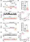
BKCa impairment in pial arteries from 5x‐FAD. (A and E) Representative traces of lumen diameter of pial arteries isolated from female (A) and male (E) WT (black, top) or 5x‐FAD (red, lower) showing constriction to BKCa blocker iberiotoxin (IbTox, 30 nM). Note blunted constriction in 5x‐FAD mice. Orange boxes: regions where data were obtained. (B and F) Summary bar graph showing blunted vasoconstriction to IbTox in female (B) and male (F) 5x‐FAD. Each data point in graph represents one pial artery from one mouse. Statistical test: unpaired two‐tailed Mann–Whitney test. (C and G) Representative traces of an inside‐out patch clamp from freshly isolated pial artery smooth muscle cells showing single‐channel BKCa currents at holding potential of +60 mV. Note the higher frequency of openings (upward deflections) in patch from female (C) WT (top trace, black) compared to 5x‐FAD (lower trace, red). Single‐channel BKCa openings were not different between male WT and 5x‐FAD (G). C: closed; 1: 1 channel; 2: 2 channels. (D and H) Summary graph showing BKCa open probability (Po) over a range of holding potentials (+20, +40, and +60 mV). Po was significantly lower in inside‐out patches from pial artery smooth muscle cells isolated from female 5x‐FAD mice (D), whereas BKCa Po was not different between male 5x‐FAD and WT littermates (H). Number of female replicates: WT n = 10 patches from five different mice; 5x‐FAD n = 7 patches from four different mice. Number of male replicates: WT n = 9 patches from five different mice; 5x‐FAD n = 9 patches from six different mice. All data are means ± SEM, two‐way ANOVA with a Šídák's post hoc test.
FIGURE 3
FIGURE 3
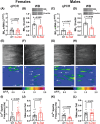
BKCa impairment in males is a consequence of lower BKα expression and reduced Ca2+ spark activity. (A and B) Expression levels of BKα mRNA (A) and protein (B) are not different between female 5x‐FAD and WT littermates. WB, Western blot. Each data point in graph is a pial artery lysate from one mouse. Statistical analysis: unpaired two‐tailed Student t test. (C and D) Expression levels of BKα mRNA are similar between male 5x‐FAD and WT littermates (C); however, BKα protein expression is significantly lower in male 5x‐FAD compared to WT littermates (D). Each data point in the graph is a pial artery lysate from one mouse. Statistical analysis: unpaired two‐tailed Student t test. (E–J) Representative grayscale (Ei, Fi) and pseudocolored (Eii, Fii) images of fields of view of pial arteries from female WT (E) and 5x‐FAD (F) prepared reverse en face for recording of Ca2+ sparks (arrows). Ca2+ sparks frequency (I) was significantly higher in pial arteries from female 5x‐FAD than in WT littermates, without differences in the number of active sites per cell (J). Each data point in the graph is one field of view from a pial artery, for a total of five fields of view per artery from three different mice of each genotype. Statistical analysis: unpaired two‐tailed Student t test. (G–L) Representative grayscale (Gi, Hi) and pseudocolored (Gii, Hii) images of fields of view of pial arteries from male WT (G) and 5x‐FAD (H) prepared reverse en face for recording of Ca2+ sparks (arrows). Ca2+ spark frequency (K) was significantly lower in pial arteries from male 5x‐FAD than in WT littermates, without differences in number of active sites per cell (L). Each data point in graph is one field of view from a pial artery, for a total of five fields of view per artery from three different mice of each genotype. Statistical analysis: unpaired two‐tailed Student t test. All data are means ± SEM.
FIGURE 4
FIGURE 4
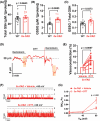
Oxidative‐dependent BKCa impairment in pial arteries from female 5x‐FAD. (A–C) Glutathione assay from brain lysates of female 5x‐FAD showing presence of oxidative environment observed as significant increase in oxidized glutathione (GSSG) (B) and ratio between GSSG and reduced glutathione (GSH) (C). Each data point represents a brain lysate from the cortex of an individual mouse. Statistical analysis: unpaired two‐tailed Student t test. (D and E) Incubation of pial arteries from isolated female 5x‐FAD with broad‐spectrum reducing agent 1,4‐dithiothreitol (DTT, 10 µM) significantly recovers sensitivity to iberiotoxin (30 nM), as observed by representative trace (D) and summary data (E). Orange boxes indicate regions where data were extracted. Each data point in the graph represents one pial artery from an individual mouse. Statistical analysis: paired two‐tailed Student t test. (F and G) Similarly, acute incubation with DTT partially recovers BKCa Po in freshly isolated pial artery smooth muscle cell from female 5x‐FAD, as observed in representative traces (F) and summary data (G). N = 16 patches from six different mice. Statistical analysis: matching mixed‐model ANOVA with a Šídák's post hoc correction for multiple comparisons. All data are means ± SEM.
FIGURE 5
FIGURE 5
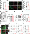
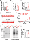
Exacerbated BKCa S‐NO in female 5x‐FAD. (A) Higher iNOS mRNA expression in lysates of pial arteries isolated from female 5x‐FAD than in WT littermates. Each data point represents one pial artery lysate from an individual mouse. Statistical analysis: unpaired two‐tailed Student t test. (B) Immunofluorescence labeling of iNOS (red) in pial arteries (collagen IV: Col IV, green). Note apparent higher fluorescence intensity in iNOS labeling in pial arteries from female 5x‐FAD compared to WT littermates. Bar = 20 µm. (C) Blinded, randomized, semi‐quantitative analysis of iNOS fluorescence intensity in pial arteries of female WT and 5x‐FAD. Each data point represents average fluorescence intensity of five pial arteries randomly imaged from an individual mouse. Statistical analysis: unpaired two‐tailed Student t test. (D and E) Quantification of global S‐NO proteins in cortical lysates of 5x‐FAD and WT littermates. Note the higher expression of S‐NO protein in lysates from 5x‐FAD cortex compared to WT in representative blot (D) and summary data (E). Each data point represents a lysate from an individual mouse. Statistical analysis: unpaired two‐tailed Mann–Whitney test. (F and G) Affinity purification (AP) of S‐NO proteins in brain lysates followed by Western blot against BKα shows a significantly higher expression of BKα S‐NO in female 5x‐FAD compared to WT littermates, as evidenced by representative blot (F) and summary data (G). Each data point represents a lysate from an individual mouse. Statistical analysis: unpaired two‐tailed Student t test. (H–K) Colocalization analysis using Mander M1 (I) and M2 (J) coefficients, as well as Pearson coefficient (K), shows a significantly higher colocalization between BKα (H, green) and global S‐NO proteins (H, red) in pial arteries from female 5x‐FAD (fluorogram in H). Bar = 20 µm. Violin plots represent distribution of colocalized pixels in each image. Imaging of pial arteries was performed in a blinded and randomized manner, a total of three arteries were imaged per mouse for a total of five mice per group. Statistical analysis: unpaired two‐tailed Student t test. Data in violin plots are median ± 95% confidence intervals; all other data are means ± SEM.
FIGURE 6
FIGURE 6
BKCa S‐NO in post mortem human brain samples. (A and B) Western blot (WB) quantification of BKα expression in post mortem brain lysates of patients without CAA/AD (non‐AD), CAA, or AD + CAA. Representative blots are shown in A and summary data in B. Each data point represents a brain lysate from an individual age‐matched woman (♀) or man (♂). Statistical analysis: Brown–Forsythe and Welch ANOVA with a Dunnett T3 correction for multiple comparisons. (C and D) Affinity purification (AP) of S‐NO proteins in brain lysates followed by Western blotting (WB) against the BKα subunit shows a trend toward an increase in BKα S‐NO in AD + CAA patients compared to non‐AD, as evidenced by the representative blots in C and summary data in D. Each data point represents a brain lysate from an individual age‐matched woman (♀) or man (♂). Statistical analysis: Brown–Forsythe and Welch ANOVA with a Dunnett T3 correction for multiple comparisons. All data are means ± SEM.
FIGURE 7
FIGURE 7
No evidence for nitro‐oxidative stress or oxidative BKCa PTM in male 5x‐FAD. (A–C) Total glutathione (GSH (A) and the ratio between GSSG/total GSH (C) in brain lysates were not significantly different between male 5x‐FAD and WT littermates, although there was a trend toward an increase in GSSG. Each data point represents one brain lysate from an individual mouse. Statistical analyses: unpaired two‐tailed Student t test. (D and E) Representative traces (D) and summary data (E) show that incubation of male excised, inside‐out membrane patches from pial artery smooth muscle cells with DTT (10 µM) does not change single‐channel BKCa Po in 5x‐FAD. Statistical analysis: matching mixed‐model two‐way ANOVA with a Šidák correction for multiple comparisons. N = 12 cells from five individual mice. (F–H) Representative blots of β‐actin (F) and global protein S‐NO (G) from brain lysates of female (♀) and male (♂) 5x‐FAD and WT littermates. The lanes for female lysates are the same as those in Figure 5 and are included here for comparison purposes. (H) Summary data show that global protein S‐NO was significantly lower in male 5x‐FAD compared to WT littermates. Each data point represents one lysate of the cortex of one mouse. Statistical analysis: unpaired two‐tailed Mann–Whitney t test. All data are means ± SEM.
FIGURE 8
FIGURE 8
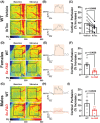
Impaired neurovascular coupling after acute BKCa inhibition and in 5x‐FAD. (A–C) Acute application of BKCa blocker iberiotoxin (IbTox, 30 nM) atop the thinned‐skull cranial window blunted neurovascular responses following somatosensory stimulation in WT mice, as evidenced by the pseudocolored perfusion maps (A), representative traces (B) of perfusion within the region of interest (black rectangle), and summary data (C). Each data point represents an individual mouse, with females squares (□) and males circles (○), following application of vehicle (□, ○) or IbTox (■, ●). Statistical analysis: paired two‐tailed Student t test. (D–I) Neurovascular coupling responses were significantly impaired in female (D–F) and showed a trend toward decrease in male (G–I) 5x‐FAD compared to WT littermates, as evidenced by the perfusion maps (D and G), representative perfusion traces (E and H), and summary data (F and I). Each data point represents an individual mouse. Statistical analysis: paired two‐tailed Student t test. PU, perfusion unit. All data are means ± SEM.

Update of

References

    1. 2023 Alzheimer's disease facts and figures. Alzheimers Dement. 2023;19(4):1598‐1695. - PubMed
    1. Collaborators GBDDF . Estimation of the global prevalence of dementia in 2019 and forecasted prevalence in 2050: an analysis for the Global Burden of Disease Study 2019. Lancet Public Health. 2022;7(2):e105‐e125. - PMC - PubMed
    1. Jellinger KA. Alzheimer disease and cerebrovascular pathology: an update. J Neural Transm (Vienna). 2002;109(5‐6):813‐836. - PubMed
    1. Mehndiratta P, Manjila S, Ostergard T, et al. Cerebral amyloid angiopathy‐associated intracerebral hemorrhage: pathology and management. Neurosurg Focus. 2012;32(4):E7. - PubMed
    1. Gireud‐Goss M, Mack AF, Mccullough LD, Urayama A. Cerebral Amyloid Angiopathy and Blood‐Brain Barrier Dysfunction. Neuroscientist. 2021;27(6):668‐684. - PMC - PubMed

MeSH terms

Substances