Review
. 2025 Jan;35(1):e23674.
doi: 10.1002/hipo.23674.

Unweaving the Cognitive Map: A Personal History

Affiliations
Review

Unweaving the Cognitive Map: A Personal History

Kate J Jeffery. Hippocampus. 2025 Jan.

Abstract

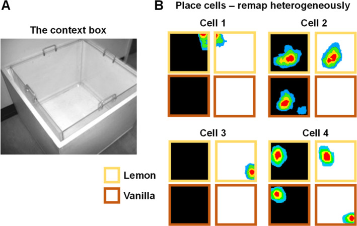
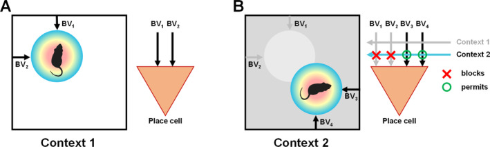
I have been incredibly fortunate to have worked in the field of hippocampal spatial coding during three of its most exciting decades, the 1990s, 2000s, and 2010s. During this time I had a ringside view of some of the foundational discoveries that were made which have transformed our understanding of the hippocampal system and its role in cognition (especially spatial cognition) and memory. These discoveries inspired me in my own lab over the years to pursue three broad lines of enquiry-3D spatial encoding, context and the sense of direction-which are outlined here. If some of my personal recollections are a little inaccurate (such is the nature of episodic memory!) I apologize in advance.

Keywords: configural learning; context; head direction cells; hippocampus; place cells; spatial cognition.


Conflict of interest statement

The author declares the following competing interests: K.J.J. is a non‐shareholding director of Axona Ltd., a company that makes and sells tetrode‐based electrophysiology products.

Figures

FIGURE 1
FIGURE 1
(A) May‐Britt and Edvard I. Moser presenting the recording system on which grid cells were first recorded to the Nobel Museum during the 2014 Nobel Laureates' get‐together on December 6, 2014. Copyright Nobel Media AB 2014. Photo: Niklas Elmehed. (B) I am pretty sure this is a grid cell.
FIGURE 2
FIGURE 2
My lab's first attempts at recording in 3D mazes. (A) The pegboard. The photo shows a rat from one of our behavioral experiments foraging over the area while standing on the pegs. The plot on the right is a spike plot from an entorhinal grid cell showing the path of the exploring rat over the course of a trial as a black line, and the spikes from the cell as red dots. Note that in the horizontal dimension the firing is interrupted, whereas in the vertical dimension it is continuous, resulting in vertical stripes. (B) The helical maze. The photo shows the assembled maze. On the right is a spike plot from a grid cell, shown from two viewpoints: overhead (top) and side‐on with the coils unwound and firing now expressed as a rate histogram (bottom). As with the pegboard, the firing seems interrupted when considered in the horizontal plane, but mostly uninterrupted in the vertical dimension (the apparent interruption is due to positional changes as the rat hugs the central pillars increasingly closely as it gets higher).
FIGURE 3
FIGURE 3
Grid cells in climbing rats. (A) The rats were raised in a large cage providing multiple climbing opportunities to ensure full 3D competence. (B) Spike plots (as in Figure 2) from three grid cells recorded on either a floor or a wall. On the floor the cells produced their characteristic regular array of firing fields (“blobs”), but on the wall they typically produced only one, or a few, fields with little evidence of a regular pattern.
FIGURE 4
FIGURE 4
The boundary vector model of Burgess and colleagues. (A) A place cell receives multiple inputs conveying information about the distance and direction of the nearby boundaries. At the point where the inputs converge, a place field (colored disk) forms. (B) If a familiar environment is stretched, pulling opposing boundary vectors apart, the field also stretches in that dimension. Note, however, that the stretching is incomplete—something else (self‐motion information) opposes the stretching.
FIGURE 5
FIGURE 5
Remapping of place cells in response to context changes. (A) The “context box” has an outer casing painted white (as in photo) or black, and an inner Plexiglas insert scented with lemon or vanilla food flavoring. (B) Four simultaneously recorded place cells tested in the four different odor‐color combinations in the experiment by Anderson et al. (2006), producing firing fields shown here as heat plots (red = max. firing). Note the heterogeneity, or “partial remapping.”
FIGURE 6
FIGURE 6
The boundary vector model of Burgess and colleagues, and our contextual gating adaptation of it. (A) The original model (see also Figure 4) proposed that a place cell receives boundary vector (BV) inputs that combine to specify a precise location of the firing field (colored disk) relative to the environment boundaries. (B) The contextual gating model addresses how the BVs could function in multiple contexts. It proposes that a cell has multiple sets of BV inputs, each specifying a field location, and these are gated (blocked or permitted) by convergent contextual inputs. Depending on which contextual inputs are active, the appropriate set of BVs will drive the cell and position its field appropriately for that context.
FIGURE 7
FIGURE 7
Contextual remapping in grid cells. (Left) Spike plots of two grid cells simultaneously recorded in the context box in the experiment by Marozzi et al. (2015). Note the very subtle shift of the grid, but no rotation, in response to the odor change. (Right) Heat plots of grid cell firing in the context boxes, painted black or white as shown in the figure, and scented lemon (yellow border) or vanilla (orange border). These cells, like the place cells in Figure 5, showed conditional remapping—the remapping induced by changing one color depended on the odor present, and vice versa.
FIGURE 8
FIGURE 8
Spatial cells in the 2‐box. (A) The 2‐box is made from two rectangular compartments connected by a central doorway, each possessing a cue‐card at one end. The boxes are visually identical so the visual scene alone cannot be used to determine which direction is which, of the two possibilities. However, the compartments are scented with lemon and vanilla so that the brain in principle has the information needed to distinguish them. (B) HD cells can use this information and produce tuning curves with the same orientation in both compartments. Bidirectional (BD) cells, by contrast, rotate their tuning curves 180° between the compartments, suggesting that they use environment layout but not odor to orient. (C) Spike plots of a place cell from the experiment by Cheng et al. shown as previously except that the paths are colored yellow and brown for lemon and vanilla compartments, respectively. Place cells remap between compartments (in this example, firing only in one compartment), suggesting sensitivity to the odor context. However, sometimes they invert the contexts, in a process we call “odor‐switching.”
FIGURE 9
FIGURE 9
Odor switching and a possible explanation for the pattern rotation. (A) Original place cell pattern in the lemon and (rotated) vanilla boxes. (B) What the BV model predicts if the HD signal disconnects from the odor context and switches directions (local rotation within each context). (C) What actually happens (global rotation across both compartments). (D) A possible model for how the three cue types (context, visual scene and direction) interact—blue denotes active signal and gray denotes inactive. (E) If the context signal drops out and the HD signal disconnects (reverses), the systems infers (reversed) the contexts based on the other two signals, and the whole pattern consequently reverses.

Similar articles

Cited by

References

    1. Anderson, M. I. , and Jeffery K. J.. 2003. “Heterogeneous Modulation of Place Cell Firing by Changes in Context.” Journal of Neuroscience 23, no. 26: 8827–8835. - PMC - PubMed
    1. Anderson, M. I. , Killing S., Morris C., et al. 2006. “Behavioral Correlates of the Distributed Coding of Spatial Context.” Hippocampus 16, no. 9: 730–742. - PubMed
    1. Barry, C. , Hayman R., Burgess N., and Jeffery K. J.. 2007. “Experience‐Dependent Rescaling of Entorhinal Grids.” Nature Neuroscience 10, no. 6: 682–684. - PubMed
    1. Barry, C. , Lever C., Hayman R., et al. 2006. “The Boundary Vector Cell Model of Place Cell Firing and Spatial Memory.” Reviews in the Neurosciences 17, no. 1–2: 71–97. - PMC - PubMed
    1. Bostock, E. , Muller R. U., and Kubie J. L.. 1991. “Experience‐Dependent Modifications of Hippocampal Place Cell Firing.” Hippocampus 1, no. 2: 193–205. - PubMed

LinkOut - more resources