. 2024 Dec 13;38(24):e70267.
doi: 10.1096/fj.202402499R.

Platelets as key cells in endometriosis patients: Insights from small extracellular vesicles in peritoneal fluid and endometriotic lesions analysis

Affiliations

Platelets as key cells in endometriosis patients: Insights from small extracellular vesicles in peritoneal fluid and endometriotic lesions analysis

Barbara Bortot et al. FASEB J. .

Abstract

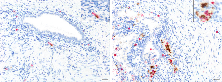
Endometriosis is a chronic inflammatory condition characterized by the presence of endometrium-like tissue outside the uterus, primarily affecting pelvic organs and tissues. In this study, we explored platelet activation in endometriosis. We utilized the STRING database to analyze the functional interactions among proteins previously identified in small extracellular vesicles (EVs) isolated from the peritoneal fluid of endometriosis patients and controls. The bioinformatic analysis indicated enriched signaling pathways related to platelet activation, hemostasis, and neutrophil degranulation. Double immunohistochemistry analysis for CD61 and MPO revealed a significant presence of neutrophils and platelets in close contact infiltrating endometriotic lesions, suggesting potential cell-cell interactions. Subsequently, we isolated small EVs from the peritoneal fluid of women diagnosed with endometriosis and from women without endometriosis who underwent surgery for non-inflammatory benign diseases. We performed single-particle phenotyping analysis based on platelet biomarkers GPIIb/IIIa and PF4 using nanoflow cytometry, as well as single-particle morphological and nanomechanical characterization through atomic force microscopy. The study demonstrated that patients with endometriosis had a notably higher proportion of particles testing positive for platelet biomarkers compared to the total number of EVs. This finding implies a potential role for platelets in the pathogenesis of endometriosis. Further research is necessary to delve into the mechanisms underlying this phenomenon and its implications for disease progression.

Keywords: GPIIb/IIIa; PF4; endometriosis; extracellular vesicles; follicular fluid; neutrophil; peritoneal fluid; platelet.


Figures

FIGURE 1
FIGURE 1
STRING protein–protein interaction (PPI) network analysis of proteins found in exosomes isolated from the peritoneal fluid during the proliferative phase in control patients (upper panel) and endometriosis patients (lower panel). The top pathways identified included the immune system, innate immune system, neutrophil degranulation, platelet degranulation, and formation of the cornified envelope. Overall, there was an increase in the observed gene count in the top pathways in the endometriosis samples. In this network, nodes represent proteins, edges indicate functional and physical protein associations, and the thickness of the lines reflects the strength of data support. Only interactions with a high confidence score of ≥0.7 are displayed. The functional pathways were obtained from the Reactome database, and the top 10 pathways are listed in ascending order based on their p‐value. FDR, false discovery rate.
FIGURE 2
FIGURE 2
Overview of the analytical workflow.
FIGURE 3
FIGURE 3
Nanoflow cytometry analysis (NanoFCM) of small EVs from peritoneal and follicular fluids in a cohort of patients. The data are reported as the means ± SDs of one experiment performed in triplicate. (A) Concentration before UC. The concentration of all particles >45 nm in diameter in precipitation‐isolated samples is reported. (B) Concentration after UC. The concentration of all particles >45 nm following ultracentrifugation (UC) is reported. (C) Size mean values. Particle size following UC is reported. (D) Cell Membrane staining positive. The percentage of all particles >45 nm, which also showed CellMask™ labeling (thus determined as EVs) is reported. Abbreviations: Endo, endometriosis sample; Con, control sample; FF, follicular fluid sample. *= p < 0.05.
FIGURE 4
FIGURE 4
Atomic force microscopy (AFM)‐based morphometry and nanomechanical screening. (A) Representative AFM micrographs of the three sample types. (B) Contact angle (CA) vs diameter plot of individual particles measured from at least 10 AFM micrographs deposited from duplicate preparations (Endo: N = 227; Con: N = 296; FF: N = 3632). The contact angle can be regarded as a measure of the mechanical stiffness of EVs; all particles above ~45 nm display the typical conserved average CA of intact vesicles, whereas smaller particles do not. (C) Normalized relative surface density of particles with diameters greater than 45 nm, showing that the FF samples are consistently more concentrated than the Endo and Con samples. (D) Box plot of the measured diameters of individual vesicles greater than 45 nm shows how FF samples contained the most significant proportion of larger EVs, while Endo samples contained the smallest. Con, control sample; Endo, endometriosis sample; FF, follicular fluid sample.
FIGURE 5
FIGURE 5
Percentage of particles positive for GPIIb/IIIa and PF4 relative to total EVs from peritoneal fluid in women with endometriosis (endometriosis), in women without endometriosis during surgery for a benign, non‐inflammatory, condition (controls), and in follicular fluid samples collected from infertile patients undergoing in vitro fertilization (follicular fluids). The samples were fluorescently labeled with FITC‐conjugated antibodies specific for GPIIb/IIIa and PF4. Additionally, the dye CellMask™ deep red plasma membrane stain (CM) was used to distinguish EVs from other small particles. **= p < 0.01
FIGURE 6
FIGURE 6
Microphotographs showing platelets in endometriotic lesions. Representative immunohistochemical staining of CD61. DAB (brown) chromogen was used to visualize the binding of the anti‐human CD61 antibody. Magnifications were 200× (up), and 400× (down); scale bar, 50 μm.
FIGURE 7
FIGURE 7
Representative microphotographs of double immunohistochemical staining for CD61 and MPO in endometriotic foci highlighting a close contact between platelet aggregates (CD61+) and neutrophils (MPO+) in the cytogenic stroma of endometriotic lesions. As shown in inset at the bottom of the pictures, this interaction occurs at tissue and intravascular sites. DAB (brown) chromogen was used to visualize the binding of the anti‐human CD61 antibody, while AEC (red) chromogen was used to visualize the binding of the anti‐human MPO antibody. The images were captured at magnifications of 200× (top) and 400× (bottom); scale bar, 50 μm.

Similar articles

Cited by

References

    1. Giudice LC, Kao LC. Endometriosis. Lancet. 2004;364:1789‐1799. doi:10.1016/S0140-6736(04)17403-5 - DOI - PubMed
    1. Zondervan KT, Becker CM, Missmer SA. Endometriosis. N Engl J Med. 2020;382:1244‐1256. doi:10.1056/NEJMra1810764 - DOI - PubMed
    1. Kennedy S, Bergqvist A, Chapron C, et al. ESHRE guideline for the diagnosis and treatment of endometriosis. Hum Reprod. 2005;20:2698‐2704. doi:10.1093/humrep/dei135 - DOI - PubMed
    1. Koninckx PR, Kennedy SH, Barlow DH. Endometriotic disease: the role of peritoneal fluid. Hum Reprod Update. 1998;4:741‐751. doi:10.1093/humupd/4.5.741 - DOI - PubMed
    1. Overton C, Fernandez‐Shaw S, Hicks B, Barlow D, Starkey P. Peritoneal fluid cytokines and the relationship with endometriosis and pain. Hum Reprod. 1996;11:380‐386. doi:10.1093/humrep/11.2.380 - DOI - PubMed