. 2025 Feb 5;16(2):e0338724.
doi: 10.1128/mbio.03387-24. Epub 2024 Dec 19.

An adenoviral vector encoding an inflammation-inducible antagonist, HMGB1 Box A, as a novel therapeutic approach to inflammatory diseases

Affiliations

An adenoviral vector encoding an inflammation-inducible antagonist, HMGB1 Box A, as a novel therapeutic approach to inflammatory diseases

Kari Ann Shirey et al. mBio. .

Abstract

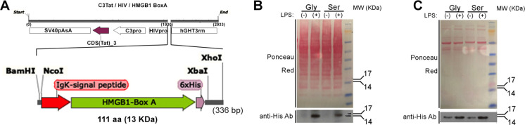
Influenza, as well as other respiratory viruses, can trigger local and systemic inflammation resulting in an overall "cytokine storm" that produces serious outcomes such as acute lung injury (ALI) or acute respiratory distress syndrome (ARDS). We hypothesized that gene therapy platforms could be useful in these cases if the production of an anti-inflammatory protein reflects the intensity and duration of the inflammatory condition. The recombinant protein would be produced and released only in the presence of the inciting stimulus, avoiding immunosuppression or other unwanted side effects that may occur when treating infectious diseases with anti-inflammatory drugs. To test this hypothesis, we developed AdV.C3-Tat/HIV-Box A, an inflammation-inducible cassette that remains innocuous in the absence of inflammation but releases HMGB1 Box A, an antagonist of high mobility group box 1 (HMGB1), in response to inflammatory stimuli such as lipopolysaccharide (LPS) or influenza virus infection. We report here that this novel inflammation-inducible HMGB1 Box A construct in a non-replicative adenovirus (AdV) vector mitigates lung and systemic inflammation therapeutically in response to influenza infection. We anticipate that this strategy will apply to the treatment of multiple diseases in which HMGB1-mediated signaling is a central driver of inflammation.IMPORTANCEMany inflammatory diseases are mediated by the action of a host-derived protein, HMGB1, on Toll-like receptor 4 (TLR4) to elicit an inflammatory response. We have engineered a non-replicative AdV vector that produces HMGB1 Box A, an antagonist of HMGB1-induced inflammation, under the control of an endogenous complement component C3 (C3) promoter sequence, that is inducible by LPS and influenza in vitro and ex vivo in macrophages (Mϕ) and protects mice and cotton rats therapeutically against infection with mouse-adapted and human non-adapted influenza strains, respectively, in vivo. We anticipate that this novel strategy will apply to the treatment of multiple infectious and non-infectious diseases in which HMGB1-mediated TLR4 signaling is a central driver of inflammation.

Keywords: Box A; HMGB1; LPS; MD-2; TLR4; adenovirus; cotton rats; influenza; mice.


Conflict of interest statement

The authors declare no conflict of interest.

Figures

Fig 1
Fig 1
(A) Structure of AdV.C3-Tat/HIV-Box A cloned under control of a two-component, inflammation-driven, AdV expression system (C3-Tat/HIV, top), our chimeric DNA construct encodes the HMGB1 Box A protein (green arrow) flanked by a secretory peptide (N-terminus, red arrow) and a C-terminal, 6x-His tag (purple arrow) (total 111 aa). (B) Detection of HMGB1 Box A produced in cell lysates of CR peritoneal Mϕ infected in vitro with AdV.C3-Tat/HIV-Box AGly or -Box ASer (MOI = 1) and then treated with LPS (10 ng/mL, 18 h). (C) Detection of secreted HMGB1 Box AGly or HMGB1 Box ASer by CR BAL Mϕ obtained 24 h after i.n. infection with 107 PFU of the indicated AdV.C3-Tat/HIV-Box A vector. BAL Mϕ were harvested 24 h after infection with AdV vectors, then cultured and treated with LPS for 24 h, followed by the analysis of culture supernatants by WB.
Fig 2
Fig 2
(A) CR peritoneal Mϕ were treated with AdV.C3-Tat/HIV-Luc for 24 h and half the samples were exposed to LPS (10 ng/mL) for an additional 24 h. Luciferase activity was measured in cell lysates (n = 4, Student’s t test, ****P < 0.0001). (B) Mice were treated with AdV.C3-Tat/HIV-Luc i.n. and challenged with saline or LPS i.t. (10 µg/mouse); mice were sacrificed at 18 h post-challenge, and lung luciferase levels were measured; P < 0.05, Student’s t test. (C) CR were treated i.n. with AdV.C3-Tat/HIV-Luc (105 PFU/CR) and challenged i.n. with PBS or influenza A(H3N2) virus (107 TCID50/animal). CR were sacrificed at the indicated times p.i. with A(H3N2) (105 TCID50/CR) and luciferase expression measured in lung homogenates. (n = 3–5/group; ANOVA, ***P < 0.0001; **P < 0.01). (D) CR peritoneal Mϕ were exposed to LPS (10 ng/mL) for the indicated times. Expression of the endogenous cotton rat C3 gene was determined by quantitative real-time (qRT)-PCR. (E) CR peritoneal Mϕ were transduced with AdV.C3-Tat/HIV-Box AGly or AdV.C3-Tat/HIV-Box ASer using an MOI = 1. Twenty-four hours post-transduction, cells were exposed to medium only (black symbols), LPS (Gly—red symbols; Ser—blue symbols) for the indicated time periods. Expression of the vector-based Box A mRNA expression was measured by qRT-PCR to show the kinetics of Adv.C3-Tat/HIV/Box A expression of the C3 mRNA post-LPS treatment.
Fig 3
Fig 3
CR were treated i.n. with PBS (A; 100 µL), or with AdV.C3-Tat/HIV-Luc (B and C; 107 PFU/CR in 100 µL) and sacrificed on days 1 (A, B) or day 3 p.i. (C), showing undetectable lung inflammation in CR treated with AdV.C3-Tat/HIV-Luc in contrast to CR infected with A(H3N2) (107 TCID50/CR) and harvested on day 3 p.i. (D). Magnification, 100×. Insets show details of bronchi, 200×.
Fig 4
Fig 4
(A) C57BL/6J mice were infected on day 0 with PR8 (LD90). Twenty-four hours later, mice were treated with saline (i.m.), AdV.C3-Tat/HIV-Luc (i.v.), or an equal mixture of AdV.C3-Tat/HIV-Box A variants (2 × 10⁷ PFU/mouse) administered either i.m or i.v. Data are from two separate experiments with 5 mice/treatment/experiment. Results from AdV-Luc-treated mice were obtained from 5 mice in a single experiment. (B) Mice were infected as in panel A. Twenty-four hours later, mice were treated i.v. with AdV.C3-Tat/HIV-Luc (2 × 10⁷ PFU/mouse), an equal mixture of AdV.C3-Tat/HIV-Box A variants, or the individual AdV.C3-Tat/HIV-Box A variants (Gly or Ser) (2 × 10⁷ PFU/mouse). Data are from two separate experiments with 5 mice/treatment/experiment. (C). Mice were infected as in panel A. Mice were treated i.v. with AdV.C3-Tat/HIV-Luc (2 × 10⁸ PFU/mouse), an equal mixture of AdV.C3-Tat/HIV-Box A variants, or the individual AdV.C3-Tat/HIV-Box A variants (Gly or Ser) (2 × 10⁸ PFU/mouse). Data are from two separate experiments with 5 mice/treatment/experiment. (D) Mice were infected as in panel A. Mice were treated i.v. with AdV.C3-Tat/HIV-Luc (2 × 10⁷ PFU/mouse) or an equal mixture of AdV.C3-Tat/HIV-Box A variants (2 × 10⁷ PFU/mouse) on day 1, day 3, or day 5 post-infection. N = 5 mice/treatment group. (E) Mice were infected as in panel A. Mice were treated i.v. with AdV.C3-Tat/HIV-Luc (2 × 10⁸ PFU/mouse) or an equal mixture of AdV.C3-Tat/HIV-Box A variants (2 × 10⁸ PFU/mouse) at day 1, day 3, or day 5 p.i. N = 5 mice/treatment group.
Fig 5
Fig 5
C57BL/6J mice were infected on day 0 with PR8 (LD90). Twenty-four hours later, mice were treated i.v. with saline (mock), AdV.C3-Tat/HIV-Luc, or an equal mixture of AdV.C3-Tat/HIV-Box A variants (2 × 10⁷ PFU). (A) Representative pathology of lung sections derived from mice treated therapeutically with AdV.C3-Tat/HIV-Luc or AdV.C3-Tat/HIV-Box A 24 h after infection with PR8 (LD90) at 5 days p.i. (B) Quantification of lung histopathology scores for the individual mice including a combined histology score. (C) Quantification of lung cytokine mRNA levels by qRT-PCR for the individual mice. (B and C) Each symbol represents one mouse.
Fig 6
Fig 6
CR were infected i.n. on day 0 with A(H3N2) (107 TCID50/CR). On day 1 p.i., CR were treated by i.v. injection with 107 PFU/CR of AdV.C3-Tat/HIV-Luc, AdV.C3-Tat/HIV-Box AGly or AdV.C3-Tat/HIV-Box ASer. CR were then sacrificed on 3 and 6 days p.i. to analyze lung cytokine mRNA expression and lung histopathology. (A) mRNA expression of TNFα and IL-10 in lung samples of CR sacrificed on day 3 p.i. (n = 4/treatment). (B) Score of alveolitis at 3 days p.i. for the different groups of CR (n = 4/treatment). (C) Representative microscopic images of lungs of CR treated with AdV.C3-Tat/HIV-Luc (a), AdV.C3-Tat/HIV-Box AGly (b), or AdV.C3-Tat/HIV-Box ASer (c) showing reduced numbers of cells in the alveolar spaces of CR treated with AdV.C3-Tat/HIV-Box A vectors. Magnification, 200×. (D) mRNA expression of TNFα, IL-1β, and IL-10 in lungs of CR harvested at day 6 p.i. (n = 5/treatment) (E) Pathology scores for lungs of individual CR for peribronchioliis, perivasculitis, interstitial pneumonia, and alveolitis (n = 5/treatment). (F) Representative microscopic images of lungs of CR treated with AdV.C3-Tat/HIV-Luc (a), AdV.C3-Tat/HIV-Box AGly (b), or AdV.C3-Tat/HIV-Box ASer (c) showing reduced peribronchiolitis in animals treated with AdV.C3-Tat/HIV-Box A vectors. Magnification, 40×. n = 4–5/group; Student t test, **P < 0.01; **P < 0.05.
Fig 7
Fig 7
Model. Intact HMGB1 contains two domains, HMGB1 Box A and HMGB1 Box B. The HMGB1 Box A domain binds to TLR4, while the HMGB1 Box B domain binds MD-2 to elicit TLR4 signaling (top image). In response to an inflammatory stimulus, the Adv.C3-Tat/HIV-Box A vector produces rBox A that competitively inhibits the binding of the intact protein HMGB1’s Box A domain to TLR4 and precludes its Box B from binding to MD-2 (bottom image).This results in disruption of TLR4-mediated signaling (modified from reference 34).

Similar articles

References

    1. Morens DM, Taubenberger JK, Fauci AS. 2008. Predominant role of bacterial pneumonia as a cause of death in pandemic influenza: implications for pandemic influenza preparedness. J Infect Dis 198:962–970. doi:10.1086/591708 - DOI - PMC - PubMed
    1. Chertow DS, Memoli MJ. 2013. Bacterial coinfection in influenza: a grand rounds review. JAMA 309:275–282. doi:10.1001/jama.2012.194139 - DOI - PubMed
    1. Rynda-Apple A, Robinson KM, Alcorn JF. 2015. Influenza and bacterial superinfection: illuminating the immunologic mechanisms of disease. Infect Immun 83:3764–3770. doi:10.1128/IAI.00298-15 - DOI - PMC - PubMed
    1. Erbelding EJ, Post DJ, Stemmy EJ, Roberts PC, Augustine AD, Ferguson S, Paules CI, Graham BS, Fauci AS. 2018. A universal influenza vaccine: the strategic plan for the National Institute of Allergy and Infectious Diseases. J Infect Dis 218:347–354. doi:10.1093/infdis/jiy103 - DOI - PMC - PubMed
    1. Rajão DS, Pérez DR. 2018. Universal vaccines and vaccine platforms to protect against influenza viruses in humans and agriculture. Front Microbiol 9:123. doi:10.3389/fmicb.2018.00123 - DOI - PMC - PubMed

LinkOut - more resources