. 2025 Jan 28;10(1):e0072124.
doi: 10.1128/msphere.00721-24. Epub 2024 Dec 19.

Development of monoclonal antibodies against P. gingivalis Mfa1 and their protective capacity in an experimental periodontitis model

Affiliations

Development of monoclonal antibodies against P. gingivalis Mfa1 and their protective capacity in an experimental periodontitis model

Mingya Cao et al. mSphere. .

Abstract

Porphyromonas gingivalis (P. gingivalis), a gram-negative, black-pigmented anaerobe, is a major etiological agent and a leading cause of periodontitis. Fimbriae protein Mfa1 is a key virulence factor of P. gingivalis and plays a crucial role in bacterial adhesion, colonization, biofilm formation, and persistent inflammation, making it a promising therapeutic target. However, the role of anti-Mfa1 antibodies and the underlying protective mechanisms remain largely unexplored. Here, we developed and characterized the monoclonal antibodies (mAbs) targeting the Mfa1 protein of P. gingivalis. Function analysis showed that anti-Mfa1 mAbs mediated bacterial agglutination and inhibited P. gingivalis adhesion to saliva-coated hydroxyapatite and host cells. Notably, anti-Mfa1 mAbs significantly reduced bacterial burden and alveolar bone loss in a P. gingivalis-induced experimental periodontitis model. These results show that anti-Mfa1 mAbs can be beneficial in alleviating P. gingivalis infections, and provide important insights for the development of adequate adjuvant treatment regimens for Mfa1-targeted therapeutics.

Importance: Fimbriae (pili) play an important role in bacterial adhesion, invasion of host cells and tissues, and formation of biofilms. Studies have shown that two types of fimbriae of Porphyromonas gingivalis, FimA and Mfa1, are important for colonization and infection through their binding to host tissues and other bacteria. While anti-FimA antibodies have been shown to improve periodontitis, the effect of anti-Mfa1 antibodies on P. gingivalis infection and periodontitis was previously unknown. In this study, we report for the first time that anti-Mfa1 monoclonal antibodies can reduce P. gingivalis infection and improve periodontitis. These findings suggest that Mfa1 represents a promising therapeutic target, and the development of anti-Mfa1 mAbs holds a potential as essential diagnostic and adjunctive therapeutic tools for managing P. gingivalis-related diseases.

Keywords: Mfa1; P. gingivalis; fimbriae; monoclonal antibodies; periodontitis.


Conflict of interest statement

The authors declare no conflict of interest.

Figures

Fig 1
Fig 1
Expression of Mfa1 protein and generation of mAbs against P. gingivalis Mfa1. (a and b) Expression and purification of recombinant Mfa1 protein. SDS-PAGE analysis of the pET28a-Mfa1 in E. coli BL21(DE3) cells. Lane M, protein molecular marker; lane 1, total protein supernatant before induction; lane 2, supernatant sample before purification; lane 3, flowthrough sample after purification; lane 4–8, purified Mfa1 was eluted with 20, 50, 70, 100, and 200 mM imidazole in equilibration buffer (pH 8.0). (c) Schematic view of immunization, hybridoma generation, and screening for obtention of anti-Mfa1 mAbs. (d) Determination of serum antibody titers by ELISA. Serum antibodies were detected using recombinant protein Mfa1-coated ELISA plates, and serum antibody titers from all three immunized mice reached 1:200,000. (e) More than 100 hybridoma clone culture supernatants were collected and screened by ELISA to finally obtain four different hybridoma cell lines capable of secreting Mfa1 monoclonal antibodies. (f) SDS-PAGE analysis of four anti-Mfa1 mAbs purified from mouse ascites via protein G affinity purification.
Fig 2
Fig 2
Analysis of mAbs binding to Mfa1 by ELISA and BLI. (a) Binding as reflected by absorbance at 450 is shown on the y axis for the mAbs concentrations shown on the x axis for each mAb. Results are representative of three independent experiments. The numerical EC50 for each mAb is indicated at the right of the panel depicting the binding curves of all mAbs. (b–e) Affinity-binding curves of anti-mAbs were measured by BLI. BLI analysis was used on the Octet R8 system to determine the association and dissociation rates by immobilizing the monoclonal antibody on an AMC-coated optical sensor. The AMC sensors were incubated with purified monoclonal antibody at set intervals to allow binding. The sensor is then moved into an antibody-free solution and allowed to dissociate at intervals of time. Curve fitting using a 1:1 interaction model allows the measurement of the affinity constant (Kd) of each nanobody. (f) Curve fitting using a 1:1 interaction model allows for the affinity constant (Kd) to be measured for each anti-Mfa1 mAbs.
Fig 3
Fig 3
Specificity and cross-reactivity analysis of anti-Mfa1 mAbs. (a) Validation of monoclonal antibodies by Western blot analysis. Total cell lysates (20 µg per lane) prepared from P. gingivalis and other bacteria were analyzed by Western blot using anti-Mfa1 mAbs; the recombinant Mfa1 protein (2 µg) was as a positive control. (b) Detection of cross-reactivity of anti-Mfa1 monoclonal antibodies by ELISA. (c) Visualization of mAbs binding to P. gingivalis cells. Nuclei DNA was labeled with Hochest, while mAb-coated bacteria were stained with 546-conjugated anti-mouse IgG antibody (scale bar: 2 µm). (d) Flow cytometric analysis of binding to P. gingivalis using anti-Mfa1 mAbs and non-specific IgG (blue). P. gingivalis was incubated with anti-Mfa1 mAbs, reacted with 488-conjugated anti-mouse IgG, and detected by flow cytometry.
Fig 4
Fig 4
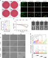
Anti-Mfa1 mAbs promote P. gingivalis agglutination. (a) Images of colony-forming assay of P. gingivalis after treatment with anti-Mfa1 mAbs and non-specific antibody. (b) Proportion of P. gingivalis inhibited after 2 h of incubation with anti-Mfa1 mAbs and non-specific IgG. (c) Images obtained from fluorescence microscopy of P. gingivalis after incubation with different anti-Mfa1 mAbs and non-specific IgG and briefly vortexing. (d) In a UV-clear cuvette, P. gingivalis was incubated with anti-Mfa1 mAb (100 µg/mL), and the absorbance at OD600 nm was continuously measured for 2.5 hours. (e) After incubation for 2.5 h, the cuvettes were photographed for visual inspection of turbidity. (f) A total of 2 × 10⁸ CFU of P. gingivalis were incubated with different purified anti-Mfa1 mAbs in PBS for 1 h. After gently vortexing, 10 µL of the mixture was dropped onto a glass slide, observed, and photographed under a microscope. Images are representative of the three independent experiments performed in duplicate for each mAb (scale bar: 20 µm). (g) More than 20 random area images were analyzed with a custom macro in ImageJ for the number of aggregates per image and the counts of bacteria in aggregates. Mean and standard error of the mean (s.e.m.) were calculated from the results of at least three independent experiments. Significant differences compared to the controls were determined using one-way ANOVA (*P < 0.05, **P < 0.01).
Fig 5
Fig 5
Anti-Mfa1 mAbs reduced the attachment of P. gingivalis to sHA. (a) The effect of anti-Mfa1 mAbs on P. gingivalis binding to sHA microspheres was observed by scanning electron microscopy. The image is representative of the triplicate experiment. (b) qPCR assay was used to evaluate the inhibitory effect of anti-Mfa1 mAbs on P. gingivalis binding sHA beads. (c) Representative pictures of biofilm formed in the presence of indicated mAbs after crystal violet staining. An anti-FimA monoclonal antibody that can increase biofilm is used as a positive control. (d) Biofilm biomass was assessed by absorbance at 600  nm. Mean and s.e.m. were calculated from the results of at least three independent experiments. (e) SEM micrographs of P. gingivalis exposed to the respective antibodies, including non-specific IgG, a FimA mAb as a positive control, and different anti-Mfa1 mAbs, for 48 h. Significant differences compared to the control IgG were determined using one-way ANOVA (*P < 0.05, **P < 0.01).
Fig 6
Fig 6
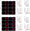
Anti-Mfa1 mAbs reduce the adhesion of P. gingivalis to host cells. (a) Immunofluorescence microscopy of the inhibitory effect of anti-Mfa1 mAbs on P. gingivalis binding hGFs. (b) Quantification of P. gingivalis adhered to hGFs after treatment with anti-Mfa1 mAbs by qPCR. (c) Antibiotic protection assay. Bars indicate the ratio of intracellular P. gingivalis recovered from hGF cells in different anti-Mfa1 mAbs treatment groups, as compared to the IgG control group. (d and e) Pro-inflammatory cytokines interleukin-1β (IL-1β) and interleukin-6 (IL-6) in hGFs infected with P. gingivalis (MOI = 10) for 24 h. (f) Immunofluorescence microscopy of the inhibitory effect of anti-Mfa1 mAbs on P. gingivalis binding hGFs. (g) Quantification of P. gingivalis adhered to CAL27 cells after treatment with anti-Mfa1 mAbs by qPCR. (h) Bars indicate the ratio of intracellular P. gingivalis recovered from hGFs cells in different anti-Mfa1 mAbs treatment groups, as compared to the IgG control group. (i and j) Relative mRNA expression of IL-1β and IL-6 in CAL27 infected with P. gingivalis (MOI = 10), treated by anti-Mfa1 mAbs for 24 h. Mean and s.e.m. were calculated from results of at least three independent experiments. *P < 0.05, **P < 0.01 as determined by one-way ANOVA.
Fig 7
Fig 7
Anti-Mfa1 mAbs reduce P. gingivalis infection and improve experimental periodontitis symptoms. (a) Schematic workflow timeline of rat periodontitis model. (b) Quantification of P. gingivalis in rat oral cavity by qPCR. (c) Representative images of alveolar bone micro-CT analysis by different treatment groups. (d) Samples of the maxilla were collected and stained with methylene blue aqueous solution (1%) to distinguish bone from teeth. (e) Rat tissue sections were stained with H&E. (f) The average distance from the ABC to the CEJ on the buccal sides and the palatal sides of the maxillary second molar was measured. (g) Representative H&E staining images showing the alveolar bone resorption and periodontal inflammation of the periodontium from each group. Data are shown as means  ±  s.e.m. (n = 6); *P < 0.05, **P < 0.01 as determined by a one-way ANOVA test.

Similar articles

Cited by

References

    1. Lamont RJ, Koo H, Hajishengallis G. 2018. The oral microbiota: dynamic communities and host interactions. Nat Rev Microbiol 16:745–759. doi:10.1038/s41579-018-0089-x - DOI - PMC - PubMed
    1. Baima G, Minoli M, Michaud DS, Aimetti M, Sanz M, Loos BG, Romandini M. 2024. Periodontitis and risk of cancer: mechanistic evidence. Periodontol 2000 96:83–94. doi:10.1111/prd.12540 - DOI - PMC - PubMed
    1. Dominy SS, Lynch C, Ermini F, Benedyk M, Marczyk A, Konradi A, Nguyen M, Haditsch U, Raha D, Griffin C, et al. . 2019. Porphyromonas gingivalis in Alzheimer’s disease brains: evidence for disease causation and treatment with small-molecule inhibitors. Sci Adv 5:eaau3333. doi:10.1126/sciadv.aau3333 - DOI - PMC - PubMed
    1. Chen WA, Dou Y, Fletcher HM, Boskovic DS. 2023. Local and systemic effects of Porphyromonas gingivalis infection. Microorganisms 11:470. doi:10.3390/microorganisms11020470 - DOI - PMC - PubMed
    1. Xu W, Zhou W, Wang H, Liang S. 2020. Roles of Porphyromonas gingivalis and its virulence factors in periodontitis. Adv Protein Chem Struct Biol 120:45–84. doi:10.1016/bs.apcsb.2019.12.001 - DOI - PMC - PubMed

LinkOut - more resources