Observational Study
. 2024 Dec;6(6):e230428.
doi: 10.1148/ryct.230428.

Prevalence of Mitral Annular Disjunction at Cardiac MRI: Results from a Multicenter Registry

Affiliations
Observational Study

Prevalence of Mitral Annular Disjunction at Cardiac MRI: Results from a Multicenter Registry

Anna Palmisano et al. Radiol Cardiothorac Imaging. 2024 Dec.

Abstract

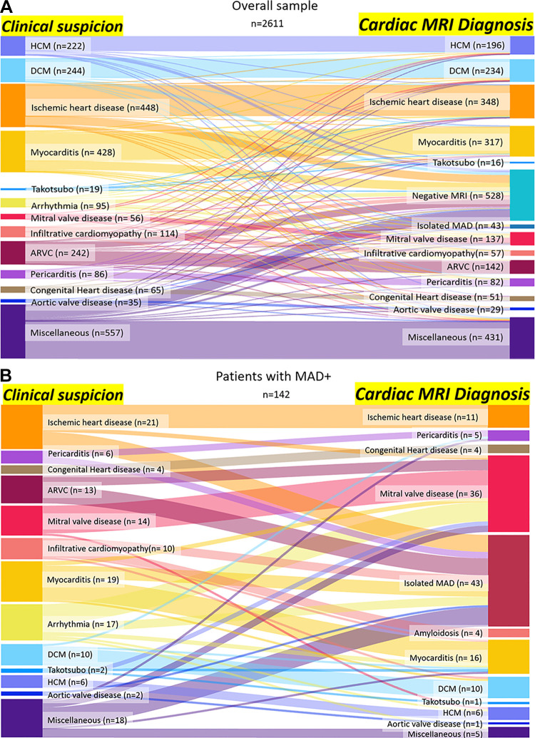
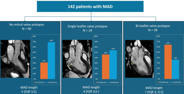
Purpose To determine the prevalence of mitral annular disjunction (MAD) in patients undergoing cardiac MRI for various clinical indications and to assess the association of MAD with arrhythmia, mitral valve prolapse (MVP), and myocardial alteration. Materials and Methods This study analyzed data from a retrospective observational registry of consecutive patients undergoing cardiac MRI for different clinical indications. Cardiac MRI examinations were performed from January 2019 to June 2019 at 13 Italian hospitals. Images underwent double reading by expert cardiac radiologists from the enrolling center and the core laboratory to assess the presence of MAD. Presence and maximum length of MAD and its association to MVP pattern, functional and structural myocardial alteration, and arrhythmia were evaluated using nonparametric and parametric tests. Logistic regression models were used to identify predictors of arrhythmia. Results Cardiac MRI studies from 2611 consecutive patients (1730, 66% male; median age, 53 years; IQR, 39-65 years) were evaluated. Prevalence of MAD was 5.44% (142 of 2611). MAD was an incidental finding in 74.6% (106 of 142) of patients. Patients with MAD had a higher prevalence of arrhythmias compared with patients without MAD (40% [57 of 142] vs 18% [444 of 2469]; P < .001). Patients with MAD and bileaflet MVP showed a longer MAD compared with patients with single-leaflet or absent MVP (median, 7 mm [IQR, 3-9.5 mm] vs 4 mm [IQR, 3-5 mm]; P < .001), a higher prevalence of systolic curling (75% [21 of 28] vs 30.7% [35 of 114]; P < .001), higher extracellular volume values (30% [IQR, 28%-32%] vs 27% [IQR, 25%-30%]; P = .04), and a higher prevalence of arrhythmia (64.2% [18 of 28] vs 34.2% [39 of 114]; P = .006). MAD length of at least 5 mm was an independent predictor of arrhythmia (odds ratio 3.96; 95% CI: 1.93, 8.15; P < .001). Conclusion MAD was a frequent incidental finding on cardiac MRI scans from a multicenter registry. MAD length of at least 5 mm and coexisting bileaflet MVP showed a higher risk of arrhythmia. Keywords: MR Imaging, Cardiac, Mitral Annular Disjunction Supplemental material is available for this article. ©RSNA, 2024.

Keywords: Cardiac; MR Imaging; Mitral Annular Disjunction.


Conflict of interest statement

Disclosures of conflicts of interest: A. Palmisano No relevant relationships. E.B. No relevant relationships. G.D.A. No relevant relationships. C.D.G. No relevant relationships. S.B. No relevant relationships. M.A. No relevant relationships. D.P. No relevant relationships. P.R. No relevant relationships. N.d.M. No relevant relationships. M.R. No relevant relationships. D.F. Consulting fees from Siemens Healthineers and support for attending meetings and/or travel from GE. A.R. No relevant relationships. S.P. No relevant relationships. V.M. No relevant relationships. L.M. No relevant relationships. G.C. No relevant relationships. N.G. No relevant relationships. D.T. No relevant relationships. M.G. No relevant relationships. R.F. No relevant relationships. P.P. No relevant relationships. E.D.C. No relevant relationships. T.D. No relevant relationships. L.R.M.L. No relevant relationships. A.B. No relevant relationships. S.D. No relevant relationships. A. Ponsiglione No relevant relationships. R.A. No relevant relationships. M.I. Member of Radiology editorial board. M.P. No relevant relationships. R.C. No relevant relationships. L.S. No relevant relationships. G.F. No relevant relationships. C.L. No relevant relationships. V.S. No relevant relationships. S.S. No relevant relationships. A.S. No relevant relationships. A.C. No relevant relationships. M.C. No relevant relationships. L.L. Payment or honoraria for lectures, presentations, speakers bureaus, manuscript writing or educational events from IeC and support for attending meetings and/or travel from Bracco. D.V. Support for attending meetings from Siemens and Philips. L.F. No relevant relationships. E.N. Payment or honoraria for lectures, presentations, speakers bureaus, manuscript writing or educational events from GE HealthCare and Bracco. A.E. No relevant relationships.

Figures

None
Graphical abstract
Flow diagram shows patient selection in this study. Positive and
negative symbols indicate if the patient has (+) or does not have (−)
MAD and/or MVP. ICD = implantable cardioverter device, LGE = late gadolinium
enhancement, MAD = mitral annular disjunction, MV = mitral valve, MVP =
mitral valve prolapse, PM = pacemaker.
Figure 1:
Flow diagram shows patient selection in this study. Positive and negative symbols indicate if the patient has (+) or does not have (−) MAD and/or MVP. ICD = implantable cardioverter device, LGE = late gadolinium enhancement, MAD = mitral annular disjunction, MV = mitral valve, MVP = mitral valve prolapse, PM = pacemaker.
Diagrams show clinical indications for cardiac MRI and diagnostic
changes after imaging in the overall study sample (A) and in patients with
evidence of MAD (B). ARVC = arrhythmogenic right ventricular cardiomyopathy,
DCM = dilated cardiomyopathy, HCM = hypertrophic cardiomyopathy, MAD =
mitral annular disjunction.
Figure 2:
Diagrams show clinical indications for cardiac MRI and diagnostic changes after imaging in the overall study sample (A) and in patients with evidence of MAD (B). ARVC = arrhythmogenic right ventricular cardiomyopathy, DCM = dilated cardiomyopathy, HCM = hypertrophic cardiomyopathy, MAD = mitral annular disjunction.
Bar graphs show the prevalence of arrhythmias in patients with MAD
according to the presence and pattern of MVP. MR images with the MVP are
marked in yellow, and the median length and IQR are shown for each type. MAD
= mitral annular disjunction, MVP = mitral valve prolapse.
Figure 3:
Bar graphs show the prevalence of arrhythmias in patients with MAD according to the presence and pattern of MVP. MR images with the MVP are marked in yellow, and the median length and IQR are shown for each type. MAD = mitral annular disjunction, MVP = mitral valve prolapse.
Bar graphs and corresponding MR images show the main cardiac MRI
findings in patients with MAD according to the presence and pattern of MVP.
bMVP = bileaflet mitral valve prolapse, ECV = extracellular volume, LAESVi =
indexed left atrium end systolic volume, LGE = late gadolinium enhancement,
LVEDVi = indexed left ventricle end diastolic volume, MAD = mitral annular
disjunction, MR = mitral regurgitation, MVP = mitral valve prolapse, sMVP =
single-leaflet mitral valve prolapse.
Figure 4:
Bar graphs and corresponding MR images show the main cardiac MRI findings in patients with MAD according to the presence and pattern of MVP. bMVP = bileaflet mitral valve prolapse, ECV = extracellular volume, LAESVi = indexed left atrium end systolic volume, LGE = late gadolinium enhancement, LVEDVi = indexed left ventricle end diastolic volume, MAD = mitral annular disjunction, MR = mitral regurgitation, MVP = mitral valve prolapse, sMVP = single-leaflet mitral valve prolapse.

References

    1. Esposito A, Gatti M, Trivieri MG, et al. . Imaging for the assessment of the arrhythmogenic potential of mitral valve prolapse . Eur Radiol 2024. ; 34 ( 7 ): 4243 – 4260 . - PMC - PubMed
    1. Gulati A, Gulati V, Hu R, et al. . Mitral Annular Disjunction: Review of an Increasingly Recognized Mitral Valve Entity . Radiol Cardiothorac Imaging 2023. ; 5 ( 6 ): e230131 . - PMC - PubMed
    1. Bennett S , Tafuro J , Duckett S , et al. . Definition, prevalence, and clinical significance of mitral annular disjunction in different patient cohorts: A systematic review . Echocardiography 2022. ; 39 ( 3 ): 514 – 523 . - PubMed
    1. Faletra FF, Leo LA, Paiocchi VL, et al. . Morphology of Mitral Annular Disjunction in Mitral Valve Prolapse . J Am Soc Echocardiogr 2022. ; 35 ( 2 ): 176 – 186 . - PubMed
    1. Dejgaard LA, Skjølsvik ET, Lie ØH, et al. . The Mitral Annulus Disjunction Arrhythmic Syndrome . J Am Coll Cardiol 2018. ; 72 ( 14 ): 1600 – 1609 . - PubMed

Publication types

LinkOut - more resources