. 2025 Jan 28;44(1):115100.
doi: 10.1016/j.celrep.2024.115100. Epub 2024 Dec 18.

Pharmacological blocking of spinal GABAA receptors in monkeys reduces sensory transmission to the spinal cord, thalamus, and cortex

Affiliations

Pharmacological blocking of spinal GABAA receptors in monkeys reduces sensory transmission to the spinal cord, thalamus, and cortex

Amr A Mahrous et al. Cell Rep. .

Abstract

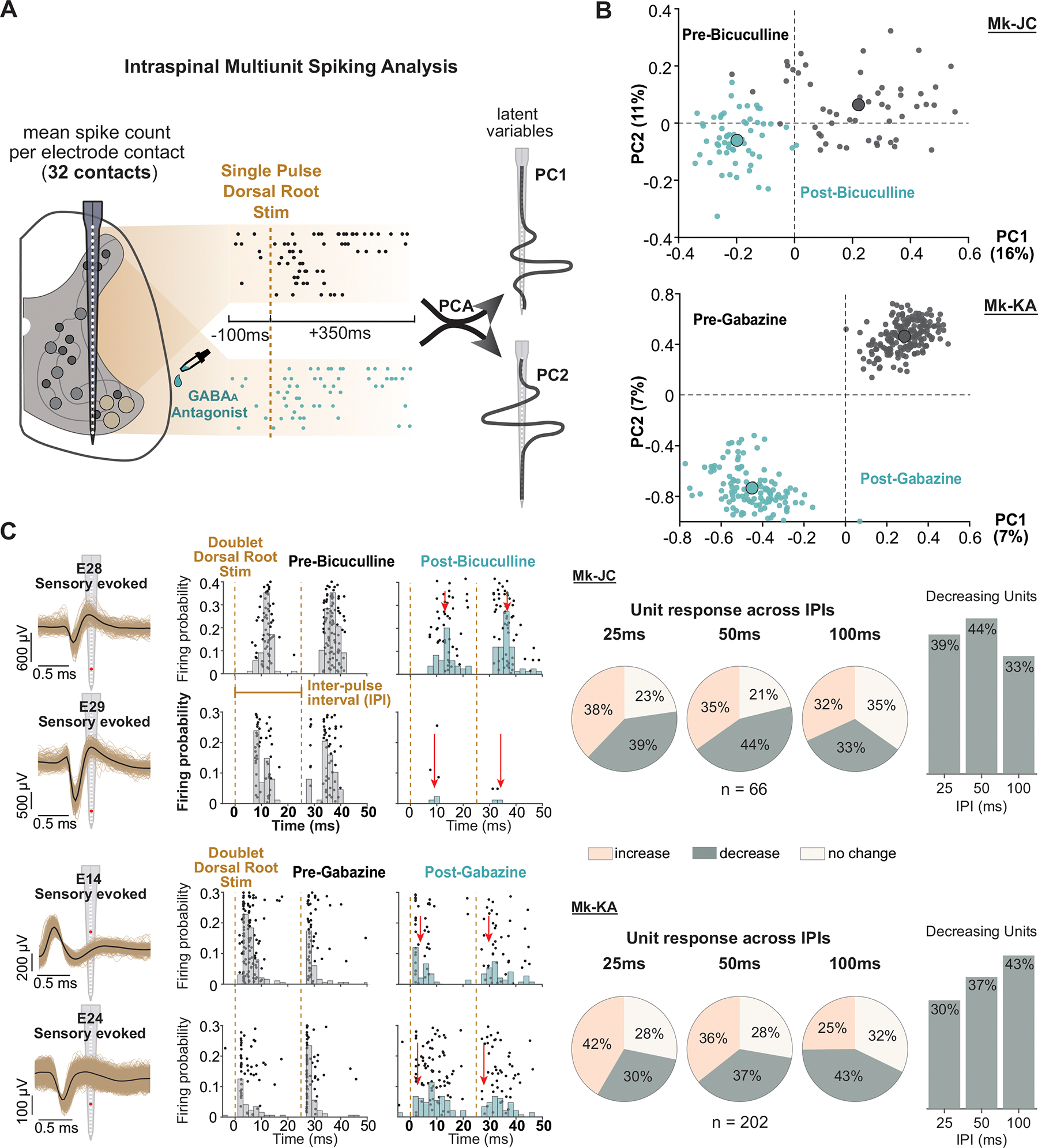
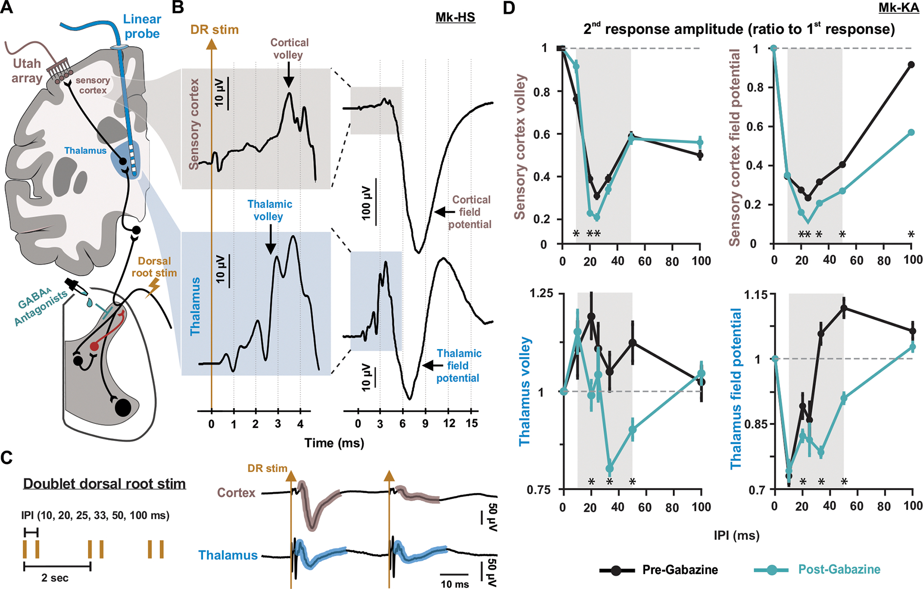
A century of research established that GABA inhibits proprioceptive inputs presynaptically to sculpt spinal neural inputs into skilled motor output. Recent results in mice challenged this theory by showing that GABA can also facilitate action potential conduction in proprioceptive afferents. Here, we tackle this controversy in monkeys, the most human-relevant animal model, and show that GABAA receptors (GABAARs) indeed facilitate sensory inputs to spinal motoneurons and interneurons and that this mechanism also influences sensory transmission to supraspinal centers. We performed causal manipulations of GABAARs with intrathecal pharmacology in anesthetized monkeys while recording electrical signals in the muscles, spinal cord, thalamus, and cortex. We show that blocking GABAARs suppresses spinal reflexes to hand muscles, sensory-evoked single-unit firing in the spinal cord, and sensory-evoked potentials in the thalamus and somatosensory cortex. Our results portray a sophisticated picture of presynaptic modulation of sensory inputs by GABA in the spinal cord.

Keywords: CP: Cell biology; CP: Neuroscience; GABA; GABA(A) receptors; hand; monkeys; presynaptic inhibition; primary afferent depolarization; sensory transmission; somatosensory cortex; spinal reflexes; thalamus.


Conflict of interest statement

Declaration of interests The authors declare no competing interests.

Figures

Figure 1 |
Figure 1 |. Primary afferent depolarization-like (PAD-like) signal in non-human primates is mediated by GABAA receptors.
A: Diagram of the tri-synaptic PAD circuit in the spinal cord. An excitatory neuron activated by primary afferents excites a GABA interneuron which forms axo-axonic synapses with primary afferents at nodes and branching points. Unlike other neurons, sensory afferents maintain a high concentration of intracellular chloride ions via expression of the NKCC1 transporter. When GABA binds to GABAA receptors on the axon, chloride ions leave the axon causing depolarization (primary afferent depolarization, PAD). B: Schematic representation of the in-vivo experimental setup in anesthetized monkeys. PAD-like signal was recorded from cervical dorsal roots using a silver hook electrode, and motor output was recorded as the EMG of hand and wrist muscles using needle electrodes. Intraspinal and supraspinal neuronal activity was recorded through multi-electrode arrays implanted in spinal cervical segments, the thalamus, and sensory cortex. The diagram on the right shows the mediolateral location and depth of the 32-channels spinal linear array. See also Tables S1 and S2. C-D: Classic PAD-like signal recorded from a C8 dorsal rootlet (blue) along with intraspinal fields across multiple spinal laminae (black traces) recorded simultaneously using the linear array inserted at C6. The PAD-like signal was evoked either by a single pulse stimulus to the ipsilateral radial nerve (C, 0.1 ms pulse) or C6 dorsal root (D, 0.4 ms pulse). Note that the field in the spinal cord is largest in the most dorsal electrode contacts, minimal around the middle, and reverses polarity at the most ventral contacts. See also Figure S1. E: Delay of the peak of PAD-like signals in the dorsal root for nerve stimulation (2 box plots on the left) and dorsal root stim (3 box plots on the right) from multiple experiments. For all boxplots, the whiskers extend to the maximum and minimum, excluding outliers. Central line, top, and bottom of the box represent median, 75th, and 25th percentile, respectively. F: PAD-like intraspinal field recorded in dorsal aspect of the cord using the array in response to a single stimulus to the dorsal root before (black) and after (cyan) local administration of GABAA antagonists at the cervical spinal segments (left: bicuculline, right: gabazine) in different monkeys. G: Mean and standard error plot of peak amplitude of the intraspinal PAD-like field (normalized to the mean before drug administration) evoked by radial nerve or dorsal root stimulation, showing reduction of the field after bicuculline or gabazine (***: P<0.001; two-tailed bootstrapping was used for pre and post drug conditions, with 258 and 121 points for Mk-HS nerve stim, 185 and 189 points for Mk-OP nerve stim, 218 and 201 points for Mk-HS DR stim, 107 and 84 points for Mk-KA DR stim). See also Figure S1–S3.
Figure 2 |
Figure 2 |. Motor responses are suppressed after blocking GABAA receptors.
A: Schematic diagram of primary afferent monosynaptic pathway to spinal motoneurons along with PAD circuit (top). EMG response of hand and arm muscles (middle) were recorded in response to stimulation of the dorsal root with doublets at different inter-pulse intervals (IPIs, bottom). B: Left: top trace (grey) shows the time course of PAD-like field in the dorsal column (recorded using the intraspinal probe) in response to a single C8 dorsal root stimulus in Mk-JC. Lower traces show, in the same animal, EMG responses of the APB muscle to dorsal root stimulation at the motor threshold with two successive pulses (doublets) at 40, 20, and 10 Hz (25, 50, 100 ms IPIs) before (black) and after (cyan) local application of bicuculline at cervical segments of the spinal cord. Right: Mean and standard error plot shows the increased failure rate of the second response in a doublet at 20 and 40 Hz, but not at 10 Hz after bicuculline. The response to the first pulse of all doublets did not show any failure before or after drug administration. *: P<0.001; two-tailed bootstrapping with 9 points for each delay, and each pre and post drug condition. C: Suprathreshold stimulation of the C7 dorsal root with doublets of different IPIs in another monkey (Mk-KA). Plots show mean and standard error of the amplitude of the 2nd EMG response normalized to its 1st response in each doublet before (black) and after (cyan) local gabazine administration at the cervical segments. After GABAA blockade with gabazine, the normalized amplitude of the second response was reduced within the time course of PAD-like field (*: P<0.001; two-tailed bootstrapping with 147 and 148 points for 10 ms delay, 146 and 143 points for 20 ms delay, 149 and 201 points for 25 ms delay, 147 and 145 points for 33 ms delay, 144 and 136 points for 50 ms delay, 137 and 136 points for 100 ms delay, for pre and post drug conditions, respectively). See also Figure S4.
Figure 3 |
Figure 3 |. Changes in sensory-evoked neuronal firing in the spinal cord caused by GABAA antagonists.
A: Schematic of intraspinal multi-unit analysis. Spiking activity across all 32 channels of the linear probe was recorded and aligned by each dorsal root stimulation pulse (100 ms pre-stim, and 350 ms post-stim), for both before and after GABAA antagonist application. With the mean firing activity of each channel as an input dimension, we performed a principal component analysis (PCA) and extracted the first 2 PCs/latent variables (waveform schematic along probe represent loading coefficient of each channel for the corresponding PC). B: Multiunit activity clustered in the PC space before and after bicuculline (top, Mk-JC) and gabazine (bottom, Mk-KA). Intraspinal multiunit activity recorded by the linear probe during single pulse SCS displayed in PC1–2. Each dot represents the mean spike counts across all the dorsoventral channels for each repetition. Centroids represent the mean spike counts across all repetitions. Top: Multi-unit spiking activity in PC1–2 in Mk-JC (1 Hz, 450 uA SCS; 54 repetitions during control and 60 after the delivery of bicuculline). Bottom: Multiunit spiking activity in PC1–2 in Mk-KA (2 Hz, 800 uA SCS; 184 repetitions during control and 118 after the delivery of gabazine). C: Left: Example single unit in channels 28 & 29 of Mk-JC (88 repetitions during control, 89 repetitions post-bicuculline) and channels 14 & 24 of Mk-KA (121 repetitions during control, 150 repetitions post-gabazine) show GABAA blocker reduced sensory input-evoked single unit activity during doublet dorsal root stimulation (25 ms IPI). The red arrows indicate the direction of change in firing probability, which is negative for both stimulation pulses for all four units shown. Right: Pie chart summarizing behavior of sensory related units in response to GABAA blocker under different doublet stimulation inter-pulse intervals (25 ms, 50 ms, 100 ms doublets, repeated at 0.5 Hz). The bar plots show the percentage of units with decreased firing evoked by both stimulation pulses after GABAA blocker application across IPIs for both Mk-JC and Mk-KA. See also Figure S5.
Figure 4 |
Figure 4 |. Spinal GABAA receptors facilitate transmission of ascending sensory inputs to the cortex.
A: Schematic diagram showing spinal and supraspinal pathways of primary afferents along with the basic experimental setup. The supraspinal pathway depicted represents the dorsal column-medial lemniscus pathway. B: Example traces recorded with the Utah array in the hand area of sensory cortex (top, nude), and a linear probe in thalamus (bottom, blue) showing evoked potentials in both areas in response to a single C6 dorsal root stimulus in Mk-HS. The shaded inset is a magnification of the early parts of the response (see scale) showing the incoming volleys that precede the evoked potentials. C: Stimulation protocols of the C7 dorsal root with doublets of different frequencies and example evoked responses in sensory cortex and thalamus. D: Mean and standard error plots summarizing the amplitude of the volley (left) and evoked potential (right) of the 2nd response normalized to its 1st response in the sensory cortex (top) and thalamus (bottom) before (black) and after (cyan) local gabazine application at the cervical segments of the spinal cord for Mk-KA (*: P<0.001; two-tailed bootstrapping with 74 and 74 points for 10 ms delay, 72 and 73 points for 20 ms delay, 101 and 75 points for 25 ms delay 73 and 74 points for 33 ms delay, 68 and 72 points for 50 ms delay, 68 and 69 points for 100 ms delay, for pre and post drug conditions, respectively). See also Figure S6–S8.

References

    1. Matthews PB (1964). Muscle Spindles and Their Motor Control. Physiol Rev 44, 219–288. 10.1152/physrev.1964.44.2.219. - DOI - PubMed
    1. Ishizuka N, Mannen H, Hongo T, and Sasaki S (1979). Trajectory of group Ia afferent fibers stained with horseradish peroxidase in the lumbosacral spinal cord of the cat: three dimensional reconstructions from serial sections. J Comp Neurol 186, 189–211. 10.1002/cne.901860206. - DOI - PubMed
    1. Fink AJ, Croce KR, Huang ZJ, Abbott LF, Jessell TM, and Azim E (2014). Presynaptic inhibition of spinal sensory feedback ensures smooth movement. Nature 509, 43–48. 10.1038/nature13276. - DOI - PMC - PubMed
    1. Eccles JC, Eccles RM, and Magni F (1961). Central inhibitory action attributable to presynaptic depolarization produced by muscle afferent volleys. J Physiol 159, 147–166. 10.1113/jphysiol.1961.sp006798. - DOI - PMC - PubMed
    1. Gotch F, and Horsley VAH (1891). VI. Croonian Lecture.—On the mammalian nervous system, its functions, and their localisation determined by an electrical method. Philosophical Transactions of the Royal Society of London. , 267–526. 10.1098/rstb.1891.0006. - DOI

Publication types

MeSH terms

LinkOut - more resources