Review
. 2025 Jan;58(1):17-23.
doi: 10.5483/BMBRep.2024-0178.

Single-molecule studies of repair proteins in base excision repair

Affiliations
Review

Single-molecule studies of repair proteins in base excision repair

Donghun Lee et al. BMB Rep. 2025 Jan.

Abstract

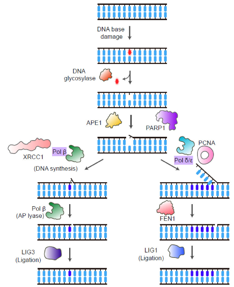
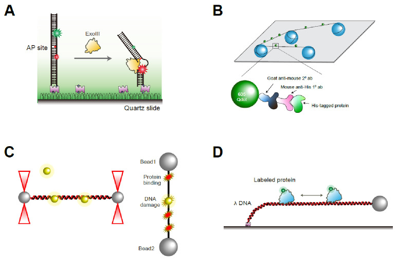
Base excision repair (BER) is an essential cellular mechanism that repairs small, non-helix-distorting base lesions in DNA, resulting from oxidative damage, alkylation, deamination, or hydrolysis. This review highlights recent advances in understanding the molecular mechanisms of BER enzymes through single-molecule studies. We discuss the roles of DNA glycosylases in lesion recognition and excision, with a focus on facilitated diffusion mechanisms such as sliding and hopping that enable efficient genome scanning. The dynamics of apurinic/apyrimidinic endonucleases, especially the coordination between APE1 and DNA polymerase β (Pol β), are explored to demonstrate their crucial roles in processing abasic sites. The review further explores the short-patch and long-patch BER pathways, emphasizing the activities of Pol β, XRCC1, PARP1, FEN1, and PCNA in supporting repair synthesis and ligation. Additionally, we highlight the emerging role of UV-DDB as a general damage sensor in BER, extending its recognized function beyond nucleotide excision repair. Single-molecule techniques have been instrumental in uncovering the complex interactions and mechanisms of BER proteins, offering unprecedented insights that could guide future therapeutic strategies for maintaining genomic stability. [BMB Reports 2025; 58(1): 17-23].


Conflict of interest statement

CONFLICTS OF INTEREST

The authors have no conflicting interests.

Figures

Fig. 1
Fig. 1
Short-patch and long-patch BER pathways. The figure illustrates the short-and long-patch pathways of BER. In both pathways, a DNA glycosylase initiates the repair by recognizing and removing a damaged base, leaving an AP site. APE1 subsequently cleaves the DNA backbone at the AP site, resulting in a single-strand break. In the short-patch pathway (left), XRCC1 forms a complex with Pol β and LIG3 to replace the damaged nucleotide and seal the nick. In the long-patch pathway (right), PCNA and PARP1 are recruited to the repair site. Pol δ/ε extends the repair patch by displacing the damaged strand. FEN1 then cleaves the displaced flap, and LIG1 seals the remaining nick to complete the repair.
Fig. 2
Fig. 2
Single-molecule techniques for studying BER. (A) single-molecule FRET is utilized to monitor conformational changes or interactions between proteins and DNA at the single-molecule level, providing real-time insights into dynamic processes by measuring fluorescence resonance energy transfer between donor and acceptor dyes. (B) Single-molecule DNA tightrope assay involves suspending DNA between two beads or surfaces, forming a “tightrope.” This setup enables the visualization of protein interactions with DNA, allowing observation of their movement and activity along the DNA strand using fluorescence microscopy. (C) Optical tweezers employ focused laser beams to manipulate single-molecules of DNA or proteins by applying picoNewton (pN) forces, making it ideal for examining mechanical properties and forces involved in BER-related processes. (D) Single-molecule flow stretch assay uses tethered DNA molecules on a surface subject to controlled flow, stretching the DNA under the exerted force to facilitate observation of protein-DNA interactions and repair activities.

Similar articles

References

    1. Lindahl T. Instability and decay of the primary structure of DNA. Nature. 1993;362:709–715. doi: 10.1038/362709a0. - DOI - PubMed
    1. Demple B, Harrison L. Repair of oxidative damage to DNA: enzymology and biology. Annu Rev Biochem. 1994;63:915–948. doi: 10.1146/annurev.bi.63.070194.004411. - DOI - PubMed
    1. Lindahl T, Wood RD. Quality control by DNA repair. Science. 1999;286:1897–1905. doi: 10.1126/science.286.5446.1897. - DOI - PubMed
    1. Krokan HE, Bjoras M. Base excision repair. Cold Spring Harb Perspect Biol. 2013;5:a012583. doi: 10.1101/cshperspect.a012583. - DOI - PMC - PubMed
    1. McCullough AK, Dodson ML, Lloyd RS. Initiation of base excision repair: glycosylase mechanisms and structures. Annu Rev Biochem. 1999;68:255–285. doi: 10.1146/annurev.biochem.68.1.255. - DOI - PubMed

MeSH terms

Substances