. 2025;17(1):56-77.
doi: 10.1159/000543274. Epub 2024 Dec 19.

Liver X Receptors and Inflammatory-Induced C/EBPβ Selectively Cooperate to Control CD38 Transcription

Affiliations

Liver X Receptors and Inflammatory-Induced C/EBPβ Selectively Cooperate to Control CD38 Transcription

Estibaliz Glaría et al. J Innate Immun. 2025.

Abstract

Introduction: Macrophages abundantly express liver X receptors (LXRs), which are ligand-dependent transcription factors and sensors of several cholesterol metabolites. In response to agonists, LXRs promote the expression of key lipid homeostasis regulators. Cross talk between LXRs and inflammatory signals exists in a cell type- and gene-specific manner. A common feature in the macrophage response to inflammatory mediators is the induction of CCAAT/enhancer-binding protein beta (C/EBPβ), a master transcriptional regulator and lineage-determining transcription factor in monocytes/macrophages.

Methods: Quantitative real-time PCR in control and C/EBPβ-deficient macrophages was used to explore the role of C/EBPβ in the cross talk between inflammatory mediators and the macrophage response to pharmacological LXR activation. The functional interaction between C/EBPβ and LXRs on selected genomic regions was further characterized by chromatin-immunoprecipitation (ChIP) and gene reporter studies.

Results: Whereas inflammatory signaling repressed several LXR-regulated genes involved in lipid metabolism, these effects were conserved after deletion of C/EBPβ. In contrast, inflammatory mediators and LXRs synergistically induced the expression of the multifunctional protein CD38 in a C/EBPβ-dependent manner. C/EBPβ and LXRs bound to several regions with enhancer activity upstream and within the mouse Cd38 gene and their functional cooperation in macrophages required intact binding sites for LXR and C/EBPβ.

Conclusion: This study reveals positive cross talk between C/EBPβ and LXRs during the macrophage inflammatory response, which selectively impacts CD38 expression.

Keywords: CCAAT/enhancer-binding protein beta; CD38; Interferon gamma; Lipopolysaccharide; Liver X receptor; Macrophage; Tumor necrosis factor alpha.


Conflict of interest statement

E.G. is currently an employee at OneChain Immunotherapeutics. J.M. is currently an employee at Avidity Biosciences. E.N.C. holds a patent on the use of CD38 inhibitors for metabolic diseases that is licensed by Elysium Health. E.N.C. is a consultant for TeneoBio, Calico, Mitobridge, and Cytokinetics. E.N.C. is on the advisory board of Eolo Pharma. E.N.C. owns stocks in TeneoBio. Dr. Chini is the head of the external research advisory board for Neolaia Bio. Research in the Chini laboratory has been conducted in compliance with the Mayo Clinic conflict of interest policies. C.C. is a consultant for Aromics. The rest of the authors declare no potential conflicts of interest.

Figures
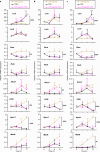
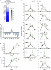
Fig. 1.
Fig. 1.
Inflammatory signals modulate LXR transcriptional responses. BMDMs were treated with the LXR agonist T1317 (1 µm) (a–c), TNFα (20 ng/mL) (a), IFNγ (5 ng/mL) (b), or LPS (100 ng/mL) (c) or with the combination of both agonist and inflammatory signal for the indicated periods of time. Cells not incubated with the LXR agonist were treated with vehicle (DMSO). The expression of LXR and RXR subtypes and of LXR-RXR target genes was determined by quantitative real time PCR and normalized by the expression levels of L14. Mean ± SD of n = 4 biological replicates obtained through 2 independent experiments. Two-way ANOVA Tukey: *p < 0.05; **p < 0.01; ***p < 0.001; and ****p < 0.0001 vs. unstimulated cells (time 0). Significant effects of inflammatory signals on the macrophage response to the LXR agonist are also indicated: ##p < 0.01; ###p < 0.001; and ####p < 0.0001. For each of the genes analyzed, the time-course data for the LXR agonist is identical in a–c and is used as reference.
Fig. 2.
Fig. 2.
Inflammatory signals and activation of the LXR/RXR pathway cooperatively induce CD38 mRNA and protein expression. BMDMs were treated with the LXR agonist T1317 (1 µm) (a–c), TNFα (20 ng/mL) (a), IFNγ (5 ng/mL) (b), or LPS (100 ng/mL) (c) or with the combination of both agonist and inflammatory signal for the indicated periods of time. d BMDMs were treated for 24 h with the same stimuli as in a and c. In a–d, cells not incubated with the LXR agonist were treated with vehicle (DMSO). The expression of Cd38 was determined by quantitative real time PCR and normalized by the expression levels of L14. The time-course data for the LXR agonist are identical in a–c. a–c Mean ± SD; n = 4 biological replicates obtained through 2 independent experiments. dn = 3 independent experiments. Two-way ANOVA Tukey: *p < 0.05; **p < 0.01; and ****p < 0.0001 vs. unstimulated cells (time 0). Additional relevant comparisons are also indicated, #p < 0.05; ##p < 0.01; and ####p < 0.0001. e BMDMs were treated for 24 h with agonists for LXR and RXR (T1317 and LG268, 1 µm each, respectively), TNFα (20 ng/mL), IFNγ (5 ng/mL), or LPS (10 ng/mL) or with simultaneous combinations of LXR/RXR agonists and inflammatory signals. Control cells were treated with vehicle (DMSO). The expression of CD38 was analyzed by Western blotting. As a control of protein loading, the expression of α-tubulin was also monitored.
Fig. 3.
Fig. 3.
Inflammatory signals induce Cebpb expression in an LXR-independent manner. BMDMs were treated with the LXR agonist T1317 (1 μm) (a), TNFα (20 ng/mL) (a), IFNγ (5 ng/mL) (b), or LPS (100 ng/mL) (c) or with simultaneous combinations of the LXR agonist and the inflammatory mediator (a–c) for the indicated periods of time. Control cells were treated with vehicle (DMSO). Cebpb and Cebpa mRNA levels were determined by quantitative real time PCR and normalized by the expression levels of L14. Mean ± SD of n = 4 biological replicates obtained through 2 independent experiments. Two-way ANOVA Tukey: ****p < 0.0001 vs. unstimulated cells (time 0). ns, nonsignificant differences between time-courses. The results from the LXR agonist time-course are only displayed in a. WT or LXRα/β-deficient (LXRα/β−/−) BMDMs were treated with TNFα (20 ng/mL, 3 h) (d), IFNγ (5 ng/mL, 6 h) (e), or LPS (100 ng/mL, 6 h) (f). Gene expression was measured by quantitative real time PCR. Mean ± SD of n = 3 biological replicates. Two-way ANOVA Tukey: ****p < 0.0001. g, h Inflammatory signals strongly increase the expression of C/EBPβ LAP. BMDMs were treated with TNFα (20 ng/mL), IFNγ (5 ng/mL), or LPS (100 ng/mL) for 6 or 12 h. Control cells were left unstimulated. The expression of C/EBPβ isoforms and β-tubulin was determined by immunoblotting. g Representative immunoblot of n = 3 independent experiments that showed similar results. h Relative protein expression for the 2 C/EBPβ isoforms detected, LAP (34 kDa) and LIP (20 kDa). The relative protein expression of each C/EBPβ isoform was calculated using the Image Studio Lite software and normalized to the expression values of β-tubulin. The graphs display the quantification data of the immunoblot shown in g.
Fig. 4.
Fig. 4.
C/EBPβ is a key regulator of CD38 expression in brain macrophages. Analysis of RNA-seq data from primary microglial cell cultures from Cebpbfl/fl and LysMCre-Cebpbfl/fl mice treated with vehicle (VEH) or LPS (100 ng/mL) for 6 h. a Heatmap of normalized expression values of LXR and RXR subtypes, and of LXR-RXR target genes. As a control, the expression of Cebpb is also included in the analysis. For better visualization of the results, expression values of LXR and RXR subtypes (b) and of selected LXR-RXR targets (c) are also represented as graphs. Two-way ANOVA-Tukey post hoc test. Comparisons between the indicated conditions: *p < 0.05; **p < 0.01; and ***p < 0.001. Comparisons vs. the same treatment in Cebpbfl/fl cells: #p < 0.05; ##p < 0.01; and ###p < 0.001.
Fig. 5.
Fig. 5.
C/EBPβ mediates the cooperative effects between inflammatory signals and LXRs on Cd38 induction. Cebpbfl/fl and LysMCre-Cebpbfl/fl BMDMs were treated with vehicle (DMSO) or an LXR agonist (T1317, 1 µm) and/or an inflammatory stimulus, TNFα (20 ng/mL), IFNγ (5 ng/mL), or LPS (100 ng/mL) for 24 h. The expression of selected genes was measured by quantitative real time PCR and normalized by L14 expression. Mean ± standard deviation (SD) of n = 4 independent experiments performed with biological duplicates or triplicates. Two-way ANOVA Tukey (for datasets with normal distribution and homogeneous variance); Kruskal-Wallis-Dunn’s test (for datasets without normal distribution). Comparisons between the indicated conditions: *p < 0.05; **p < 0.01; and ***p < 0.001. Comparisons vs. the same treatment in Cebpbfl/fl cells: #p < 0.05; ##p < 0.01; and ###p < 0.001. For some genes, the effect of the LXR agonist was also compared using a Student’s t-test (if normal distribution) or a Mann-Whitney test (otherwise): ^p < 0.05; ^ ^p < 0.01; and ^ ^ ^p < 0.001.
Fig. 6.
Fig. 6.
C/EBPβ and LXR co-occupy several regions with enhancer activity upstream and within the Cd38 gene. a Analysis of ChIP-Seq data to test chromatin occupancy of H3K27ac (orange), LXRα (green), RXR (gray), and C/EBPβ (red) in the vicinity of the Cd38 gene. The occupancy of H3K27ac was analyzed in BMDMs treated with LPS for the indicated times (2 h and 4 h in database GSE56123 and 24 h in database GSE38377). LXRα ChIP-Seq was carried out using an antibody against LXRα/β in LXRβ-deficient BMDMs stimulated for 24 h with the LXR agonist GW3965 (1 μm) either alone or in combination with LPS (100 ng/mL) (database GSE200922). Binding events of RXR were determined in BMDMs treated with an LXR agonist (GW3965) for 1 h (database SRP019970). The occupancy of CEBPβ was analyzed in BMDMs from C57BL/6 mice stimulated with Kdo2-Lipid A (KLA) for 1 h (database GSE109965). Control cells were left untreated in all datasets. Three regions (R1, R2, and R3) with pronounced H3K27ac marks and LXRE and C/EBPβ binding motifs are displayed. b BMDMs were treated with an LXR agonist (T1317, 1 μm), LPS (100 ng/mL), or both stimuli simultaneously during 24 h. Control cells were treated with vehicle (DMSO). The binding of LXRs, RXRβ, and C/EBPβ to enhancer regions R1, R2, and R3 was measured by ChIP assay. One-way ANOVA: *p < 0.05; **p < 0.01; and ***p < 0.001 between the indicated conditions.
Fig. 7.
Fig. 7.
C/EBPβ and LXRs functionally cooperate in a genomic region with enhancer activity. a–f Luciferase reporter studies using a pGL3 luciferase reporter vector containing the enhancer region R2 (pGL3-R2). Raw264.7 macrophages (a–e) or COS-7 kidney cells (f) were transiently co-transfected with pGL3-R2 containing either a WT sequence (a–f) or a mutated (MUT) LXRE (b, e, f) or C/EBPβ binding site (c–e). In addition, some transfections included pMSV-C/EBPβ (for C/EBPβ overexpression) (a–f), and/or a combination of pcDNA3-RXRα and either pcDNA3-LXRα (a, e) or pcDNA3-LXRβ (a, b, d, f) (for LXR/RXR overexpression). In a, control cells were transfected with pGL3-R2 and an empty plasmid (empty pcDNA3 without overexpression of transcription factors). All transfections included pRL-TK (for constitutive Renilla expression) and the total amount of plasmid DNA was equilibrated using empty pcDNA3. The cells were then stimulated with an LXR agonist (GW3965, 1 µm) or vehicle for 24 h. The enhancer activity is represented as luciferase activity normalized by Renilla activity. Mean ± SD of biological triplicates (a, c, f) or of n = 3 independent experiments each performed with biological duplicates or triplicates (b, d–e). a–f Two-way ANOVA: *p < 0.05; **p < 0.01; and ***p < 0.001 between the indicated conditions; in addition, #p < 0.05; ##p < 0.01; and ###p < 0.001 in comparison with the same condition in control cells not overexpressing transcription factors (empty plasmid) (a) or in comparison with the same condition in cells transfected with the WT pGL3-R2 sequence (b, d–f).
Fig. 8.
Fig. 8.
CD38 is dispensable for the induction of C/EBPβ-dependent genes, but contributes to fine-tuning macrophage cytokine production in response to LPS. a RNA-Seq data from Cebpbfl/fl and LysMCre-Cebpbfl/fl brain macrophages treated with vehicle or LPS (100 ng/mL) for 6 h were analyzed to identify LPS-induced genes that are highly dependent on C/EBPβ expression. Heatmap displaying a list of filtered genes ordered by the log2 fold change between LPS-treated LysMCre-Cebpbfl/fl vs. LPS-treated Cebpbfl/fl macrophages. b Graph representing log2 fold changes in the expression of selected cytokines and inflammatory mediators in LPS-treated LysMCre-Cebpbfl/fl vs. LPS-treated Cebpbfl/fl macrophages. ce BMDMs obtained from WT or CD38-deficient mice were treated with LPS (100 ng/mL) during the indicated periods of time. In c, the expression of selected genes, including C/EBPβ-dependent genes identified in a, was analyzed by quantitative real time PCR. The levels of secreted IL-6 (d) and IL-12 (e) were measured by ELISA. Mean ± SD; n = 3 experiments. Two-way ANOVA Tukey: *p < 0.05; **p < 0.01; ***p < 0.001; and ****p < 0.0001 vs. unstimulated cells (time 0). Significant changes between WT- and CD38-deficient cells are also indicated: #p < 0.05; ##p < 0.01; and ###p < 0.001.

References

    1. Okabe Y, Medzhitov R. Tissue biology perspective on macrophages. Nat Immunol. 2016;17(1):9–17. - PubMed
    1. Wynn TA, Chawla A, Pollard JW. Macrophage biology in development, homeostasis and disease. Nature. 2013;496(7446):445–55. - PMC - PubMed
    1. Schulman IG. Liver X receptors link lipid metabolism and inflammation. FEBS Lett. 2017;591(19):2978–91. - PMC - PubMed
    1. A-González N, Castrillo A. Liver X receptors as regulators of macrophage inflammatory and metabolic pathways. Biochim Biophys Acta. 2011;1812(8):982–94. - PubMed
    1. Hu X, Li S, Wu J, Xia C, Lala DS. Liver X receptors interact with corepressors to regulate gene expression. Mol Endocrinol. 2003;17(6):1019–26. - PubMed

Publication types

Substances