. 2024 Dec 19;30(1):248.
doi: 10.1186/s10020-024-01031-2.

Denatonium inhibits RANKL-induced osteoclast differentiation and rescues the osteoporotic phenotype by blocking p65 signaling pathway

Affiliations

Denatonium inhibits RANKL-induced osteoclast differentiation and rescues the osteoporotic phenotype by blocking p65 signaling pathway

Sheunghun Lee et al. Mol Med. .

Abstract

Background: Bone remodeling is a critical process that maintains skeletal integrity, orchestrated by the balanced activities of osteoclasts, which resorb bone, and osteoblasts, which form bone. Osteoclastogenesis, the formation of osteoclasts, is primarily driven by NFATc1, a process activated through c-Fos and NF-κB signaling pathways in response to receptor activator of nuclear factor κB ligand (RANKL). Dysregulation of RANKL signaling is a key contributor to pathological bone loss, as seen in conditions such as osteoporosis.

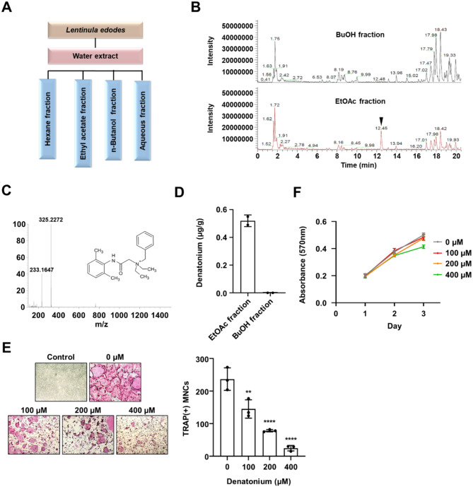
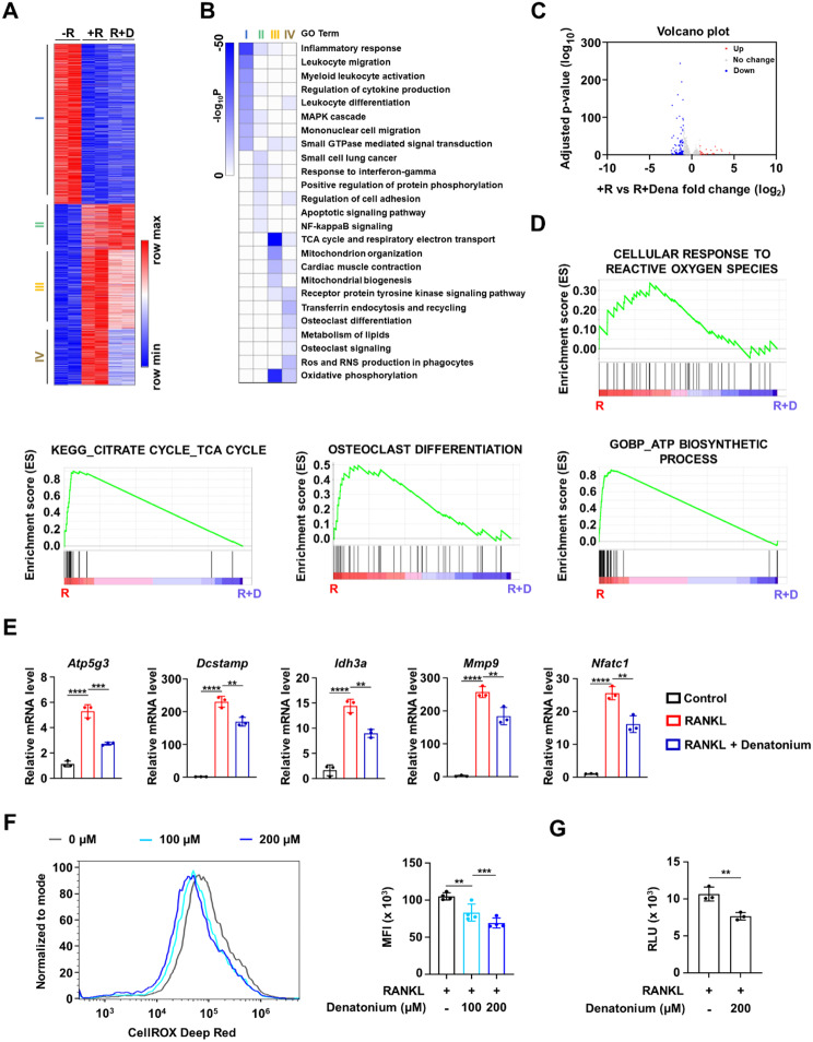
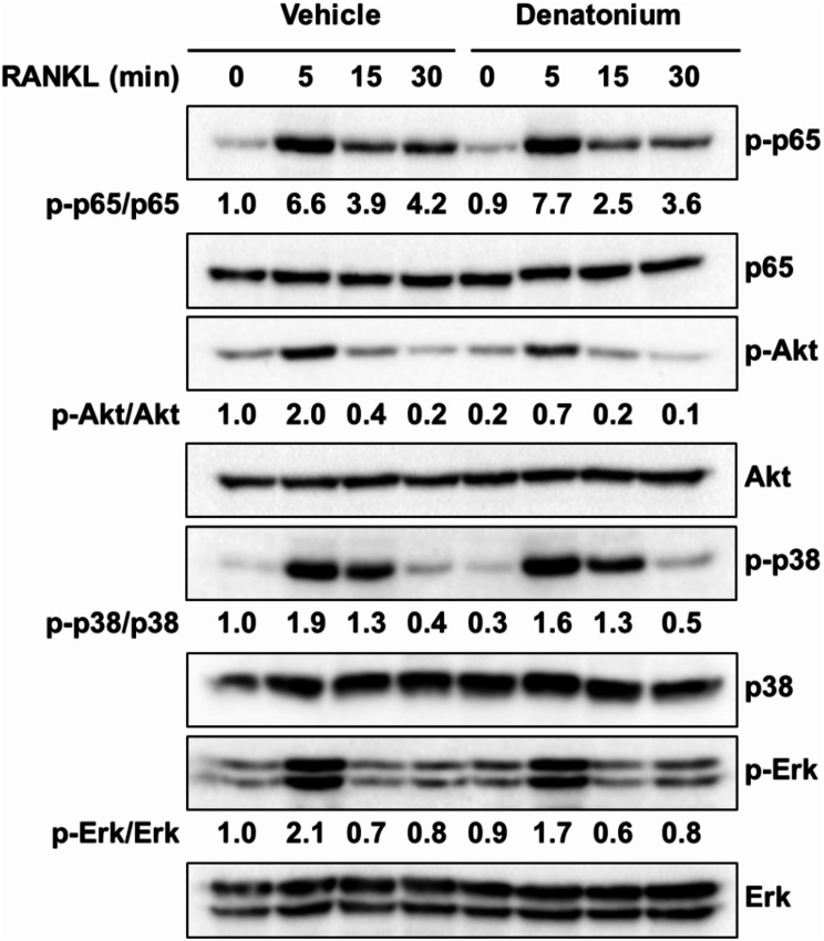
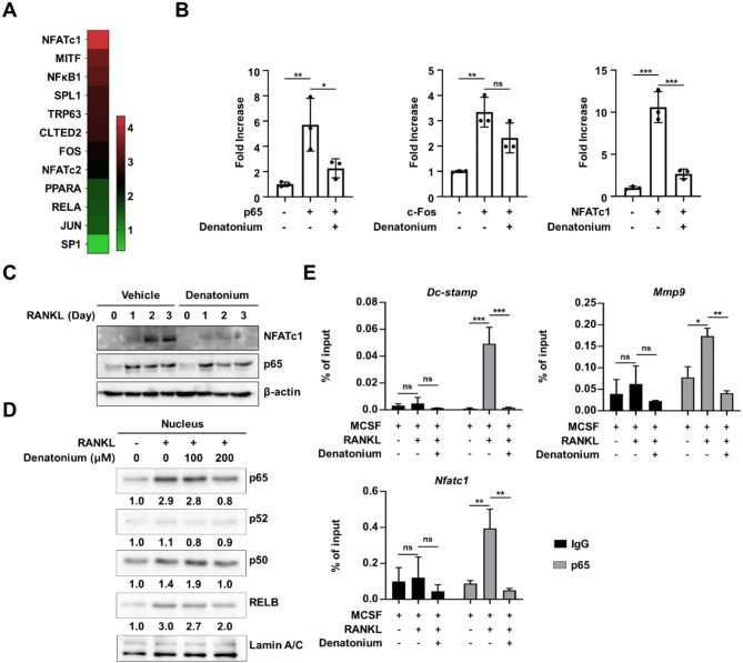
Methods: We investigated the effects of denatonium, a known bitter compound, on RANKL-induced osteoclast differentiation. We used RNA sequencing (RNA-seq) to analyze gene expression profiles in osteoclast precursors treated with denatonium. Transcription factor prediction analysis was conducted to identify key targets of denatonium action. Additionally, we performed Western blotting to examine the phosphorylation status of AKT and p65, crucial components of the NF-κB pathway. Chromatin immunoprecipitation (ChIP) assays were employed to assess the binding of p65 to promoter regions of osteoclast-related genes. Finally, we tested the therapeutic potential of denatonium in a mouse model of osteoporosis.

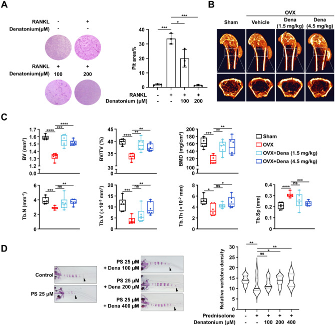
Results: Our findings demonstrated that denatonium significantly inhibited RANKL-induced osteoclastogenesis by targeting the p65 pathway. RNA-seq analysis revealed a downregulation of osteoclast-related genes following denatonium treatment, corroborated by transcription factor prediction analysis, which highlighted p65 as a key target. Denatonium effectively blocked the phosphorylation of AKT and p65, key steps in NF-κB activation. ChIP assays further confirmed that denatonium reduced the enrichment of p65 at promoter regions critical for osteoclast differentiation. In vivo, denatonium treatment in an osteoporosis animal model led to a significant restoration of bone health, demonstrating its potential as a therapeutic agent.

Conclusions: This study identifies denatonium as an inhibitor of RANKL-induced osteoclast differentiation, potentially acting through suppression of the p65 signaling pathway. The ability of denatonium to downregulate osteoclast-related genes and inhibit key signaling events highlights its potential as a candidate for further investigation in the context of bone loss and osteoporosis.

Keywords: Denatonium; Osteocastogenesis; Osteoporosis; p65.


Conflict of interest statement

Declarations. Ethics approval and consent to participate: This study was approved by the Institutional Animal Management and Use Committee of Chungbuk National University (CBNUA-1245-19-02 and CBNUA-1391-20-01). Consent for publication: All the authors have read this manuscript and would like to have it considered exclusively for publication. Competing interests: The authors declare no competing interests.

Figures

Fig. 1
Fig. 1
Denatonium, a bioactive molecule isolated from Lentinula erodes, inhibits osteoclast differentiation. A A schematic representation outlining the procedure for extracting and fractionating antiosteoclastic compounds from L. edodes. B LC-MS chromatograms of the butanol and ethyl acetate fractions. C Identification of denatonium from an EtOAc fraction using LC-MS/MS. D Comparison of denatonium content in two fractions (BuOH and EtOAc). E Representative images of TRAP-stained cells treated with various concentrations of denatonium. One-way ANOVA followed by Dunnett’s multiple comparisons test, Mean ± SD of three independent experiments. **P < 0.01; ****P < 0.0001. F Effect of denatonium on OCPs proliferation
Fig. 2
Fig. 2
Denatonium alters gene expression profiling in osteoclast precursor (OCP) cells. A K-means (K = 4) clustering of 2,687 DEGs for any pairwise comparison among three conditions [− R; no RANKL, +R; RANKL (100ng/ml), R + D; RANKL with denatonium (400µM)]. OCP cells were exposed to RANKL (100ng/ml) for 72 h, with or without the presence of denatonium. B Heatmap showing the p-value significance of GO term enrichment for genes in each cluster. C Volcano plot of transcriptomic changes between + R and R + D. Genes with increased (red) or decreased (blue) expression in R + D-treated cells relative to RANKL-treated cells defined based on an FDR-adjusted p < 0.05 and > 2-fold expression change. D GSEA of 2,687 genes as in A shows the enrichment of genes associated with osteoclast differentiation, ATP biosynthetic process, TCA cycle, and cellular response to ROS. E qRT-PCR was performed to quantitate relative mRNA levels of representative genes for D. One-way ANOVA followed by Tukey’s multiple comparisons test, Mean ± SD of three independent experiments. *P < 0.05; **P < 0.01; ***P < 0.001; ****P < 0.0001; ns, not significant. F Flow cytometry histograms were used to measure cellular ROS levels using CellROX deep red. The bar graphs represent the MFI of OCP cells stimulated by RANKL (100 ng/ml, 10 min) with or without denatonium (200 µM). One-way ANOVA followed by Dunnett’s multiple comparisons test, Mean ± SD of three independent experiments. **P < 0.01; ***P < 0.001. G OCP cells were incubated with denatonium (200 µM) in the presence of RANKL (100 ng/ml). Cellular ATP levels were measured. Two-tailed t-test, Mean ± SEM of three independent experiments. **P < 0.01
Fig. 3
Fig. 3
Denatonium inhibits the phosphorylation of AKT and p65. Effect of denatonium on the AKT, MAPK, and NF-κB signaling pathways in response to RANKL treatment. OCP cells were pretreated with denatonium (200 µM) alongside RANKL (100 ng/ml) for the specified durations. Whole cell lysates were subjected to western blot analysis using the appropriate antibodies. The intensity of the western blot bands was quantified using ImageJ software, and results are presented as the relative ratio of each protein band intensity normalized to the total protein band intensity
Fig. 4
Fig. 4
Denatonium blocks the nuclear translocation and recruitment of p65 at osteoclastogenic genes. A Analysis of transcription factors governing the expression of genes in cluster 4 as shown in Fig. 2A, using TRRUST v2. B 293T cells were transfected with the reporter plasmid Nfatc1-Luc along with p65, c-Fos, or Nfatc1, with or without denatonium (200 µM). Each bar represents the Means ± S.D. of three independent experiments, one-way ANOVA followed by Tukey’s multiple comparisons test. *P < 0.05; **P < 0.01; *** p < 0.001; ns = not significant. C Effect of denatonium on NFATc1 expression. OCO cells were cultured with or without denatonium (200 µM) in the presence of M-CSF (30 ng/ml) and RANKL (100 ng/ml). Whole-cell lysate were analyzed by immunoblotting with NFATc1 and p65 antibodies. β-Actin served as the loading control. D Effect of denatonium on NF-κB nuclear translocation. Nuclear fractions of OCPs were prepared after RANKL (100 ng/ml, 30 min) treatment with vehicle or denatonium and nuclear translocation of NF-κB was measured by western blot analysis with the indicated antibodies. The western blot band was quantified using Image J software and shown as the relative ratio of each protein band intensity normalized to Lamin A/C band intensity. E ChIP-qPCR of p65 enrichment at the Mmp9 (− 0.5 kb), Nfatc1 (− 0.7 kb), and Dcstamp (− 0.6 kb) promoters following RANKL treatment (100 ng/ml, 30 min) with or without denatonium (200 µM). Two-way ANOVA followed by Tukey’s multiple comparisons test, Mean ± SD of three independent experiments. *P < 0.05; **P < 0.01; ***P < 0.001; ns, not significant
Fig. 5
Fig. 5
Denatonium prevents bone loss in osteoporotic models. A Bone-resorption activity of OCPs treated with vehicle or denatonium (100 µM and 200 µM) with or without RANKL (100 ng/mL) for 10 d. One-way ANOVA analysis, Mean ± SD of three independent experiments. *P < 0.05; ***P < 0.001. B–C Micro-CT analysis of the femurs of a two-month-old sham-operated, saline-treated (Sham), saline-treated OVX (OVX), and denatonium-treated OVX (OVX + Dena 1.5 mg/kg or 4.5 mg/kg) mice (n = 6). BMD, bone mineral density; BV, bone volume; BV/TV, trabecular bone volume per tissue volume; Tb.Th, trabecular thickness; Tb. Sp, trabecular spacing; Tb.N, trabecular number; Tb.V, trabecular volume. Two-tailed t-test, Mean ± SEM. *P < 0.05; **P < 0.01; ***P < 0.001; ****P < 0.0001. D The larvae at 10 d post-fertilization were treated with 25 µM prednisolone (PS) and varying denatonium concentrations (0, 100, 200, and 400 µM) for 3 d. Whole-mount Alizarin red staining was performed to analyze the mineralized bone. The relative density of vertebral bones was evaluated by measuring the areas of the initial five stained vertebrae (indicated by arrowhead). One-way ANOVA followed by Tukey’s multiple comparisons test, Mean ± SEM (n = 36). *P < 0.05; **P < 0.01; ns, not significant

Similar articles

Cited by

References

    1. Abu-Amer Y. NF-kappaB signaling and bone resorption. Osteoporos Int. 2013;24:2377–86. - PMC - PubMed
    1. Aceto J, et al. Zebrafish bone and General Physiology are differently affected by hormones or changes in gravity. PLoS ONE. 2015;10:e0126928. - PMC - PubMed
    1. Alles N, et al. Suppression of NF-kappaB increases bone formation and ameliorates osteopenia in ovariectomized mice. Endocrinology. 2010;151:4626–34. - PubMed
    1. An D, Kim K, Lu W. Defective entry into mitosis 1 (Dim1) negatively regulates osteoclastogenesis by inhibiting the expression of nuclear factor of activated T-cells, cytoplasmic, calcineurin-dependent 1 (NFATc1). J Biol Chem. 2014;289:24366–73. - PMC - PubMed
    1. Asagiri M, Takayanagi H. The molecular understanding of osteoclast differentiation. Bone. 2007;40:251–64. - PubMed

LinkOut - more resources