. 2024 Dec 20;11(1):83.
doi: 10.1186/s40779-024-00584-x.

Dietary methionine supplementation promotes mice hematopoiesis after irradiation

Affiliations

Dietary methionine supplementation promotes mice hematopoiesis after irradiation

Wei-Wei Zhang et al. Mil Med Res. .

Abstract

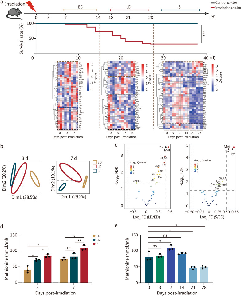
Background: With the increasing risk of nuclear exposure, more attention has been paid to the prevention and treatment of acute radiation syndrome (ARS). Although amino acids are key nutrients involved in hematopoietic regulation, the impacts of amino acids on bone marrow hematopoiesis following irradiation and the associated mechanisms have not been fully elucidated. Hence, it is of paramount importance to study the changes in amino acid metabolism after irradiation and their effects on hematopoiesis as well as the related mechanisms.

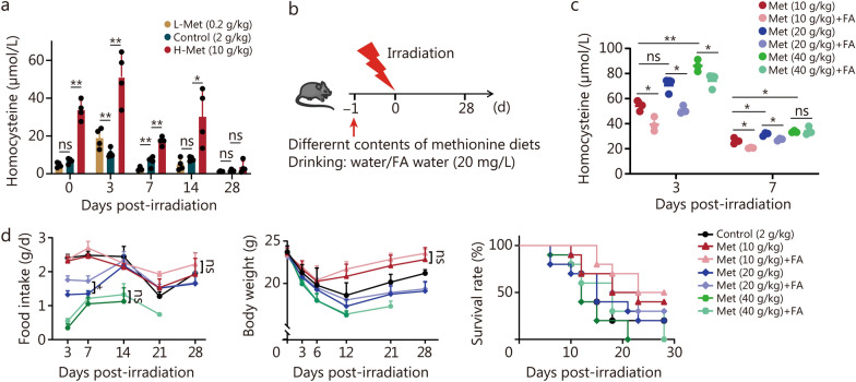
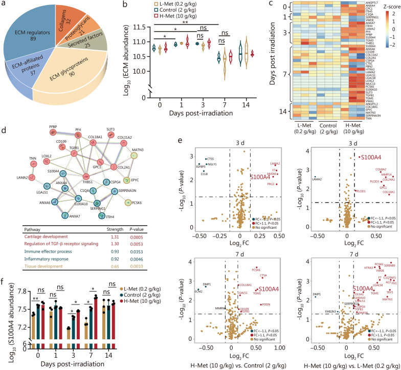
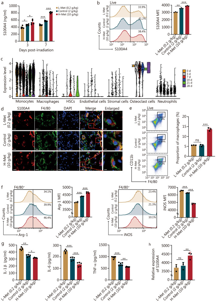
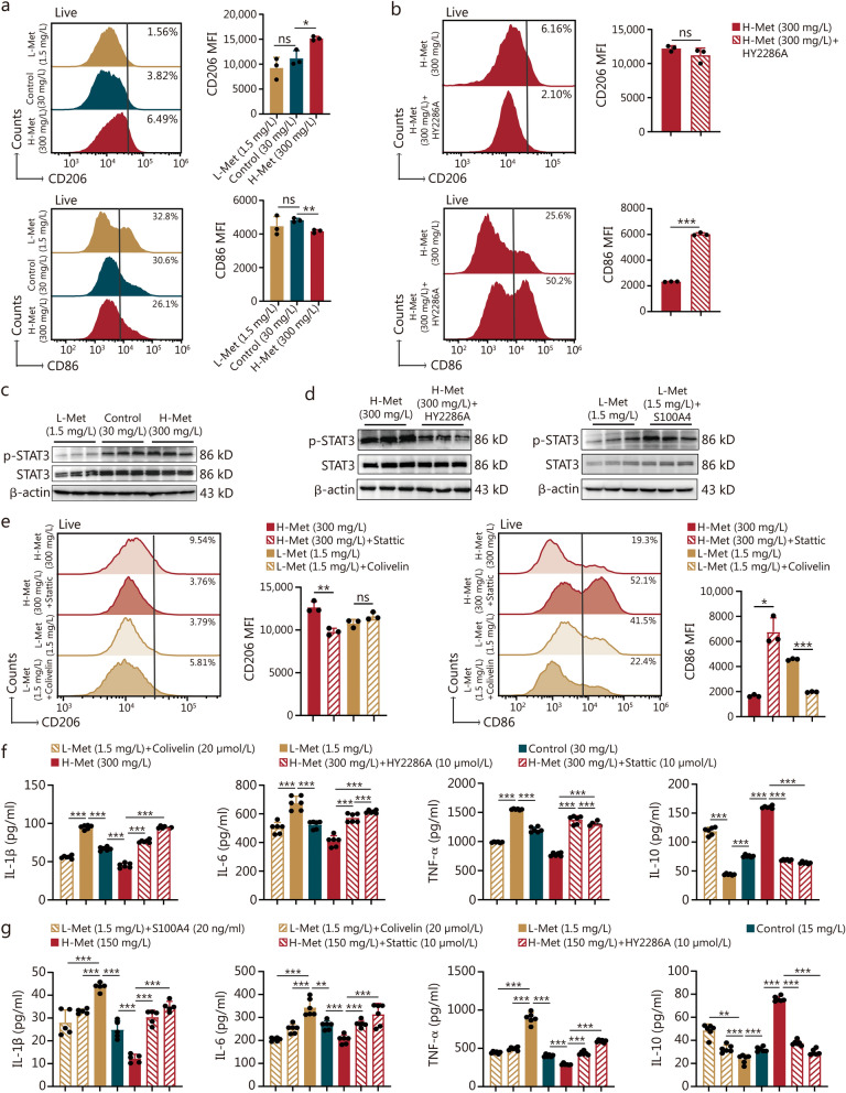
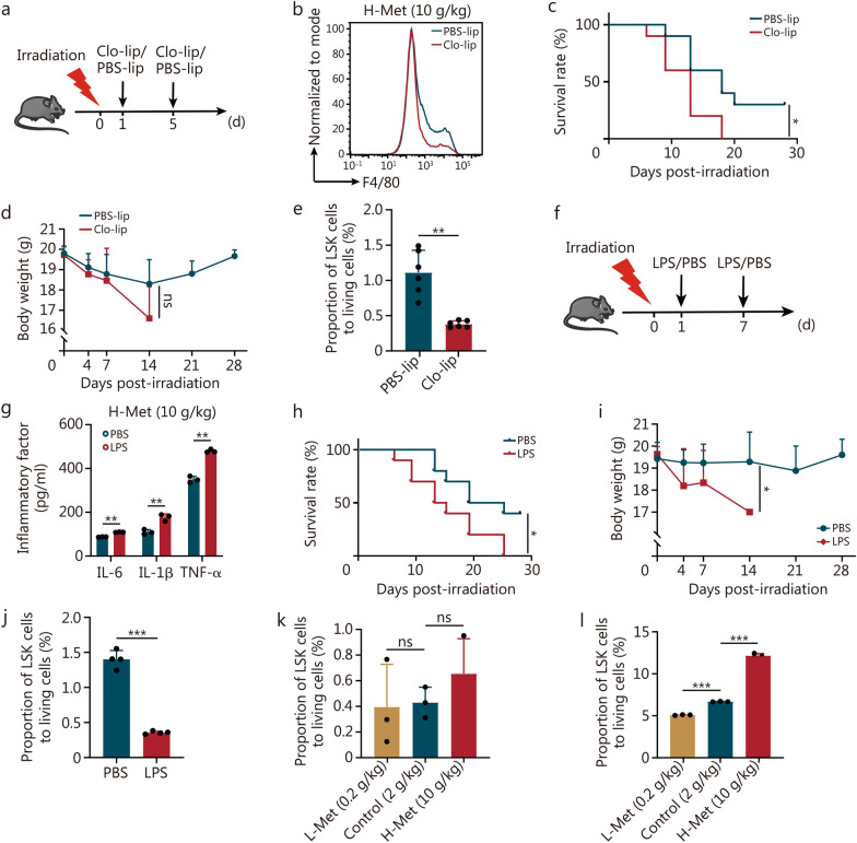
Methods: The content of serum amino acids was analyzed using metabolomic sequencing. The survival rate and body weight of the irradiated mice were detected after altering the methionine content in the diet. Extracellular matrix (ECM) protein analysis was performed via proteomics analysis. Inflammatory factors were examined by enzyme-linked immunosorbent assay (ELISA). Flow cytometry, Western blotting, and immunofluorescence were employed to determine the mechanism by which S100 calcium-binding protein A4 (S100A4) regulates macrophage polarization.

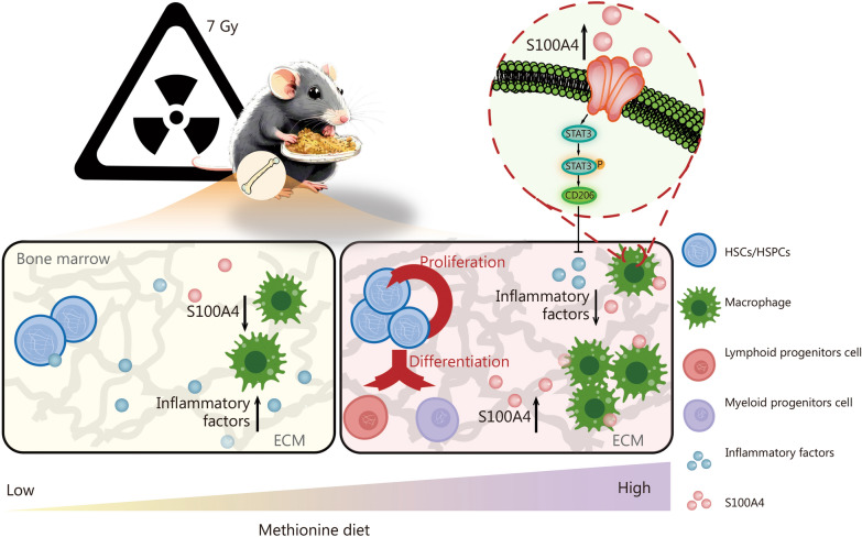
Results: The survival time of irradiated mice was significantly associated with alterations in multiple amino acids, particularly methionine. A high methionine diet promoted irradiation tolerance, especially in the recovery of bone marrow hematopoiesis, yet with dose limitations. Folate metabolism could partially alleviate the dose bottleneck by reducing the accumulation of homocysteine. Mechanistically, high methionine levels maintained the abundance of ECM components, including collagens and glycoproteins, in the bone marrow post-irradiation, among which the level of S100A4 was significantly changed. S100A4 regulated macrophage polarization via the STAT3 pathway, inhibited bone marrow inflammation and facilitated the proliferation and differentiation of hematopoietic stem/progenitor cells.

Conclusions: We have demonstrated that an appropriate elevation in dietary methionine enhances irradiation tolerance in mice and explains the mechanism by which methionine regulates bone marrow hematopoiesis after irradiation.

Keywords: Bone marrow hematopoiesis; Irradiation; Macrophage; Methionine; S100A4.


Conflict of interest statement

Declarations. Ethics approval and consent to participate: This study was approved by the Laboratory Animal Welfare and Ethics Committee of the Army Medical University (AMUWEC2020122, AMUWEC2020055, AMUWEC20211415, AMUWEC20224721). All experiments involving animals were performed following relevant policies. Consent for publication: No applicable. Competing interests: The authors declare that they have no competing financial interests.

Figures

Fig. 1
Fig. 1
The survival time of irradiated mice is significantly correlated with changes in multiple amino acids, particularly methionine. a Schematic diagram of the experiment, survival rate of 8-week-old male C57BL/6 mice irradiated with 7 Gy (n = 10 in the control group and n = 40 in the IR group), and heatmap of the serum amino acid contents of the mice at different times of death after irradiation (n = 3). b Principal component analysis (PCA) plot of the three groups at 3 d or 7 d after irradiation. c Volcano plot depicting the significantly different amino acids among the three groups at 3 d after irradiation. d Changes in methionine content among the three groups at 3 d and 7 d after irradiation (n = 3). e Methionine content in the S group at different time points after irradiation (n = 3). The error bars indicate the standard deviation from three or more independent experimental replicates, *P < 0.05, **P < 0.01, ***P < 0.001, ns non-significant, as determined by the log-rank (Mantel-Cox) test (a) and Student’s t-test (d, e). ED early death, LD late death, S survival, FDR false discovery rate, Dim dimension, FC fold change, 1MHis 1-methylhistidine, 3MHis 3-methylhistidine, 6AHC 6-aminocaproic acid, Aad α-aminoadipate, Abu 2-aminobutyric acid, Ala alanine, Allolle valisoleucine, Arg arginine, Asn asparagine, Asp-AA aspartic acid, bAib 3-aminoisobutyric acid, bAla β-alanine, Car carnosine, Cit-AA citrulline, GABA γ-aminobutyric acid, Gln glutamine, Glu glutamic acid, Gly glycine, Harg homocysteine, Hcit homocitrulline, His histidine, Hpro homotypic proline, Hyp hydroxy-proline, Ile isoleucine, KC kynurenine, Leu leucine, Lys lysine, Met methionine, Orn ornithine, Phe phenylalanine, Pro proline, Sar sarcosine, Ser serine, Thr threonine, Trp tryptophan, Tyr tyrosine, Val valine
Fig. 2
Fig. 2
Dietary methionine supplementation enhances irradiation tolerance in mice, particularly for bone marrow hematopoiesis, but with dose limitation. a Schematic representation of the experiment, 8-week-old male C57BL/6 mice were irradiated with 7 Gy, and the mice were provided with diets supplemented with different concentrations of methionine after irradiation. b Survival rate of mice after irradiation by being fed with different methionine concentrations (n = 20). c Methionine content in the serum and bone marrow of mice fed with diets of different methionine concentrations at 7 d after 7 Gy irradiation (n = 3). d Changes in white blood cells, red blood cells, and platelets in the peripheral blood by routine analysis after 7 Gy irradiation (n = 6). e H&E staining of femurs at 14 d after irradiation with diets of different methionine contents. Scale bar = 50 μm (left)/20 μm (right). f The number of nucleated cells in bone marrow at 7 d and 14 d after irradiation with diets of different methionine contents (n = 5). g Bone marrow cells proliferation on day 7 after irradiation with diets of different methionine contents (n = 5). The error bars indicate the standard deviation from three or more independent experimental replicates, *P < 0.05, **P < 0.01, ***P < 0.001, ns non-significant, as determined by log-rank (Mantel-Cox) test (b) and Student’s t-test (c, d, f, g). Met methionine, L-Met low methionine diet, H-Met high methionine diet
Fig. 3
Fig. 3
Folate (FA) supplementation can partially rescue the excess homocysteine produced by a high methionine diet. a Changes in the homocysteine content in the serum after irradiation (n = 4). b Schematic illustration of the experiment, 8-week-old male C57BL/6 mice were irradiated with 7 Gy, and the mice were provided with diets supplemented with different concentrations of methionine and FA in the water. c Serum homocysteine content of mice at 3 d and 7 d following FA supplementation after irradiation (n = 3). d Survival rate (n = 10), food intake (n = 3) and body weight (n = 10) of the mice after irradiation. The error bars indicate the standard deviation from three or more independent experimental replicates, *P < 0.05, **P < 0.01, ns non-significant, as determined by log-rank (Mantel-Cox) test (survival rate in d) and Student’s t-test (a, c, food intake and body weight in d). Met methionine, L-Met low methionine diet, H-Met high methionine diet
Fig. 4
Fig. 4
Methionine maintained the abundance of the bone marrow ECM after irradiation, and S100A4 was significantly elevated. a Pie chart of the number of proteins in all ECM categories. b The total abundance of ECM in the context of different methionine diets (n = 3). c Heatmap of differentially expressed proteins associated with different methionine diets (n = 3). d Protein–protein interaction (PPI) network of differentially expressed proteins associated with different methionine diets. e Volcano plot of the significantly different proteins associated with different methionine diets at 3 d and 7 d post-irradiation. f The abundance of S100A4 in response to different methionine diets (n = 3). The error bars indicate the standard deviation from three or more independent experimental replicates, *P < 0.05, **P < 0.01, ns non-significant, determined by Student’s t-test. ECM extracellular matrix, FC fold change, ANGPTL7 angiopoietin-like 7, ANGPTL2 angiopoietin-like 2, ANXA4 annexin A4, GPC1 glypican 1, ITIH5 inter-alpha (globulin) inhibitor H5, C1QA complement component 1Q subcomponent alpha polypeptide, SERPING1 serine (or cysteine) peptidase inhibitor clade G member 1, VWDE von Willebrand factor D and EGF domains, ANXA7 annexin A7, ITIH4 inter-alpha (globulin) inhibitor H4, LAMA2 laminin, alpha 2, PF4 platelet factor 4, PPBP pro-platelet basic protein, S100A4 S100 calcium binding protein A4, TGM3 transglutaminase 3, THBS1 thrombospondin 1, ANXA6 annexin A6, COL11A2 collagen type XI alpha 2, COL2A1 collagen type II alpha 1, CSPG4 chondroitin sulfate proteoglycan 4, CTSC cathepsin C, FBN2 fibrillin 2, HTRA3 HtrA serine peptidase 3, LGALS1 lectin galactose binding soluble 1, LGALS8 lectin galactose binding soluble 8, LOXL2 lysyl oxidase-like 2, MUC13 mucin 13, PCSK6 proprotein convertase subtilisin/kexin type 6, S100A10 S100 calcium binding protein A10, SLIT3 slit homolog 3, TGFB1 transforming growth factor β1, TGM1 transglutaminase 1, VWA1 von Willebrand factor A domain containing 1, COL18A1 collagen type XVIII alpha 1, EPYC epiphycan, MATN3 matrilin 3, SERPINA3N serine (or cysteine) peptidase inhibitor clade A member 3N, TNN tenascin N, CTSS cathepsin S, NGLY1 N-glycanase 1, IGFBP5 insulin-like growth factor binding protein 5, CCL8 chemokine (C–C motif) ligand 8, PRG3 proteoglycan 3, F7 coagulation factor VII, PLOD1 procollagen-lysine 2-oxoglutarate 5-dioxygenase 1, PLOD3 procollagen-lysine 2-oxoglutarate 5-dioxygenase 3, COL8A2 collagen type VIII alpha 2, COL24A1 collagen type XXIV alpha 1, MMP14 matrix metallopeptidase 14, COL10A1 collagen type XX alpha 1, DMP1 dentin matrix protein 1, EMILIN3 elastin microfibril interfacer 3, C1QTNF5 C1q and tumor necrosis factor related protein 5, AGT angiotensinogen, FN1 fibronectin 1, TRY5 trypsin 5, MMRN multimerin 1, ECM2 extracellular matrix protein 2, PODN podocan, CTSA cathepsin A, SERPINB9 serine (or cysteine) peptidase inhibitor clade B member 9, L-Met low methionine diet, H-Met high methionine diet
Fig. 5
Fig. 5
The high methionine diet enhanced S100A4 expression in bone marrow macrophages and suppressed inflammation. a The concentration of S100A4 in the bone marrow supernatant after irradiation with diverse methionine diets was determined via ELISA (n = 5). b The expression of S100A4 in bone marrow at 7 d after irradiation with different methionine diets was detected by flow cytometry (n = 3). c Transcriptional expression of S100A4 in bone marrow cells at various time points after irradiation. d Immunofluorescence results demonstrated that macrophages colocalized with S100A4. Scale bar = 20 μm. e Proportion of macrophages at 7 d after irradiation with different methionine diets (n = 3). f Expression of Arg-1 and iNOS in bone marrow macrophages at 7 d after irradiation in the setting of different methionine diets (n = 4). g Levels of inflammatory factors including IL-1β (n = 5), IL-6 (n = 5), and TNF-α (n = 6) in the bone marrow supernatant at 7 d of irradiation with different methionine concentrations. h The relative expression of S100A4 detected by transcriptome sequencing in macrophages at 7 d after irradiation with different methionine diets (n = 3). The error bars indicate the standard deviation from three or more independent experimental replicates, *P < 0.05, **P < 0.01, ***P < 0.001, ns non-significant, as determined by Student’s t-test. S100A4 S100 calcium-binding protein A4, MFI mean fluorescent intensity, HSCs hematopoietic stem cells, DAPI 4',6-diamidino-2-phenylindole, Arg-1 arginase 1, iNOS inducible nitric oxide synthase, TNF-α tumor necrosis factor-α, IL interleukin, L-Met low methionine diet, H-Met high methionine diet
Fig. 6
Fig. 6
S100A4 regulates macrophage polarization to be involved in the bone marrow inflammatory response via STAT3. a The expression of CD206 and CD86 in RAW264.7 cells at 1 d after irradiation with different methionine media (n = 3). b The expression of CD206 and CD86 in RAW264.7 cells at 1 d after irradiation with the S100A4 inhibitor (HY2286A) in 300 mg/L methionine media (n = 3). c STAT3 protein expression was determined by Western blotting in RAW264.7 cells cultured in different methionine media. d STAT3 protein expression in RAW264.7 cells detected by Western blotting in the context of changes in S100A4 expression. e The expression of CD206 and CD86 in RAW264.7 cells at 1 d after irradiation with an STAT3 agonist (Colivelin) or inhibitor (Stattic) (n = 3). f The levels of inflammatory factors (IL-1β, IL-6, TNF-α, and IL-10) in the culture medium of RAW264.7 cells were measured via ELISA (n = 6). g The levels of inflammatory factors (IL-1β, IL-6, TNF-α, and IL-10) in the culture medium of BMDMs were measured via ELISA (n = 5). The error bars indicate the standard deviation from three or more independent experimental replicates, *P < 0.05, **P < 0.01, ***P < 0.001, ns non-significant, as determined by Student’s t-test. S100A4 S100 calcium-binding protein A4, MFI mean fluorescent intensity, TNF-α tumor necrosis factor-α, IL interleukin, L-Met low methionine diet, H-Met high methionine diet
Fig. 7
Fig. 7
Methionine promotes HSC/HSPC proliferation and differentiation by alleviating bone marrow inflammation. a Schematic representation of the experiment with Clo-lip/PBS-lip; 8-week-old male C57BL/6 mice were irradiated with 7 Gy, and the mice were provided with diets supplemented with different concentrations of methionine and injected with Clo-lip/PBS-lip. b Flow cytometry was employed for the analysis of the expression of F4/80 (macrophage) in bone marrow with Clo-lip/PBS-lip on day 10 post-irradiation. Survival rates (c) and body weights (d) of the mice after irradiation with Clo-lip/PBS-lip (n = 10). e Proportion of LSK cells in the bone marrow of mice injected with Clo-lip/PBS-lip on day 10 post-irradiation (n = 6). f Schematic illustration of the experiment with LPS/PBS; 8-week-old C57BL/6 mice were irradiated with 7 Gy, and the mice were provided with diets supplemented with different concentrations of methionine and injected with LPS/PBS after irradiation. g ELISA was conducted for the determination of inflammatory factor levels in bone marrow supernatants after LPS/PBS injection on day 12 post-irradiation (n = 3). Survival rates (h) and body weights (i) of the mice after irradiation with LPS/PBS (n = 10). j Proportion of LSK cells in the bone marrow of mice injected with LPS/PBS on day 12 post-irradiation (n = 4). k Proportion of LSK cells in the bone marrow of mice fed different methionine diets on day 7 after irradiation (n = 3). l Proportion of LSK cells in the bone marrow of mice fed different methionine diets on day 14 after irradiation (n = 3). The error bars indicate the standard deviation from three or more independent experimental replicates, *P < 0.05, **P < 0.01, ***P < 0.001, ns non-significant, as determined by the log-rank (Mantel-Cox) test (c, h) and Student’s t-test (d, e, g, i, j, k, l). PBS phosphate buffer saline, Clo-lip clodronate liposomes, PBS-lip control liposomes, LPS lipopolysaccharide, HSC/HSPC hematopoietic stem/progenitor cell, TNF-α tumor necrosis factor-α, IL interleukin, L-Met low methionine diet, H-Met high methionine diet
Fig. 8
Fig. 8
The mechanism by which dietary methionine supplementation promotes hematopoiesis in mice after irradiation. IR irradiation, HSCs/HSPCs: hematopoietic stem/progenitor cells, ECM extracellular matrix, S100A4 S100 calcium-binding protein A4

References

    1. Waselenko JK, Macvittie TJ, Blakely WF, Pesik N, Wiley AL, Dickerson WE, et al. Medical management of the acute radiation syndrome: recommendations of the strategic national stockpile radiation working group. Ann Intern Med. 2004;140(12):1037–51. - PubMed
    1. Weiss W. Radiological protection issues arising during and after the fukushima nuclear reactor accident-memorandum of TG84 of ICRP. Radiat Prot Dosimetry. 2016;171(1):20–2. - PubMed
    1. Eaton EB Jr, Varney TR. Mesenchymal stem cell therapy for acute radiation syndrome: innovative medical approaches in military medicine. Mil Med Res. 2015;2:2. - PMC - PubMed
    1. Kiang JG, Olabisi AO. Radiation: a poly-traumatic hit leading to multi-organ injury. Cell Biosci. 2019;9:25. - PMC - PubMed
    1. Mendelson A, Frenette PS. Hematopoietic stem cell niche maintenance during homeostasis and regeneration. Nat Med. 2014;20(8):833–46. - PMC - PubMed