. 2024 Dec 19;10(1):107.
doi: 10.1038/s41523-024-00714-7.

Breast organoid suspension cultures maintain long-term estrogen receptor expression and responsiveness

Affiliations

Breast organoid suspension cultures maintain long-term estrogen receptor expression and responsiveness

Kung-Chi Chang et al. NPJ Breast Cancer. .

Abstract

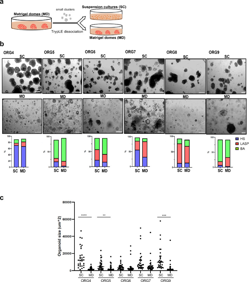
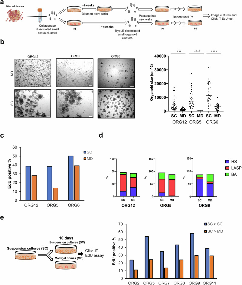
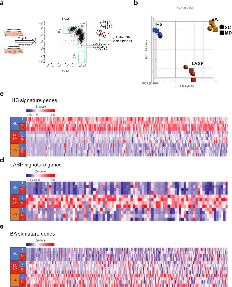
Organoid cultures offer a powerful technology to investigate many different aspects of development, physiology, and pathology of diverse tissues. Unlike standard tissue culture of primary breast epithelial cells, breast organoids preserve the epithelial lineages and architecture of the normal tissue. However, existing organoid culture methods are tedious, difficult to scale, and do not robustly retain estrogen receptor (ER) expression and responsiveness in long-term culture. Here, we describe a modified culture method to generate and maintain organoids as suspension cultures in reconstituted basement membrane (™Matrigel). This method improves organoid growth and uniformity compared to the conventional Matrigel dome embedding method, while maintaining the fidelity of the three major epithelial lineages. Using this adopted method, we are able to culture and passage purified hormone sensing (HS) cells that retain ER responsiveness upon estrogen stimulation in long-term culture. This culture system presents a valuable platform to study the events involved in initiation and evolution of ER-positive breast cancer.


Conflict of interest statement

Competing interests: J.S.B. is a scientific advisory board (SAB) member of Frontier Medicines and eFFECTOR Therapeutics. J.E.G. is a paid consultant for Helix and an uncompensated consultant for Konica Minolta and Earli. D.D. receives research funding from Canon, Inc (not related to work in this manuscript).

Figures

Fig. 1
Fig. 1. Propagation of normal human breast organoids as suspension cultures.
a Established breast organoid cultures derived using the conventional Matrigel dome method (passage 5–6) were dissociated and either re-cultured in Matrigel domes or as suspension cultures in 5% Matrigel for more than two months. b Representative brightfield images of six matched organoid lines grown as Matrigel domes (MD) or suspension cultures (SC) and their corresponding percentage of HS, LASP and BA cells based on FACS sorting using EpCAM and CD49f antibodies (4× magnification, scale bar 200 μm). c Mean organoid size of the indicated matched cultures grown as domes or in suspension. A total of 30 organoids per line per culture condition were measured using Image J. **p value < 0.01, ***p value < 0.001, ****p value < 0.0001, paired t-Test, two-tailed.
Fig. 2
Fig. 2. Breast organoid suspension cultures have a higher fraction of proliferating cells than Matrigel dome cultures.
a Minced normal breast tissues from three reductive mammoplasty samples were dissociated and used to derive matched Matrigel dome and suspension organoid cultures. Scheme was created with BioRender.com b Representative brightfield images of the three matched organoid lines grown as Matrigel domes (MD) or suspension cultures (SC) for 5 passages and their corresponding mean organoid size. Scale bar 200 μm. A total of 30 organoids per line per culture condition was measured using Image J. ***p value < 0.001, ****p value < 0.0001, paired t-Test, two-tailed. c Percentage of EdU-positive cells in the three organoid lines at passage 5. d Percentage of HS, LASP and BA cells based on FACS analyses of EpCAM/CD49f expression. e Percentage of EdU-positive cells in the indicated organoid lines 10 days after changing the culture condition from suspension to Matrigel domes (SC - > MD) or reseeding as suspension culture (SC - > SC).
Fig. 3
Fig. 3. Organoid suspension cultures preserve breast epithelial lineage fidelity.
a Organoid cultures were dissociated into single cells, FACS sorted into HS, LASP and BA cells based on differential expression of EpCAM/CD49f and total RNA was extracted for bulk RNA sequencing. b Principal Component Analysis (PCA) plot showing the HS, LASP and BA cell clusters isolated from organoids in Matrigel domes and suspension cultures. ce Heatmaps showing the breast epithelial subtype-specific gene expression across the samples. (Gene list in Supplementary Table 2).
Fig. 4
Fig. 4. Hormone sensing cells can be isolated and propagated in suspension cultures in the absence of other cell types.
a Representative brightfield images of four suspension cultures derived from sorted single HS cells based on expression of EpCAM/CD49f and cultured more than 2 weeks. 4× magnification, scale bar 200 μm. b Representative flow cytometry plot of HS cultures isolated from ORG6. c Percentage of HS, LASP and BA cells based on EpCAM/CD49f expression of HS cultures isolated from ORG4 (n = 4) and ORG6 (n = 3) and cultured for over 2 months, mean ± SD. d Percentage of HS cells based on expression EpCAM/CD49f in HS cultures isolated from eight different organoid lines and cultured for more than two months. e Representative immunofluorescence confocal images of HS cultures isolated from ORG2. Fluorescently conjugated antibodies targeting FOXA1, CD133 and α-SMA were used to detect the three different epithelial lineages and DAPI for nuclei staining. 20× magnification, scale bar 100 μm.
Fig. 5
Fig. 5. Organoids and HS cells cultured in suspension can activate ER signaling upon estrogen stimulation.
a Expression of the indicated ER target genes in triplicate cultures from ORG2 and ORG5 suspension cultures after estrogen treatment for seven days, assessed by qRT-PCR (n = 3, mean ± SD). *p value < 0.05, **p value < 0.01, ***p value < 0.001, ****p value < 0.0001, paired t-Test, two-tailed. b Heatmap showing early and late estrogen response gene expression in HS cells isolated from three organoid lines in suspension cultures then treated with or without estrogen for seven days (duplicate) with p value < 0.05 as the cutoff. (Gene list in Supplementary Table 4). c Expression of the indicated ER target genes in triplicate HS cultures isolated from three organoid lines and treated with or without estrogen for seven days (n = 3, mean ± SD), assessed by qRT-PCR. *p value < 0.05, **p value < 0.01, paired t-Test, two-tailed. d Representative immunofluorescence confocal images of HS cultures from ORG1 and ORG2 treated with or without estrogen (E2) and stained for estrogen receptor (ER) and progesterone receptor (PR), scale bar 100 μm.

Update of

References

    1. Guillen, K. P. et al. A human breast cancer-derived xenograft and organoid platform for drug discovery and precision oncology. Nat. Cancer3, 232–250 (2022). - PMC - PubMed
    1. Lukasiewicz, S. et al. Breast Cancer-Epidemiology, Risk Factors, Classification, Prognostic Markers, and Current Treatment Strategies-An Updated Review. Cancers13, 4287 (2021). - PMC - PubMed
    1. Sorlie, T. et al. Gene expression patterns of breast carcinomas distinguish tumor subclasses with clinical implications. Proc. Natl Acad. Sci. USA98, 10869–10874 (2001). - PMC - PubMed
    1. Waks, A. G. & Winer, E. P. Breast Cancer Treatment: A Review. JAMA321, 288–300 (2019). - PubMed
    1. Lumachi, F., Brunello, A., Maruzzo, M., Basso, U. & Basso, S. M. Treatment of estrogen receptor-positive breast cancer. Curr. Med Chem.20, 596–604 (2013). - PubMed