. 2024 Dec 19;7(1):1672.
doi: 10.1038/s42003-024-07357-1.

Enhancement of adult liver regeneration in mice through the hepsin-mediated epidermal growth factor receptor signaling pathway

Affiliations

Enhancement of adult liver regeneration in mice through the hepsin-mediated epidermal growth factor receptor signaling pathway

Yu-Fei Tsai et al. Commun Biol. .

Abstract

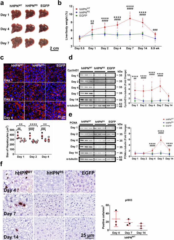
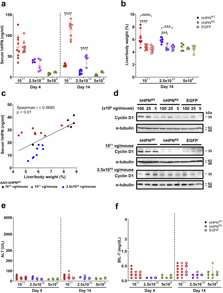
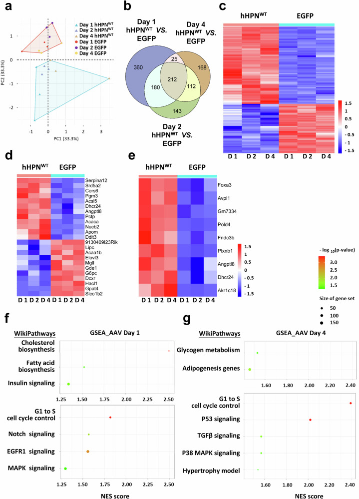
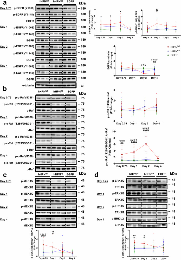
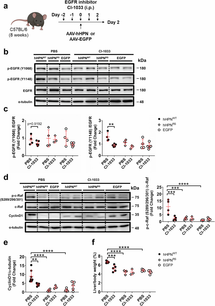
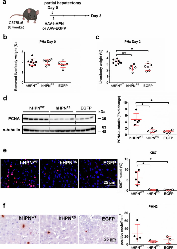
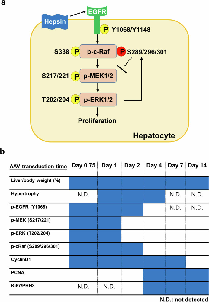
Given the widespread use of partial hepatectomy for treating various liver pathologies, understanding the mechanisms of liver regeneration is vital for enhancing liver resection and transplantation therapies. Here, we demonstrate the critical role of the serine protease Hepsin in promoting hepatocyte hypertrophy and proliferation. Under steady-state conditions, liver-specific overexpression of Hepsin in adult wild-type mice triggers hepatocyte hypertrophy and proliferation, significantly increasing liver size. This effect is predominantly driven by the catalytic activity of Hepsin, engaging the EGFR-Raf-MEK-ERK signaling pathway. Significantly, administering Hepsin substantially enhances hepatocyte proliferation and facilitates liver regeneration following a 70% partial hepatectomy. Crucially, the proliferation induced by Hepsin is a transient event, without leading to long-term adverse effects such as liver fibrosis or hepatocellular carcinoma, as evidenced by extensive observation. These results offer substantial potential for future clinical applications and translational research endeavors in the field of liver regeneration post-hepatectomy.


Conflict of interest statement

Competing interests: The authors declare no competing interests.

Figures

Fig. 1
Fig. 1. Induction of hepatocyte hypertrophy and proliferation in adult mice upon overexpression of human hepsin.
a The gross anatomy of the liver was examined on Days 1, 4 and 7 after administering AAV-hHPNWT, AAV-hHPNRS, or AAV-EGFP (control AAV). b The ratio of liver to body weight was determined at the indicated time points. c Representative fluorescent-tagged phalloidin staining of liver sections was used to visualize hepatocytes. Hepatocyte size was quantified. d, e Immunoblotting analysis and quantification of cyclin D1 and PCNA were conducted from Days 1 to 14. f Representative immunohistochemistry photomicrographs of mouse livers stained for phosphorylated histone H3 (pHH3). The pHH3-positive hepatocytes were quantified in the liver of mice that had been administered AAV-hHPNWT. At least n = 3 biologically independent animals were used per group. In the line graphs, data are presented as mean ± SD with individual data points included. In the dot plots, each dot represents the value for an individual mouse, and red lines indicate group means ± SD. Statistical significance was assessed using two-way ANOVA with Tukey’s post-test in (be) and Kruskal–Wallis test with Dunn’s post-test in (f). Significance levels are denoted by asterisks as follows: **p < 0.01, ****p < 0.0001 between the AAV-hHPNWT and AAV-EGFP groups, ##p < 0.01, ###p < 0.001, ####p < 0.0001 between the AAV-hHPNWT and AAV-hHPNRS groups.
Fig. 2
Fig. 2. Low levels of hepsin result in increased liver size in adult mice by inducing hepatocyte proliferation while maintaining normal liver function.
a Levels of human hepsin in serum samples on Days 4 and 14 in mice after administering a low dose of AAV-hHPNWT, AAV-hHPNRS, or AAV-EGFP (control AAV). b The ratio of liver to body weight were determined on Day 14. c The correlation between the liver-to-body weight ratio and serum human hepsin levels on Day 14 following AAV administration at a dosage of 1 × 10¹² vg/mouse (black), 1 × 10¹¹ vg/mouse (red), and 2.5 × 10¹⁰ vg/mouse (blue). d Immunoblotting analysis of cyclin D1 on Day 14. e, f Serum alanine aminotransferase (ALT) and total bilirubin (BIL-T) levels on Days 4 and 14. At least n = 5 biologically independent animals were used per group. Dots represent values for individual mice, and red lines represent group means ± SD. Statistical significance was assessed using two-way ANOVA with Sidak’s post-test at each time point in (a, b, e, f). Spearman correlation analysis was used in (c). Significance levels are denoted by asterisks as follows: ***p < 0.001, ****p < 0.0001.
Fig. 3
Fig. 3. Expression pattern of the liver transcriptome after overexpression of human hepsin.
a Principal component (PC) analysis plots depicting the integrated gene expression data matrix for mouse liver on Days 1, 2, and 4 post-administration of AAV-EGFP (control AAV) or AAV-hHPNWT. b Venn diagram illustrating the overlap and differential expression of transcripts on Days 1, 2, and 4 between the AAV-EGFP (control AAV) and AAV-hHPNWT groups. c Unsupervised clustering analysis of the AAV-EGFP (control AAV) and AAV-hHPNWT groups based on the common expressed transcripts across Days 1, 2, and 4. d, e Clustering analysis of transcripts associated with metabolism and cell proliferation from the shared expressed transcripts on Days 1, 2, and 4. f, g Results of Gene Set Enrichment Analysis (GSEA) in the AAV-hHPNWT and AAV-EGFP groups on Days 1 and 4, respectively. Each group consists of three mice. For the clustering analysis to generate a heatmap, each column corresponds to a specific time point, and the values within each column represent the average probe intensity of the transcriptomes obtained from three individual mice.
Fig. 4
Fig. 4. Hepsin-induced hepatocyte proliferation is activated by the EGFR-Raf-MEK-ERK signaling pathway.
Immunoblot analysis and quantification of mouse liver lysates at the indicated time points after administration of AAV-hHPNWT, AAV-hHPNRS, or AAV-EGFP (control AAV). a Cellular levels of phosphorylated EGFR (p-EGFR; Tyr1068 and Tyr1148) and total EGFR. b Levels of phospho-c-Raf (Ser338 and Ser289/Ser296/Ser301) and total c-Raf. c Levels of phospho-MEK1/2 (Ser217/221) and total MEK1/2. d Levels of phospho-ERK1/2 (Thr202/Tyr204) and total ERK1/2. At least n = 5 biologically independent animals were used per group. Data are presented as mean ± SD with individual data points included. Statistical significance was assessed using two-way ANOVA with Tukey’s post-test. Significance levels are denoted by asterisks as follows: *p < 0.05, **p < 0.01, ***p < 0.001, ****p < 0.0001 between the AAV-hHPNWT and AAV-EGFP groups, #p < 0.05, ##p < 0.01, ####p < 0.0001 between the AAV-hHPNWT and AAV-hHPNRS groups.
Fig. 5
Fig. 5. Effect of an EGFR inhibitor on hepatocyte proliferation induced by hepsin overexpression in adult mice.
a Study design: The irreversible inhibitor of EGFR (CI-1003) was administered daily starting 2 days before the administration of AAV-hHPNWT, AAV-hHPNRS, or AAV-EGFP (control AAV). Immunoblot analysis and quantification of mouse liver lysates were performed on Day 2 after AAV administration. b, c Levels of phosphorylated EGFR (p-EGFR; Tyr1068 and Tyr1148) and total EGFR. d, e Levels of p-c-Raf (Ser289/Ser296/Ser301), total c-Raf, and cyclin D1. f Ratio of liver weight to body weight. At least n = 3 biologically independent animals were used per group. Dots represent individual mouse values, and red lines represent group means ± SD. Statistical significance was assessed using two-way ANOVA with Tukey’s post-test. Significance levels are denoted by asterisks as follows: **p < 0.01, ***p < 0.001, ****p < 0.0001.
Fig. 6
Fig. 6. Enhanced liver regeneration in adult mice following partial hepatectomy induced by hepsin-mediated hepatocellular proliferation.
a Study design: Immediately after partial hepatectomy (PHx), adult mice were administered AAV-hHPNWT, AAV-hHPNRS, or AAV-EGFP (control AAV). b, c Liver-to-body weight ratio was determined based on the amount of liver tissue removed during surgery and then re-evaluated after a subsequent 3-day recovery period. d Immunoblot analysis and quantification of PCNA in mouse liver lysates. e Representative immunofluorescence micrographs of mouse liver tissue stained for Ki67, with quantification of Ki67-positive hepatocytes. f Representative immunohistochemistry photomicrographs of mouse livers stained for phosphorylated histone H3 (pHH3) with quantification of pHH3-positive hepatocytes.on Day 3 after PHx. At least n = 5 biologically independent animals were used per group. Dots represent values for individual mice, and red lines represent group means ± SD. Statistical significance was assessed using Kruskal–Wallis test with Dunn’s post-test. Significance levels are denoted by asterisks as follows: *p < 0.05, **p < 0.01.
Fig. 7
Fig. 7. Characteristics and experimentally verified signaling pathway of hepsin-induced hepatocyte proliferation.
a Mechanism of hepatocyte proliferation induced by the hepsin-mediated EGFR (epidermal growth factor receptor) signaling pathway. b Summary of the time course of hepsin-induced hepatocyte proliferation in adult mice via AAV-mediated overexpression. The upregulated signaling upon hepsin overexpression is highlighted in a blue box. N.D. not detected.

References

    1. Jiang, M., Ren, J., Belmonte, J. C. I. & Liu, G. H. Hepatocyte reprogramming in liver regeneration: biological mechanisms and applications. FEBS J.290, 5674–5688 (2023). - PubMed
    1. Gilgenkrantz, H. & Collin de l’Hortet, A. Understanding liver regeneration: from mechanisms to regenerative medicine. Am. J. Pathol.188, 1316–1327 (2018). - PubMed
    1. Abu Rmilah, A. et al. Understanding the marvels behind liver regeneration. Wiley Interdiscip. Rev. Dev. Biol.8, e340 (2019). - PMC - PubMed
    1. Rodimova, S. et al. Effect of Hepatic Pathology on Liver Regeneration: The Main Metabolic Mechanisms Causing Impaired Hepatic Regeneration. Int. J. Mol. Sci.24, 9112 (2023) - PMC - PubMed
    1. Min, J. S. et al. Systems analysis of the complement-induced priming phase of liver regeneration. J. Immunol.197, 2500–2508 (2016). - PMC - PubMed