. 2024 Dec 19;9(1):68.
doi: 10.1038/s41525-024-00457-1.

Functional assessment of IDUA variants of uncertain significance identified by newborn screening

Affiliations

Functional assessment of IDUA variants of uncertain significance identified by newborn screening

Seok-Ho Yu et al. NPJ Genom Med. .

Abstract

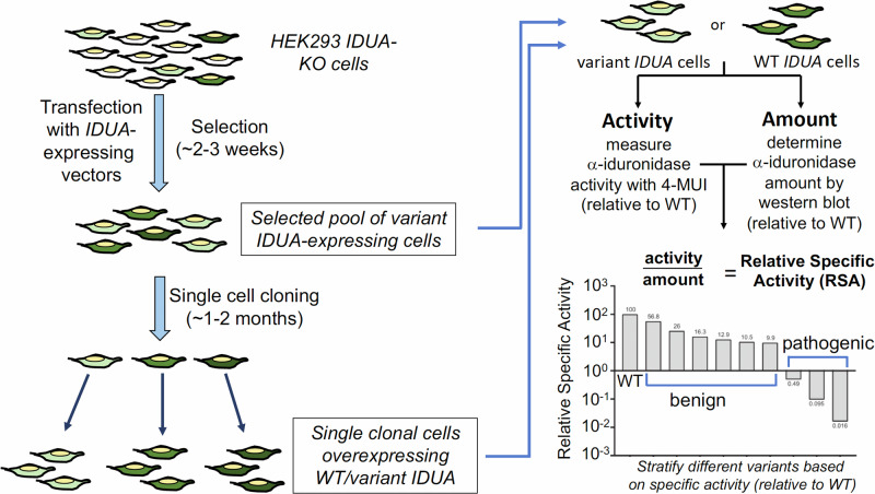
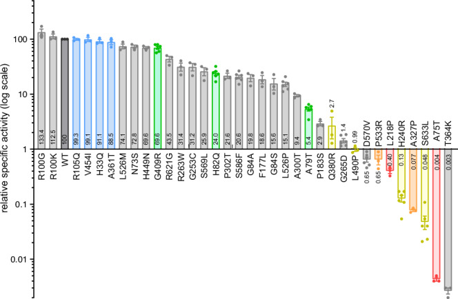
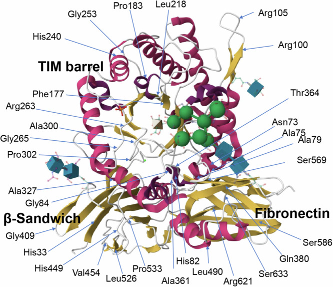
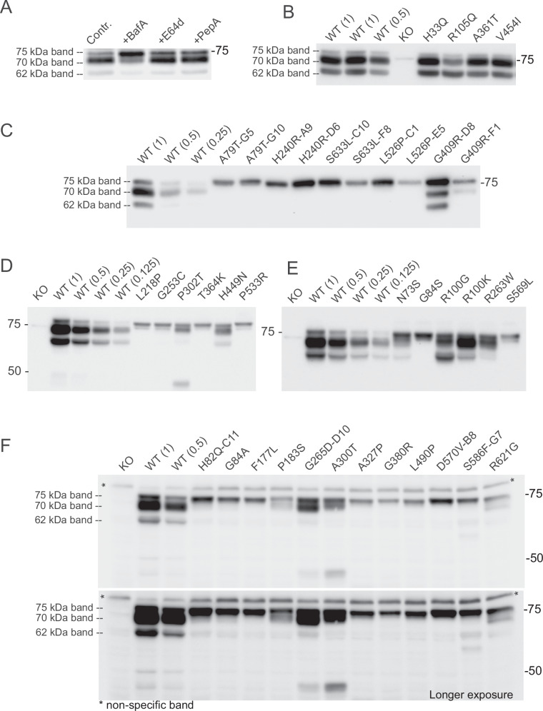
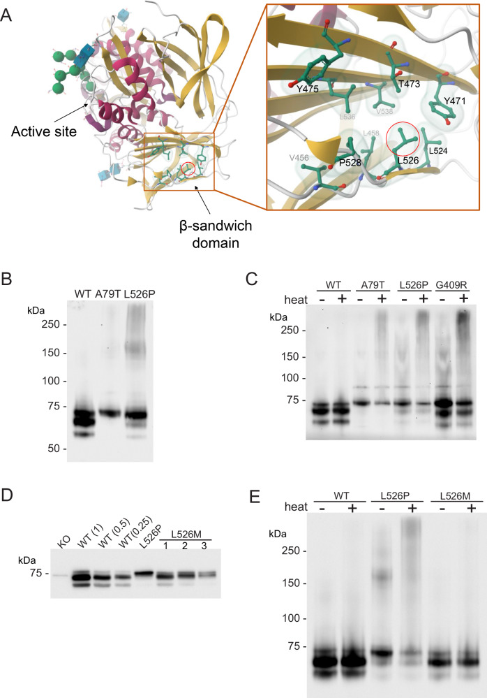
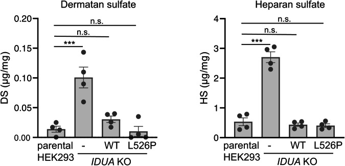
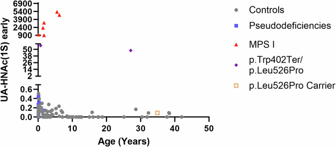
With the expansion of newborn screening efforts for MPS disorders, the number of identified variants of uncertain significance in IDUA continues to increase. To better define functional consequences of identified IDUA variants, we developed a HEK293-based expression platform that can be used to determine the relative specific activity of variant α-iduronidases by combining a fluorescence-based activity assay and semi-quantitative western blotting. We employed the current platform to characterize over thirty different IDUA variants, including known benign and pathogenic variants, as well as multiple variants of uncertain significance identified through newborn screening. This analysis allowed the stratification of variant enzymes based on their relative specific activity, and uncovered distinct effects of the different variants on enzyme folding, processing, and stability. While relative specific activity serves as a useful first-level test for enzyme function, our observations reinforce the need for secondary analyses of enzyme function to fully assess variant pathogenicity.


Conflict of interest statement

Competing interests: The authors declare no competing interests.

Figures

Fig. 1
Fig. 1. Overview of the IDUA platform and experimental workflow.
HEK293 IDUA KO cells are transfected with WT or variant-bearing IDUA expression vectors followed by antibiotic selection and, in some cases, single-cell cloning. Activity measurements using a fluorescent substrate for α-iduronidase are performed in the resulting cells along with quantitative western blot analysis to determine enzyme amount. These values are determined relative to the WT enzyme and combined to provide the relative specific activity (RSA) for each variant enzyme. This value represents the primary readout for each variant enzyme studied, but other secondary analyses can be performed to probe aggregation potential and ability to clear GAG accumulation.
Fig. 2
Fig. 2. Summary of relative specific activity values for IDUA variants tested in this study.
Graph showing the relative specific activity (% RSA) values (plotted on a log 10 scale) for every variant characterized in this study. Replicate analysis (n = 3–6) was performed on either cell populations after initial antibiotic selection, or 2-3 different single cell clones. Average RSA values are shown within the bars and standard error of the mean is depicted with the error bars. Bar colors denote the following classifications: dark gray – WT; light gray – VUS; blue – benign; green – known pseudodeficiency; yellow – associated with attenuated MPS I; orange – associated with attenuated or severe MPS I; red – associated with severe MPS I.
Fig. 3
Fig. 3. Position of amino acids within the α-iduronidase protein structure for which variants were characterized.
The location of specific amino acid residues corresponding to the variants we studied in this paper is shown. Domains within the crystal structure (PDB: 3W82; RSCB PDB), of the enzyme are also labeled.
Fig. 4
Fig. 4. Impaired lysosomal processing is observed in certain variant α-iduronidase enzymes.
Representative western blots for variant α-iduronidases in this study. A WT IDUA-expressing cells were treated with bafilomycin A (BafA), or the protease inhibitors, E64d or pepstatin A (PepA) prior to analysis by western blot. Note that only bafilomycin A blocked proteolytic processing of the enzyme. B Comparison of WT to the four known benign variants in this study. Note all four variant α-iduronidases are processed similarly to the WT enzyme. C Western blot analysis of various single-cell clones expressing the variant enzymes. Despite differences in enzyme amount within the different clones, the extent of proteolytic processing is comparable. D, E, F Representative blots for different variant-expressing selected cell populations showing variable impact on the proteolytic processing of the enzymes.
Fig. 5
Fig. 5. p.Leu526Pro increases aggregation of the resulting variant α-iduronidase enzyme.
A Protein structure of α-iduronidase (PDB: 3W82; RSCB PDB), inset depicts the location of leucine 526. B Representative blot of WT, p.Ala79Thr and p.Leu526Pro α-iduronidase following native gel electrophoresis. C Western blot showing accumulation of high molecular weight forms of α-iduronidase with certain variants in the presence or absence of heating at 40 °C. D Western blot analysis of WT, p.Leu526Pro, and p.Leu526Met α-iduronidase following denaturing gel electrophoresis. Note that substitution of Leu526 with methionine permits proteolytic processing while proline does not. E Western blot of WT, p.Leu526Pro, and p.Leu526Met α-iduronidase on a native gel with and without prior heating at 40 °C.
Fig. 6
Fig. 6. Overexpression of WT or p.Leu526Pro α-iduronidase reduces accumulation of dermatan and heparan sulfate.
Graph depicting abundance of total GAGs (dermatan and heparan sulfate) in WT parental and IDUA-KO HEK293 cells, and IDUA-KO HEK293 cells-expressing either WT or p.Leu526Pro IDUA. Measurements were made in four independent replicates. Error bars represent the standard error of the mean. Statistical analyses were performed using a Student’s t-test; *** denotes P < 0.001, n.s. denotes not significant.
Fig. 7
Fig. 7. Endogenous, non-reducing end GAG semi-quantitative analysis by tandem mass spectrometry on urine of patients bearing the p.Leu526Pro shows very modest elevation of UA-HNAc(1S).
Graph depicted NRE levels in different individuals as a function of age. Samples from two patients carrying the p.Leu526Pro were analyzed. Levels of the MPS I NRE marker UA-HNAc(1S) are expressed in apparent μmoles/mole of creatinine. Patient 1 is 27 years and 10 months old, with a genotype of p.Trp402Ter (pathogenic)/p.Leu526Pro (VUS). This patient had 48 apparent μmoles/mole of creatinine. Patient 2 (same genotype as patient 1) is 7 month-old male with 57 apparent μmoles/mole of creatinine. Their levels of UA-HNAc(1S) early are above the normal range (≤0.5 apparent μmoles/mole of creatinine), however they are not in the affected range seen for patients with MPS I (range: 906–5326 apparent μmoles/mole of creatinine).

References

    1. Fillman, T. et al. First three years’ experience of mucopolysaccharidosis type-I newborn screening in California. J. Pediatr.263, 113644 (2023). - PubMed
    1. Fuller, M. Laboratory diagnosis of lysosomal diseases: newborn screening to treatment. Clin. Biochem. Rev.41, 53–66 (2020). - PMC - PubMed
    1. Arunkumar, N. et al. Newborn screening of mucopolysaccharidoses: past, present, and future. J. Hum. Genet.65, 557–567 (2020). - PubMed
    1. Donati, M. A., Pasquini, E., Spada, M., Polo, G. & Burlina, A. Newborn screening in mucopolysaccharidoses. Ital. J. Pediatr.44, 126 (2018). - PMC - PubMed
    1. Stapleton, M. et al. Clinical presentation and diagnosis of mucopolysaccharidoses. Mol. Genet. Metab.125, 4–17 (2018). - PubMed

LinkOut - more resources