. 2025 Aug;43(8):1337-1347.
doi: 10.1038/s41587-024-02516-5. Epub 2024 Dec 19.

NIS-Seq enables cell-type-agnostic optical perturbation screening

Affiliations

NIS-Seq enables cell-type-agnostic optical perturbation screening

Caroline I Fandrey et al. Nat Biotechnol. 2025 Aug.

Abstract

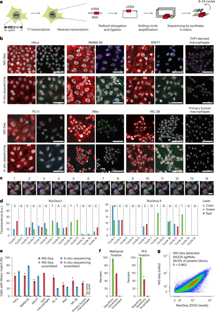
Optical pooled screening offers a broader-scale alternative to enrichment-based perturbation screening, using fluorescence microscopy to correlate phenotypes and perturbations across single cells. Previous methods work well in large, transcriptionally active cell lines, because they rely on cytosolic detection of endogenously expressed barcoded transcripts; however, they are limited by reliable cell segmentation, cytosol size, transcriptional activity and cell density. Nuclear In-Situ Sequencing (NIS-Seq) expands this technology by creating bright sequencing signals directly from nuclear genomic DNA to screen nucleated cells at high density and high library complexity. By inserting an inverted phage promoter downstream of the single guide RNA (sgRNA), many RNA copies of the sgRNA can be generated and sequenced independently of cellular transcription. In this study, we benchmarked NIS-Seq across eight cell types from two species and performed four genome-scale optical perturbation screens, identifying key players of inflammation-related cellular pathways. Finally, we performed a small-scale pooled optical screen in primary human macrophages from blood of healthy donors and demonstrated barcode identification in lentivirally transduced human skin tissue.


Conflict of interest statement

Competing interests: C.I.F., P.K. and J.L.S.-B. are inventors on a patent application related to nuclear in situ sequencing and pooled optical screening. J.L.S.-B. is a co-founder and shareholder of LAMPseq Diagnostics and ions.bio. E.L. and F.I.S. are co-founders and consultants of Odyssey Therapeutics. E.L. is a co-founder of Beren Therapeutics, Metaimmune Therapeutics and IFM Therapeutics. The other authors declare no competing interests.

Figures

Fig. 1
Fig. 1. NIS-Seq enables optical barcode identification in nucleated cells.
a, Outline of NIS-Seq reaction steps, adding a T7 transcription step to previously established in situ sequencing of barcoded mRNA. b, First-cycle NIS-Seq imaging results in comparison to cytosolic in situ sequencing results obtained across eight cell types. Live cells were stained for nuclei (gray) and membrane (red) before fixation (left panels). Scale bar, 50 µm. Representative images from two experimental replicates are shown. c, Raw images of 14 cycles of NIS-Seq barcode sequencing from THP1 cells. Nuclear staining was performed at cycles 1, 4, 7, 10 and 13. Scale bar, 10 µm. Representative data from four experimental replicates are shown. d, Quantitative spot intensities obtained from THP1 cell nuclei I and II highlighted in c. Indicated on top is the base calling result, matching two members of the pooled lentiviral library used. e, Fraction of cells mapping to known library member sequences. Scrambled controls indicate analysis results using a permutated reference library with equal base distribution. Dots indicate analysis results from two halves of a 0.56-cm2 surface. Two rows of edge tiles were excluded from analysis to avoid empty or distorted images. f, Specificity and sensitivity of NIS-Seq was quantified using a mixed population of cells either expressing GFP or containing NIS-Seq-compatible genomic insertions. The standard NIS-Seq protocol was compared to a modified version using PFA fixation and subsequent de-crosslinking (Methods). Dots indicate analysis results from two independent wells. g, Library coverage in transduced THP1 macrophages, measured by PCR-based NGS and NIS-Seq. Each library member covered in PCR-based sequencing is represented by one dot; dropping out sgRNAs—for example, those targeting essential genes—are not shown. Jitter was added to values to visualize spot density at low integer values.
Fig. 2
Fig. 2. Genome-scale optical perturbation screening for mediators of NF-κB activation.
a, Genome-wide NIS-Seq perturbation screening in HeLa–Cas9–p65–mNeonGreen cells stimulated with IL-1β. Fold changes are calculated based on the mean pixel-wise Pearson correlation between mNeonGreen and nuclear staining signals across cells with the same targeted gene versus non-targeting (NT) control cells. Deviation of their correlation value distributions was tested using a two-sided Wilcoxon–Mann–Whitney test followed by the Benjamini–Hochberg procedure. b, Collages of cellular images from a mapped to perturbed genes indicated. Shown is the mNeonGreen signal. c, Arrayed hit validation in HeLa–Cas9–p65–mNeonGreen cells using alternative sgRNA sequences from the TKOv3. Top panel, exemplary mNeonGreen images of IL-1β-stimulated cells. Bottom panel, distribution of activation states, quantified by pixel-wise Pearson correlation between mNeonGreen and nuclear staining signals. Scale bar, 50 µm. Representative data from two experimental replicates are shown. d, Genome-wide NIS-Seq perturbation screening in HeLa–Cas9–p65–mNeonGreen cells stimulated with TNF. Fold changes were calculated based on the mean pixel-wise Pearson correlation between mNeonGreen and nuclear staining signals across cells with the same targeted gene versus NT control cells. Deviation of their correlation value distributions was tested using a two-sided Wilcoxon–Mann–Whitney test followed by the Benjamini–Hochberg procedure. e, Collages of cellular images from d mapped to perturbed genes indicated. Shown is the mNeonGreen signal. f, Arrayed hit validation in HeLa–Cas9–p65–mNeonGreen cells using alternative sgRNA sequences from the TKOv3. Top panel, exemplary mNeonGreen images of TNF-stimulated cells. Bottom panel, distribution of activation states, quantified by pixel-wise Pearson correlation between mNeonGreen and nuclear staining signals. Scale bar, 50 µm. Representative data from two experimental replicates are shown.
Fig. 3
Fig. 3. Genome-scale optical perturbation screening in THP1-derived macrophages for inflammasome activation.
a, Genome-wide NIS-Seq perturbation screening in THP1–Cas9–ASC–GFP–CASP1/8DKO cells stimulated with nigericin, which is a known trigger of the NLRP3 inflammasome. Fold changes were calculated based on the high-frequency filtered GFP signal relative to the overall GFP signal per cell across cells with the same targeted gene versus non-targeting control cells. Deviation of gene-wise value distributions from controls was tested using a two-sided Wilcoxon–Mann–Whitney test followed by the Benjamini–Hochberg procedure. b, Collages of cellular images from a mapped to perturbed genes indicated. Shown are membrane stain (red) and ASC–GFP (green) signals. c, Arrayed hit validation in THP1–Cas9–ASC–GFP cells using alternative sgRNA sequences from the TKOv3. Shown are fractions of cells with an ASC speck upon nigericin stimulation from four independent replicate viral transductions. **P < 0.01 and ***P < 0.001, two-sided t-test. d, Cytokine secretion in response to two inflammasome triggers in wild-type or clonal gene-deficient THP1–ASC–GFP cells, measured by IL-1β ELISA. Shown is the mean of two technical replicate measurements from two biological replicates. e, Genome-wide NIS-Seq perturbation screening in THP1–Cas9–ASC–GFP–CASP1/8DKO cells stimulated with PrgI+PA, which is a known trigger of the NLRC4 inflammasome. Fold changes were calculated based on the high-frequency filtered GFP signal relative to the overall GFP signal per cell across cells with the same targeted gene versus non-targeting control cells. Deviation of gene-wise value distributions from controls was tested using a two-sided Wilcoxon–Mann–Whitney test followed by the Benjamini–Hochberg procedure. f, Collages of cellular images from e mapped to perturbed genes indicated. Shown are membrane stain (red) and ASC-GFP (green) signals. g, Arrayed hit validation in THP1–Cas9–ASC–GFP cells using alternative sgRNA sequences from the TKOv3. Shown are fractions of cells with an ASC speck upon PrgI+PA stimulation from four independent replicate viral transductions. ***P < 0.001, two-sided t-test. h, Cytokine secretion in response to two inflammasome triggers in wild-type or clonal gene-deficient THP1–ASC–GFP cells, measured by IL-1β ELISA. Shown is the mean of three technical replicate measurements from two biological replicates.
Fig. 4
Fig. 4. NIS-Seq perturbation screening in primary human macrophages.
a, Lentiviral vector co-expressing an sgRNA and C1C–EGFP for monitoring inflammasome activation (top) and a VPX–VPR expression plasmid for efficient lentiviral transduction of primary human macrophages (bottom). b, Outline of NIS-Seq perturbation screening in primary human macrophages. c, EGFP expression in primary human M-CSF macrophages transduced with CROPseq-iT7 C1C–EGFP and analyzed by FACS. FSC, forward scatter. d, Confocal imaging of C1C–EGFP localization in primary human macrophages. Cells were primed with 20 ng ml−1 LPS for 3 h and activated after 30 min of caspase inhibition (40 µM VX-765 and 50 µM Z-VAD) by 1 h of stimulation with 1 µg ml−1 PA and 10 ng ml−1 PrgI. Nuclei were stained with Hoechst 33342 (blue). Scale bars, 100 µm. Representative data from two experimental replicates are shown. e, Image analysis results of FACS-sorted EGFP+ cells stimulated as in c. f, Genome editing efficiencies in primary human macrophages transduced with the lentiviral vector depicted in a and electroporated with SpCas9 protein. Genomic editing rates were quantified by NGS in cells from two healthy donors. g, Reporter activation and four cycles of NIS-Seq in primary human macrophages after stimulation as described in d. Highlighted are two cells assigned to either a non-targeting control or an ANTXR2-targeting sgRNA by NIS-Seq. Scale bars, 50 μm. Representative data from two biological replicates are shown. h, Pooled optical perturbation screen in primary human macrophages. Shown are relative fractions of cells with blunted reporter activation for each of five perturbed genes targeted by four sgRNAs each or eight non-targeting control sgRNAs. Data points indicate results from two independent screening wells, each containing a mixture of macrophages from two healthy donors. In total, 2,663 macrophage cells were mapped to both a library sgRNA and a phenotype. i, Arrayed validation using alternative sgRNAs delivered as RNP complexes to primary human macrophages. Knockout rates were determined by sequencing (blue bars), and PrgI+PA-induced ASC specking rates were determined by anti-ASC antibody staining (red bars). Data from four biological replicates based on different human donors are shown. **P < 0.01; NS P > 0.05; two-sided t-test. NS, not significant.
Fig. 5
Fig. 5. NIS-Seq optical barcode identification in human epidermal tissue sheets.
a, Outline of preparing, transducing and sequencing human epidermal sheets using NIS-Seq. b, Six cycles of NIS-Seq barcode sequencing in human epidermal sheets transduced with a CROPseq-iT7 lentiviral library. Nuclear staining (gray) was performed at each cycle. Colors correspond to excitation lasers used to image NIS-Seq spots. Shown are maximum projections of eight tissue z-stacks starting at the bottom of the well distanced by 5 µm. Scale bar, 25 μm. Representative data from two experimental replicates are shown. c, Epidermal sheet shown in b stained for F-actin by phalloidin-AF670 (red). Scale bar, 25 μm. d, Epidermal sheet shown in b with indicated NIS-Seq signals matching members of the library used (green) or a scrambled library with the same base composition (yellow). Nuclei images from the first cycle (red) and sixth cycle (cyan) were overlayed in the background to visualize local tissue deformation artifacts. Instead of high-pass frequency filtering NIS-Seq data for sequence calling, images were low-pass filtered to compensate for imperfect alignments. Scale bar, 25 μm. e, One cycle of NIS-Seq barcode sequencing in human epidermal sheets as in a and b without prior lentiviral library transduction. Scale bar, 25 μm.
Fig. 6
Fig. 6. Zero-knowledge pooled optical screening analysis.
a, p65–mNeonGreen channel images of 712,146 HeLa cells stimulated with IL-1β were embedded using the SwinV2-T computer vision model pre-trained on a general image dataset, k-means clustered based on 768 penultimate layer activations and visualized using UMAP dimensionality reduction. b, Cluster representation among top-ranking perturbed genes, identified by zero-knowledge image clustering. Perturbed genes covered by at least 15 cells were ranked by the maximum over representation of any cluster as compared to non-targeting control cells. c, Single-cell localizations of top-ranking outlier genes visualized on the same UMAP embedding as used in a.

References

    1. Cong, L. et al. Multiplex genome engineering using CRISPR/Cas systems. Science339, 819–823 (2013). - PMC - PubMed
    1. Mali, P. et al. RNA-guided human genome engineering via Cas9. Science339, 823–826 (2013). - PMC - PubMed
    1. Carette, J. E. et al. Haploid genetic screens in human cells identify host factors used by pathogens. Science326, 1231–1235 (2009). - PubMed
    1. Shalem, O. et al. Genome-scale CRISPR–Cas9 knockout screening in human cells. Science343, 84–87 (2014). - PMC - PubMed
    1. Parnas, O. et al. A genome-wide CRISPR screen in primary immune cells to dissect regulatory networks. Cell162, 675–686 (2015). - PMC - PubMed

LinkOut - more resources