. 2024 Dec 19;14(1):30564.
doi: 10.1038/s41598-024-83206-9.

Histone variant H3.5 in testicular cell differentiation and its interactions with histone chaperones

Affiliations

Histone variant H3.5 in testicular cell differentiation and its interactions with histone chaperones

Patrick Philipp Weil et al. Sci Rep. .

Abstract

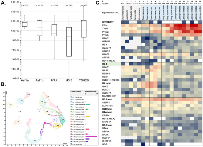
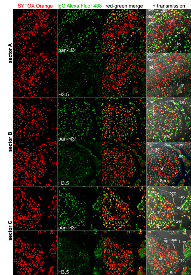
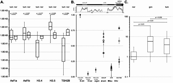
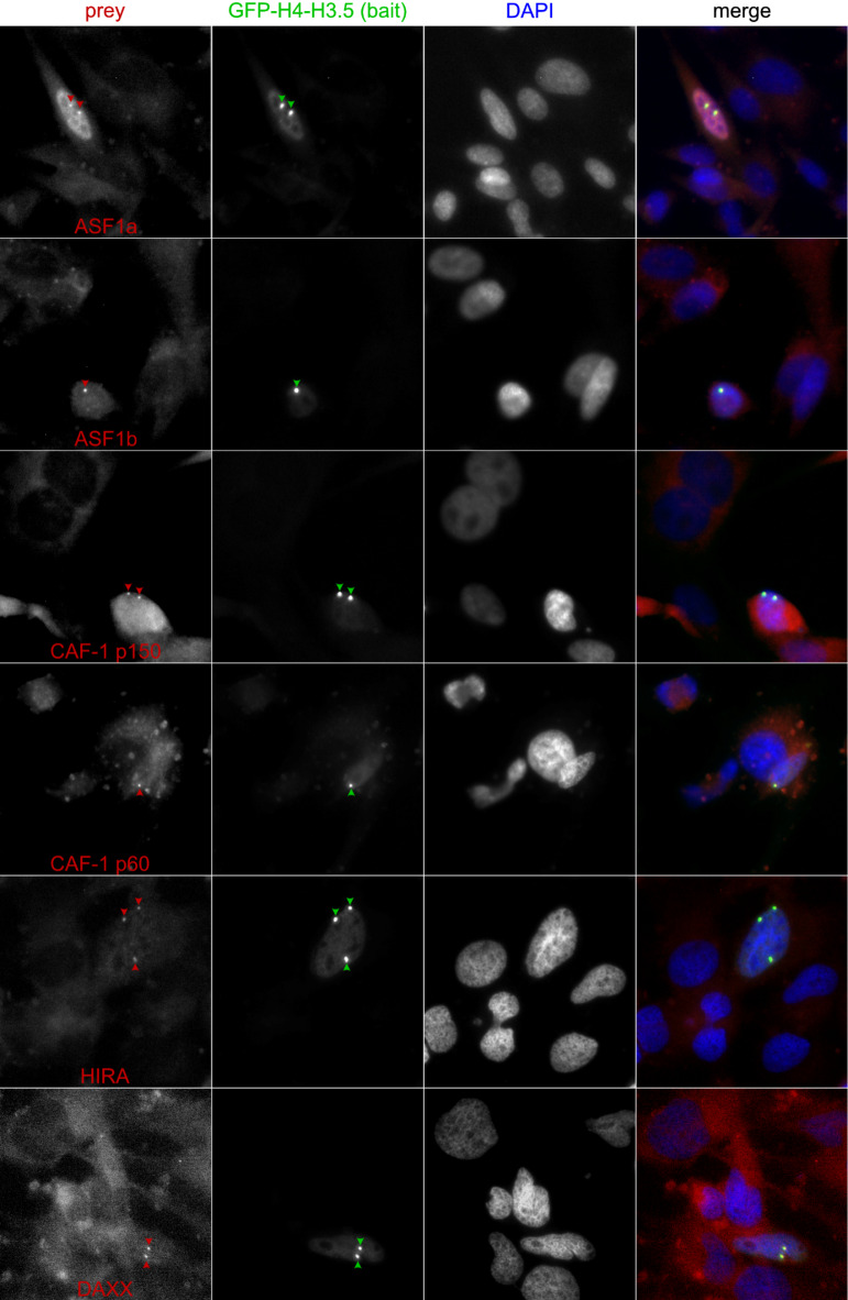
Testicular cell differentiation is a highly regulated process, essential for male reproductive health. The histone variant H3.5 is apparently a critical player in this intricate orchestra of cell types, but its regulation and function remains poorly understood. To elucidate its role, we fractionized testicular cells using c-Kit/CD117 as a separation marker and analyzed H3.5 expression. Further, we investigated the regulation of H3.5 expression using public data repositories. We explored DNA methylation patterns in specific regions of the H3-5 gene and assessed H3-5 copy number gain in seminoma specimens. Additionally, we examined the testicular localization of H3.5 and its histone chaperone interactions to understand its regulation at the protein level. We used qRT-PCR, MeDIP, and qPCR to study H3.5 expression and DNA methylation in various cell types. H3-5 copy number gain was analyzed using qPCR. Protein interactions were investigated through fluorescence-2-hybrid assays in baby hamster kidney cells. H3.5 is primarily enriched in spermatocytes. DNA methylation of a CpG island overlapping the H3-5 promoter appeared to be involved in the tissue-specific regulation of H3.5 expression. Elevated H3.5 expression was observed in seminoma specimens, suggesting a potential link to testicular tumors. H3-5 copy number gain was associated with elevated H3.5 expression in seminoma specimens. Furthermore, we identified physical interactions between H3.5 and histone chaperones Asf1a and Asf1b, HIRA, CAF p150 and DAXX, shedding light on the protein-level regulation of H3.5. These findings provide valuable insights into the molecular mechanisms governing testicular cell differentiation and the potential role of H3.5 in testicular pathologies.

Keywords: Chromatin remodeling; Epigenetic Plasticity; Epigenetic regulation; Histone chaperones; Histone variants; Spermatogenesis.


Conflict of interest statement

Declarations. Competing interests: The authors declare no competing interests. Ethics approval: This study was conducted with approval of the Witten/Herdecke University Ethics board (No’s. 90/2011 and 138/2013). In addition, for investigations involving human subjects, informed consent has been obtained from all participants. Patients or the public were not involved in the design, or conduct, or reporting, or dissemination plans of our research. Research involving human research participants were performed in accordance with the Declaration of Helsinki. Informed consent: Informed written consent was obtained by all involved donors.

Figures

Fig. 1
Fig. 1
Evolutionary distance analyses of human H3 variants. (A) The number of amino acid substitutions per site from between sequences are shown. Standard error estimate(s) are shown above the diagonal. Analyses were conducted using the Poisson correction model. The analysis involved 16 amino acid sequences. All ambiguous positions were removed for each sequence pair. There were a total of 105 positions in the final dataset. Evolutionary analyses were conducted in MEGA7. (B) The evolutionary history was inferred by using the Maximum Likelihood method and JTT matrix-based model. The tree with the highest log likelihood (-530.27) is shown. Initial tree(s) for the heuristic search were obtained automatically by applying Neighbor-Join and BioNJ algorithms to a matrix of pairwise distances estimated using the JTT model, and then selecting the topology with superior log likelihood value. This analysis involved 10 amino acid sequences. There were a total of 100 positions in the final dataset. Evolutionary analyses were conducted in MEGA X,. (C) Alignment of 11 histone H3 variants.
Fig. 2
Fig. 2
(A) Box plot diagram of the expression analysis after cell type separation by MACS technology. Y-axis: Fold change of mRNA expression in c-Kit/CD117-negative vs. positive cells, whereby fold changes > 1.0E + 00 mean relative upregulation in c-Kit/CD117-negative cells and fold changes < 1.0E + 00 mean relative upregulation in c-Kit/CD117-positive cells. (B) H3-5 expression (scRNAseq-data) associated with 20 testicular cell type-clusters (c-0 – c-19). (C) The heat map illustrates the enrichment of H3.5 mRNA (scRNAseq-data) and mRNAs from other available histone variant genes, multiple histone chaperones, protamines, and other genes of interest associated with 20 testicular cell type-clusters. With respect to spermatogenesis, non-germline cells were separated on the left side of the heat map, and spermatogenesis stages were serially ordered from left to right. Shades of red represent high expression levels, shades of yellow represent moderate levels, and shades of blue represent low expression levels. White indicates the absence of expression data.
Fig. 3
Fig. 3
Immunofluorescence microscopy analyses on the localization of pan-H3 or H3.5 in human normal testis sections. In order to compare similar areas (sectors A-C), we analyzed consecutive microscopic thin sections, each loaded with the anti-H3.5 antibody or a polyclonal anti-pan-H3 antibody, which cannot distinguish between different H3 variants. Red: DNA counterstaining using Sytox Orange dye. Green: H3 staining using goat anti-rabbit pAb conjugated with Alexa Fluor 488. Abbreviations: pcc, peritubular contractile cells/myoid cells; Ser, Sertoli cells; Ley, Leydig cells; sg, spermatogonia; sc, (primary) spermatocytes; st, spermatids.
Fig. 4
Fig. 4
(A) Box plot diagram of the expression analysis. The expression of the genes Asf1a, Asf1b, H2A.Bbd, H3.4, H3.5, and TSH2B (x-axis) in tumor tissue (tum) and normal testis (nor), normalized to ACTB, GAPDH and RPS27 (y-axis), is depicted. (B) DNA of leukocytes, liver cells, normal testicular cells, germ cell neoplasia in situ (gcn) cells and testicular tumor cells (Seminoma) exhibited overlapping amounts of intermediately methylated DNA at the distant CpG island. In contrast, the CpG island adjacent and overlapping to H3-5 exhibited hypermethylation in DNA from leukocytes and liver cells. In contrast, the same CpG island exhibited hypomethylated DNA in normal and tumor testicular cells and CIS. DNA methylation levels within the AT-rich segment were almost indistinguishable from unmethylated and hydroxymethylated control DNA. These observations suggest that DNA methylation of the CpG island adjacent and overlapping with H3-5 could play a role in the tissue-specific differential regulation of H3.5 expression. However, DNA methylation did not seem to be involved in the elevation of H3.5 expression in a fraction of seminoma-type testicular tumors. (C) Boxplots of gene copy number results. Graphical representation of the fold changes of H3F3C in the three examined tissue types. nor = healthy testicular tissue, gcn = germ cell neoplasia in situ, tum = testicular tumor tissue.
Fig. 5
Fig. 5
To understand, how H3.5 is regulated by interactions with histone chaperones on the protein level, we performed fluorescence-2-hybrid assays. We interrogated the nuclear localization, chromatin patterns and interactions of GFP-H3.5 with Asf1, HIRA, CAF as well as DAXX. The latter were fused with RFP. We observed that H3.5 physically interacted with Asf1, HIRA or DAXX, but not with CAF, when interrogated proteins-of-interest were expressed under CMV promoter control in baby hamster kidney (BHK) cells.
Fig. 6
Fig. 6
To understand, how H3.5 is regulated by interactions with histone chaperones on the protein level, we performed fluorescence-2-hybrid assays. We interrogated the nuclear localization, chromatin patterns and interactions of GFP-H3.5 with Asf1, HIRA, CAF as well as DAXX. The latter were fused with RFP. We observed that H3.5 physically interacted with Asf1, HIRA or DAXX, but not with CAF, when interrogated proteins-of-interest were expressed under CMV promoter control in baby hamster kidney (BHK) cells.

References

    1. Gaspa-Toneu, L. & Peters, A. H. Nucleosomes in mammalian sperm: conveying paternal epigenetic inheritance or subject to reprogramming between generations? Curr. Opin. Genet. Dev.79, 102034 (2023). - PMC - PubMed
    1. Borg, M., Jiang, D. & Berger, F. Histone variants take center stage in shaping the epigenome. Curr. Opin. Plant. Biol.61, 101991 (2021). - PubMed
    1. Gurard-Levin, Z. A. & Almouzni, G. Histone modifications and a choice of variant: a language that helps the genome express itself. F1000Prime Rep.6, 76 (2014). - PMC - PubMed
    1. Kamakaka, R. T. & Biggins, S. Histone variants: deviants? Genes Dev.19(3), 295–310 (2005). - PubMed
    1. Postberg, J., Forcob, S., Chang, W. J. & Lipps, H. J. The evolutionary history of histone H3 suggests a deep eukaryotic root of chromatin modifying mechanisms. BMC Evol. Biol.10, 259 (2010). - PMC - PubMed

LinkOut - more resources