. 2024 Dec 19;7(1):1669.
doi: 10.1038/s42003-024-07342-8.

Protogenin facilitates trunk-to-tail HOX code transition via modulating GDF11/SMAD2 signaling in mammalian embryos

Affiliations

Protogenin facilitates trunk-to-tail HOX code transition via modulating GDF11/SMAD2 signaling in mammalian embryos

Yu-Sheng Hung et al. Commun Biol. .

Abstract

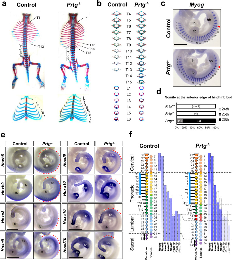
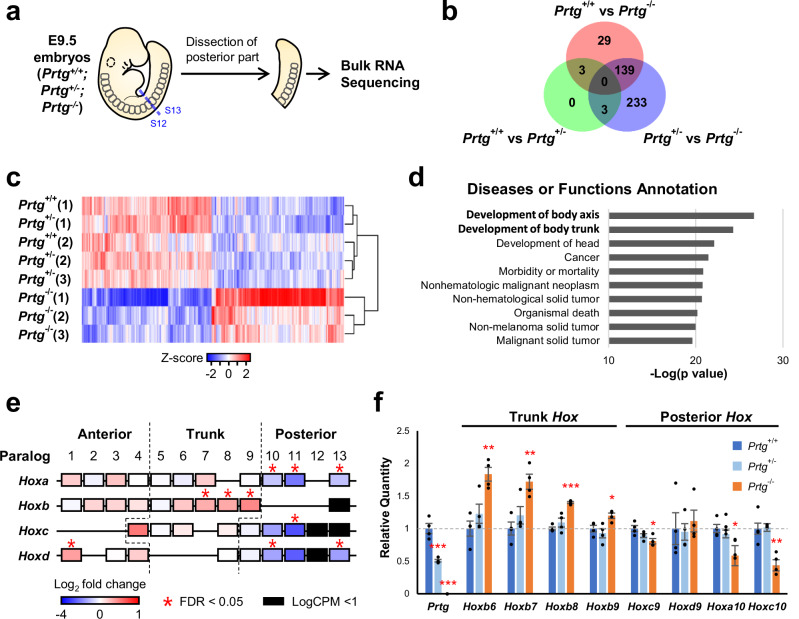
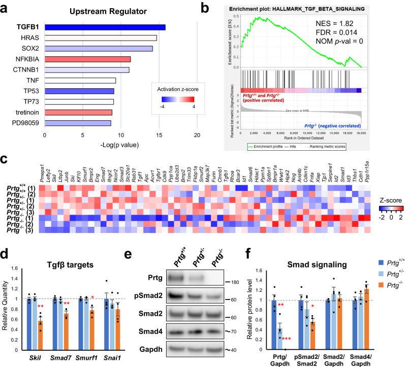
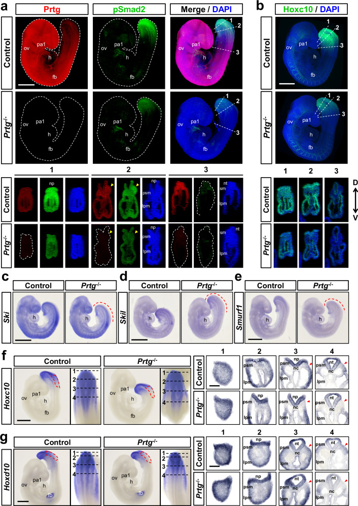
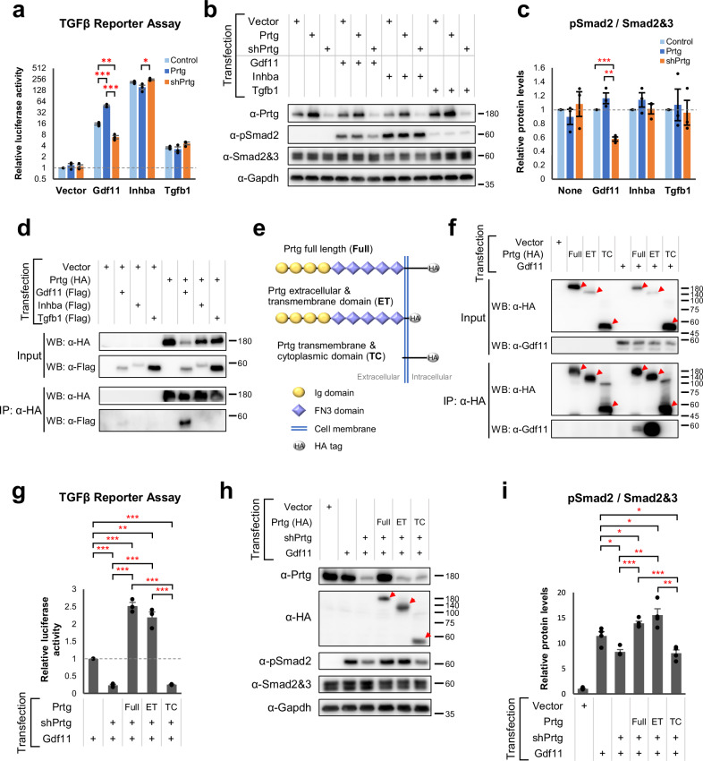
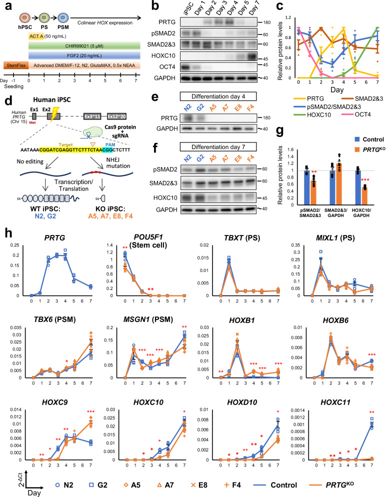
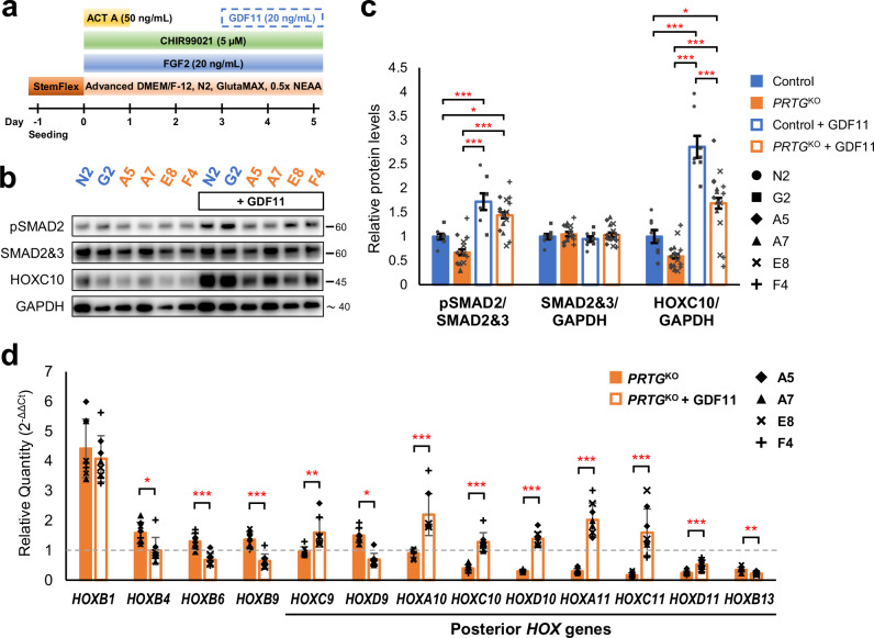
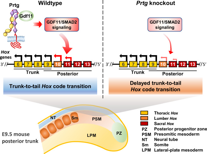
During embryogenesis, vertebral axial patterning is intricately regulated by multiple signaling networks. This study elucidates the role of protogenin (Prtg), an immunoglobulin superfamily member, in vertebral patterning control. Prtg knockout (Prtg-/-) mice manifest anterior homeotic transformations in their vertebral columns and significant alterations in homeobox (Hox) gene expression. Transcriptomic profiling of Prtg-/- mouse embryos highlights Prtg-regulated genes involved in axial development, particularly within the transforming growth factor beta (TGFβ) signaling pathway. Reduced TGFβ signaling in Prtg-/- mouse embryos is evidenced by decreased phosphorylated Smad2 (pSmad2) levels and its downstream target genes in the developing tail. We further show that Prtg interacts with growth differentiation factor 11 (GDF11) to enhance GDF11/pSmad2 signaling activity. Using human-induced pluripotent stem cell-derived presomitic mesoderm-like (hiPSC-PSM) cells, we demonstrate delayed posterior HOX gene expression upon PRTG knockout, which is rescued by GDF11 supplementation. These findings provide compelling evidence that PRTG regulates HOX genes through the GDF11/SMAD2 signaling pathway.


Conflict of interest statement

Competing interests: The authors declare no competing interests.

Figures

Fig. 1
Fig. 1. Anterior homeotic transformation in the vertebrae of Prtg/ mice.
a Skeletal and cartilaginous tissue staining of control and Prtg/ neonatal mice. Two additional thoracic vertebrae (top) and three more sternum-attached rib pairs (bottom) were observed in Prtg/ mice. b Isolated thoracic and lumbar vertebrae from control and Prtg/ mice. In Prtg/ mice, T8 to T10 were transformed into a T7-like shape, while T11 to T13 were transformed into a T8-like shape (n = 32). c Whole-mount in situ hybridization of Myog mRNA in E11.0 control and Prtg/ embryos. The somite at the anterior edge of the hindlimb bud is indicated by a red arrowhead. The scale bar represents 1 mm; fb forelimb bud, h heart, hb hindlimb bud. d Quantification results of the somite number located at the anterior edge of the hindlimb bud, which has been visualized and manually enumerated as shown in (c), in E11.0 Prtg+/+ (n = 2), Prtg+/− (n = 4), and Prtg/ (n = 6) embryos from 2 litters. e Whole-mount in situ hybridization of Hoxb6 (n = 10), Hoxb9 (n = 10), Hoxc8 (n = 5), Hoxc9 (n = 8), Hoxd9 (n = 5), Hoxa10 (n = 5), Hoxc10 (n = 4), and Hoxd10 (n = 4) in control and Prtg/ embryos. In situ hybridization of Hoxa10 was performed on E11.0 and that of others on E10.0. Dashed red lines indicate the regions where Prtg/ embryos displayed differential expression compared with control embryos. Scale bars represent 1 mm. f Summary of the vertebral phenotypes and Hox expression patterns in control and Prtg/ mice. Dashed boxes delineate the regions of Hox gene expression in the control group. The intensity of the blue color indicates the level of Hox gene expression observed by in situ hybridization. C cervical vertebra (orange), L lumbar vertebra (light blue), S sacral vertebra (purple), T thoracic vertebra (yellow and green). Red-colored vertebrae indicate additional vertebrae in Prtg/ mice.
Fig. 2
Fig. 2. Transcriptome analysis of the posterior trunks of E9.5 Prtg/ embryos.
a Schematic representation of the experimental design for transcriptome analysis. E9.5 embryos were dissected between the 12th and 13th somite, and the posterior trunks were subjected to bulk RNA-seq analysis. b Numbers of differentially expressed genes (DEGs) observed in Prtg+/+ vs Prtg+/, Prtg+/+ vs Prtg/, and Prtg+/ vs Prtg/ embryos. DEGs were identified using edgeR with FDR < 0.05 as the cutoff. c Heatmap of DEGs in the Prtg+/+, Prtg+/, and Prtg/ samples. A total of 529 DEGs were identified between the control (Prtg+/+ and Prtg+/) and Prtg/ samples, among which 267 and 262 genes were up-regulated and down-regulated in the Prtg/ samples, respectively. d Diseases or Functions Annotation generated via Ingenuity Pathway Analysis (IPA) illustrating the significant association of Prtg-regulated genes with body axis development. e Heatmap illustrating the fold change in Hox gene expression observed in Prtg/ samples relative to control embryos (Prtg+/+ and Prtg+/), derived from RNA-seq data. f The expression levels of Prtg, Hoxb6, Hoxb7, Hoxb8, Hoxb9, Hoxc9, Hoxd9, Hoxa10, and Hoxc10 in the posterior trunk samples of Prtg+/+, Prtg+/, and Prtg/ embryos were quantified using RT-qPCR. Tbp was used as the reference gene. Data are presented as the mean ± SEM. Statistical significance relative to Prtg+/+ is indicated (n = 4 for each bar; *p < 0.05; **p < 0.01; ***p < 0.001, by one-way ANOVA).
Fig. 3
Fig. 3. Down-regulation of TGFβ signaling activity in the posterior trunks of E9.5 Prtg/ embryos.
a Upstream regulator analysis via IPA demonstrated that the DEGs in the Prtg/ samples were significantly associated with TGFβ1. b GSEA revealed that TGFβ signaling was significantly altered in Prtg/ embryos. c Heatmap of the expression levels of genes within the TGFβ signaling gene set. d The expression levels of TGFβ signaling target genes in the Prtg+/+, Prtg+/, and Prtg/ posterior trunk samples were quantified using qRT-PCR. Tbp was used as the reference gene. Data are presented as the mean ± SEM. Statistical significance relative to Prtg+/+ is indicated (n = 4 for each bar; *p < 0.05; **p < 0.01; ***p < 0.001, by one-way ANOVA). e Western blot analysis of the Prtg, phosphorylated Smad2 (pSmad2), total Smad2, and Smad4 levels in Prtg+/+, Prtg+/, and Prtg/ posterior trunk samples. Gapdh was used as an internal control. The molecular weight ladders are labeled, and the estimated molecular weight size is indicated by a tilde. f Quantitative results of (e). The level of pSmad2 was significantly decreased in the Prtg/ samples. Data are presented as the mean ± SEM. Statistical significance relative to Prtg+/+ is indicated (n = 4 for each bar; *p < 0.05; **p < 0.01; ***p < 0.001, by one-way ANOVA).
Fig. 4
Fig. 4. Decreased TGFβ signaling activity in the PSM of Prtg/ embryos.
a Whole-mount immunostaining of Prtg (red), pSmad2 (green), and DAPI (blue) in E9.5 control and Prtg/ embryos. Digital transverse sections at the indicated levels in the whole-mount embryos are shown at the bottom (n = 8 embryos per group). Scale bars represent 500 µm. b Whole-mount immunostaining of Hoxc10 (green) and DAPI (blue) in E9.5 control and Prtg/ embryos (n = 4 embryos per group). Digital transverse sections at the indicated levels in the whole-mount embryos are shown at the bottom. Arrowheads indicate the PSM regions. Scale bars represent 500 μm. D dorsal, V ventral. ce Whole-mount in situ hybridization of Ski (n = 2) (c), Skil (n = 2) (d), and Smurf1 (n = 2) (e) in E9.5 control and Prtg/ embryos. Dashed lines indicate regions where the expression levels are decreased in Prtg/ embryos. Scale bars represent 500 µm. f, g Whole-mount in situ hybridization of Hoxc10 (n = 2) (f) and Hoxd10 (n = 2) (g) in E9.5 embryos. Red dashed boxes indicate regions of reduced expression levels of the indicated gene in Prtg/ embryos. Dashed lines indicate the positions of sections in the right panels. Scale bars represent 500 µm in whole-mount images and 100 µm in sections. fb forelimb bud, h heart, lpm lateral plate mesoderm, nc notochord, np neural plate, nt neural tube, ov otic vesicle, pa1 1st pharyngeal arch, psm presomitic mesoderm, sm somitic mesoderm.
Fig. 5
Fig. 5. Prtg interacts with Gdf11 and modulates its signaling activity.
a (CAGA)12-MLP-Luc reporter activities in P19 cells transfected with Gdf11, Inhba, Tgfb1, or a control, along with either Prtg overexpression or knockdown (shPrtg) vectors. Control and Prtg overexpression vectors contain a shRNA with a scrambled shPrtg sequence (shCtrl). Data are presented as the mean ± SEM. Statistical significance is indicated (n = 3 for each bar; *p < 0.05; **p < 0.01; ***p < 0.001, by one-way ANOVA). b Protein levels of Prtg, pSmad2, and Smad2&3 were measured by western blot. Gapdh serves as an internal control. c Quantification of pSmad2 levels in (b). Data are presented as the mean ± SEM. Statistical significance is indicated (n = 3 for each bar; *p < 0.05; **p < 0.01; ***p < 0.001, by one-way ANOVA). d Co-immunoprecipitation of P19 cells transfected with HA-tagged Prtg and flag-tagged TGFβ ligands. IP immunoprecipitation, WB western blot. e Schematic illustration of Prtg variants: full-length Prtg (Full), Prtg lacking the intracellular domain (ET), and Prtg lacking the extracellular domain (TC). f Co-immunoprecipitation of Prtg Full, Prtg ET, and Prtg TC with Gdf11 in P19 cells. Arrowheads indicate Prtg variants in the immunoblot images. g (CAGA)12-MLP-Luc reporter activities in P19 cells expressing Gdf11 with Prtg Full, Prtg ET, or Prtg TC. Data are presented as the mean ± SEM. Statistical significance is indicated (n = 3 for each bar; *p < 0.05; **p < 0.01; ***p < 0.001, by one-way ANOVA). h Levels of pSmad2 in P19 cells expressing Gdf11 with Prtg variants were analyzed by western blot. i Quantification of pSmad2 levels (h). Data are presented as the mean ± SEM. Statistical significance is indicated (n = 3 for each bar; *p < 0.05; **p < 0.01; ***p < 0.001, by one-way ANOVA).
Fig. 6
Fig. 6. Recapitulation of PRTG knockout (PRTGKO) phenotypes observed in embryos using an in vitro hiPSC-derived PSM model.
a Schematic illustration of the differentiation protocol for generating induced pluripotent stem cell-derived presomitic mesoderm-like (iPSC-PSM) cells. PS, primitive streak; PSM, presomitic mesoderm. b The protein levels of PRTG, pSMAD2, SMAD2/3, HOXC10, and OCT4 at different time points in differentiated iPSC-PSM cells from control clones were measured using a western blot. The molecular weight of nearby ladders is labeled. The estimated molecular weight size is labeled using a tilde. c Quantification results of Fig. 5b. Data are presented as the mean ± SEM (n = 4). d Schema illustrating the generation of PRTGKO (A5, A7, E8, and F4) and non-edited (G2 and N2) hiPSC clones via CRISPR/Cas9 gene editing. e PRTG expression in each hiPSC clone was validated using a western blot on day 4 of the iPSC-PSM model. f The protein levels of pSMAD2, SMAD2/3, and HOXC10 on day 7 of the hiPSC-PSM model were measured using a western blot. g Quantification results of Fig. 5f. Data are presented as the mean ± SEM (n = 4 for control group and n = 8 for PRTGKO group) (**p < 0.01; ***p < 0.001; by student’s t-test). h The expression levels of PRTG, stem cell marker (POU5F1, also known as OCT4), PS markers (TBXT and MIXL1), PSM markers (TBX6 and MSGN1), HOXB1, HOXB6, HOXC9, HOXD9, HOXC10, HOXD10, and HOXC11 from day 0 to day 7 of the differentiated hiPSC-PSM model were measured using RT-qPCR. RPL13A was used as the reference gene. Data are presented as the mean ± SEM (n = 4 for control group and n = 8 for the PRTGKO group) (*p < 0.05; **p < 0.01; ***p < 0.001; by two-way ANOVA).
Fig. 7
Fig. 7. GDF11 administration rescues the delayed expression of posterior HOX genes in PRTG-deficient hiPSC-PSM cells.
a Schematic illustration of the experimental design. b Protein levels of pSMAD2, total SMAD2/3, and HOXC10 on day 5 of the PRTGKO hiPSC-PSM model, with or without GDF11 treatment, were measured using western blot. c Quantification results of (b). Data are presented as the mean ± SEM (n = 8 in the control group and n = 16 in the PRTGKO group; *p < 0.05; ***p < 0.001; by one-way ANOVA). d The expression levels of HOXB1, HOXB4, HOXB6, HOXB9, HOXC9, HOXD9, HOXA10, HOXC10, HOXD10, HOXA11, HOXC11, HOXD11, and HOXB13 on day 5 of the PRTGKO and GDF11-treated PRTGKO iPSC-PSM cells were measured using RT-qPCR. RPL13A was used as the reference gene. Expression levels were normalized to the levels of control iPSC-PSM cells (N2 and G2 clones). Data are presented as the mean ± SEM (n = 8 in each group; *p < 0.05; **p < 0.01; ***p < 0.001; by student’s t-test).
Fig. 8
Fig. 8. Delayed transition of thoracic-to-lumbar Hox code in Prtg knockout mice.
A schematic representation illustrating the process of thoracic-to-lumbar Hox code transition in wild-type and Prtg knockout embryos. Arrows above the Hox genes denote the direction of Hox code translation. In wild-type mice, Prtg interacts with Gdf11 to enhance TGFβ signaling, thereby facilitating the transition of the Hox code from thoracic to lumbar identity, which occurs in the tail region of E9.5 embryos. In Prtg knockout mice, decreased TGFβ signaling results in a delayed transition of the Hox code.

References

    1. Mallo, M., Vinagre, T., & Carapuco, M. The road to the vertebral formula. Int J. Dev. Biol.53, 1469–1481 (2009). - PubMed
    1. Martin, B. L. Mesoderm induction and patterning: insights from neuromesodermal progenitors. Semin Cell Dev. Biol.127, 37–45 (2022). - PMC - PubMed
    1. Tzouanacou, E., Wegener, A., Wymeersch, F. J., Wilson, V. & Nicolas, J. F. Redefining the progression of lineage segregations during mammalian embryogenesis by clonal analysis. Dev. Cell17, 365–376 (2009). - PubMed
    1. Henrique, D., Abranches, E., Verrier, L. & Storey, K. G. Neuromesodermal progenitors and the making of the spinal cord. Development142, 2864–2875 (2015). - PMC - PubMed
    1. Hubaud, A. & Pourquie, O. Signalling dynamics in vertebrate segmentation. Nat. Rev. Mol. Cell Biol.15, 709–721 (2014). - PubMed

MeSH terms

LinkOut - more resources