

USP8-Dependent Family Tyrosine Kinase Promotes the Malignant Progression of Esophageal Squamous Cell Carcinoma by Upregulating Protein Tyrosine Kinase 2 Expression
- PMID: 39702934
- PMCID: PMC11739124
- DOI: 10.1111/1759-7714.15489
USP8-Dependent Family Tyrosine Kinase Promotes the Malignant Progression of Esophageal Squamous Cell Carcinoma by Upregulating Protein Tyrosine Kinase 2 Expression
Abstract
Background: Esophageal squamous cell carcinoma (ESCC) is a lethal malignancy, and the molecular underpinnings of its aggressive behavior are not fully understood. FYN proto-oncogene, Src family tyrosine kinase (FYN) has been linked to cancer progression, yet its role in ESCC remains elusive. This study investigated the influence of FYN on ESCC malignancy.
Methods: Quantitative real-time polymerase chain reaction was used to assess the mRNA expression of FYN, while western blotting and immunohistochemistry (IHC) assays were performed to detect the protein expression of FYN, ubiquitin specific peptidase 8 (USP8) and protein tyrosine kinase 2 (PTK2). Cell viability was measured with a cell counting kit-8 assay, and cell apoptosis was evaluated using flow cytometry.
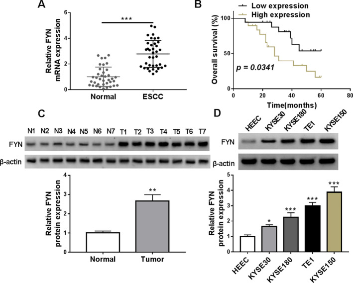
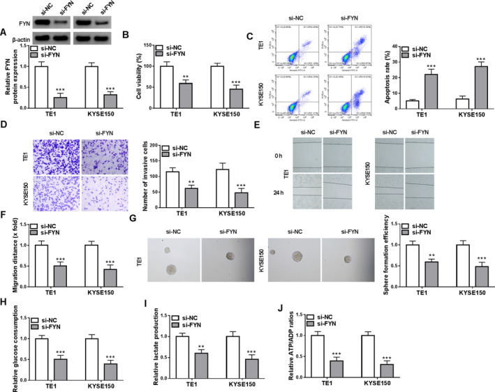
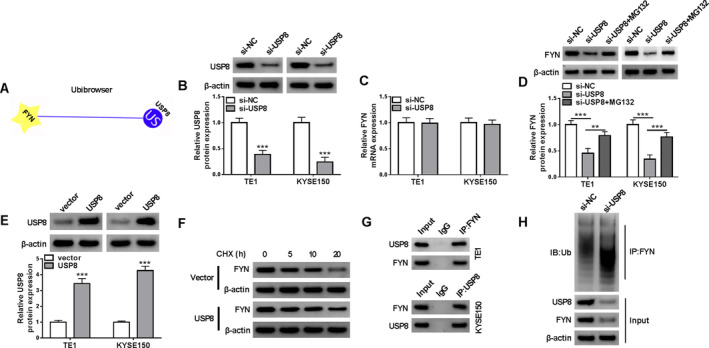
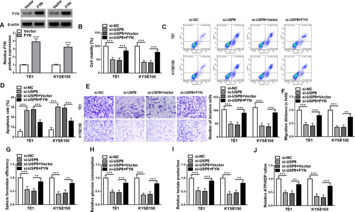
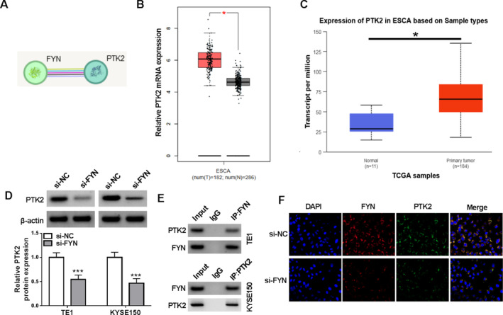
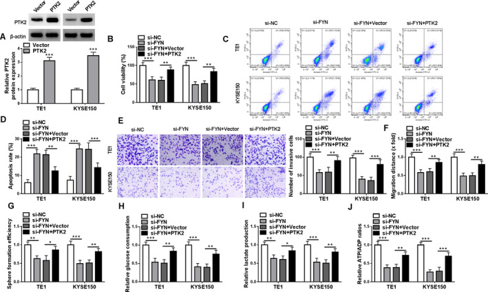
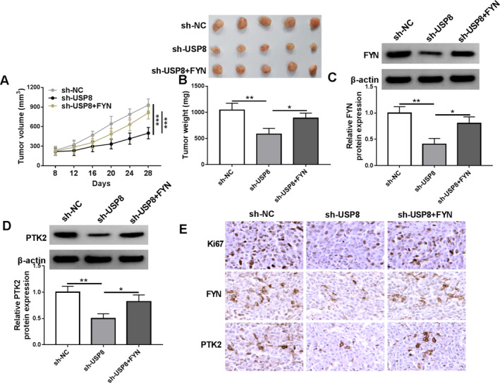
Results: FYN expression was increased in ESCC tissues and cells when compared with normal esophageal tissues and normal esophageal epithelial cells. Knockdown of FYN inhibited cell invasion, migration, stem-like traits, and glycolysis, while promoting apoptosis. USP8 was shown to stabilize FYN protein expression through its deubiquitinating activity in ESCC cells. Overexpression of FYN reversed the effects of USP8 silencing on the malignant phenotypes of ESCC cells in vitro and in vivo. FYN upregulated PTK2 expression in both TE1 and KYSE150 cell lines. Furthermore, PTK2 overexpression reversed the effects of FYN silencing on the malignant phenotypes of ESCC cells. Further, USP8 silencing-induced inhibitory effect on PTK2 protein expression was counteracted after FYN overexpression.
Conclusion: USP8-dependent FYN contributed to the malignant progression of ESCC by interacting with PTK2. Targeting this pathway may offer a novel therapeutic strategy for ESCC treatment.
Keywords: ESCC; FYN; PTK2; USP8.
© 2024 The Author(s). Thoracic Cancer published by John Wiley & Sons Australia, Ltd.
Conflict of interest statement
The authors declare no conflicts of interest.
Figures
Similar articles
-
Deubiquitinase USP8 increases ID1 stability and promotes esophageal squamous cell carcinoma tumorigenesis.Cancer Lett. 2022 Aug 28;542:215760. doi: 10.1016/j.canlet.2022.215760. Epub 2022 May 27. Cancer Lett. 2022. PMID: 35636624
-
MiR-106b-5p regulates esophageal squamous cell carcinoma progression by binding to HPGD.BMC Cancer. 2022 Mar 22;22(1):308. doi: 10.1186/s12885-022-09404-8. BMC Cancer. 2022. PMID: 35317779 Free PMC article.
-
Luteolin enhances drug chemosensitivity by downregulating the FAK/PI3K/AKT pathway in paclitaxel‑resistant esophageal squamous cell carcinoma.Int J Mol Med. 2024 Sep;54(3):77. doi: 10.3892/ijmm.2024.5401. Epub 2024 Jul 12. Int J Mol Med. 2024. PMID: 38994756 Free PMC article.
-
STK3 kinase activation inhibits tumor proliferation through FOXO1-TP53INP1/P21 pathway in esophageal squamous cell carcinoma.Cell Oncol (Dordr). 2024 Aug;47(4):1295-1314. doi: 10.1007/s13402-024-00928-8. Epub 2024 Mar 4. Cell Oncol (Dordr). 2024. PMID: 38436783 Free PMC article.
-
Fyn: a novel molecular target in cancer.Cancer. 2010 Apr 1;116(7):1629-37. doi: 10.1002/cncr.24879. Cancer. 2010. PMID: 20151426 Free PMC article. Review.
References
-
- Rogers J. E., Sewastjanow‐Silva M., Waters R. E., and Ajani J. A., “Esophageal cancer: Emerging Therapeutics,” Expert Opinion on Therapeutic Targets 26 (2022): 107–117. - PubMed
-
- Bray F., Laversanne M., Sung H., et al., “Global cancer Statistics 2022: GLOBOCAN Estimates of Incidence and Mortality Worldwide for 36 Cancers in 185 Countries,” CA: A Cancer Journal for Clinicians 74 (2024): 229–263. - PubMed
-
- Hagymási K. and Tulassay Z., “Risk Factors for Esophageal Cancer, and Possible Genetic Background,” Orvosi Hetilap 150 (2009): 407–413. - PubMed
MeSH terms
Substances
LinkOut - more resources
Full Text Sources
Medical
Miscellaneous
