. 2025 Feb;292(3):602-618.
doi: 10.1111/febs.17364. Epub 2024 Dec 19.

Cryo-EM structures of the full-length human contactin-2

Affiliations

Cryo-EM structures of the full-length human contactin-2

Zhenzhen Zhang et al. FEBS J. 2025 Feb.

Abstract

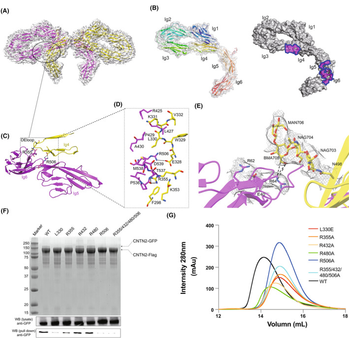
Contactin-2 (CNTN2), an immunoglobulin cell adhesion molecule (IgCAM) expressed on the neural cell surface, regulates the formation of myelin sheaths, facilitates communication between neurons and axoglial cells, and coordinates the migration of neural cells. However, the assembly of full-length CNTN2 is still not fully elucidated. Here, we found that the full-length human CNTN2 forms a concentration-dependent homodimer. We further determined the cryo-EM structures of the full-length CNTN2, revealing a novel bowknot-shaped scaffold constituted of the Ig1-6 repeats from two protomers, with the flexible ribbon-like FNIII repeats extending outward in opposite directions. The Ig1-6 domains, rather than the previously proposed Ig1-4 domains, have an indispensable role in mediating CNTN2-dependent cell adhesion and clustering. Moreover, structure-guided mutagenesis analyses supported the idea that CNTN2 homodimerization observed in our structure is essential for cell adhesion. Our findings offer novel insights into the mechanism through which CNTN2 forms a homodimer to maintain cell-cell contacts in the nervous system.

Keywords: cell adhesion; contactin‐2; cryo‐EM; dimerization; full‐length.


Conflict of interest statement

The authors affirm that the research was conducted without any commercial or financial relationships that might be perceived as a potential conflict of interest.

Figures

Fig. 1
Fig. 1
Cryo‐EM (Cryogenic electron microscopy) analysis of human CNTN2 (Contactin‐2) Ig1‐6. (A) Schematic of CNTN2 full‐length domain organization. The yellow color represents the Ig domain, which is the structure we studied. The gray represents the FNIII domain. (B, C) Overall cryo‐EM map and structure of CNTN2 Ig1‐6 homodimer shown in two views. One monomer is colored yellow, and the other is colored magenta. (D) Distribution diagram of average sedimentation coefficient of samples at three concentrations (ls‐g (s) model). The experiment was conducted once. (E) CNTN2 mature full‐length gel filtration analysis shows a concentration‐dependent peak shift. The gel filtration analysis was conducted in duplicate. The structural figures were generated utilizing UCSF Chimera software.
Fig. 2
Fig. 2
CNTN2 (contactin‐2) dimerization is mediated through two small interfaces between the Ig3‐6 interface. (A) A broad view of the CNTN2 Ig 3, 4, 5, and 6 interface interactions underlying the characteristic architecture. One monomer is colored yellow, and the other is colored magenta. (B) CNTN2 Ig1‐6 monomer (domain rainbow colored) and monomer of the contactin 2 Ig1‐6 in surface representation colored according to various properties and with dimerization surfaces outlined (homodimer surfaces purple colored). (C, D) The molecular details of CNTN2 Ig 4 with lg 5 and lg 6 interface interactions. The key residues of dimerization interface are shown as sticks. The black dotted lines represent hydrogen bonding. (E) Close‐up and overlay of cryo‐EM density (gray) of the N498‐glycan on Ig5, which is part of the extensive interface. The N498‐glycan contacts with E40, R62, R64 of Ig1, shown as sticks. The black dotted lines represent hydrogen bonding. (F) Pull‐down assay of Flag‐tagged CNTN2 wild‐type protein and GFP‐tagged CNTN2 point mutations from transgenic cell lysates, utilizing Coomassie Blue staining and western blot analysis. The western blot experiments were conducted in duplicate. (G) Gel filtration/mutagenesis studies of the dimerization interface of CNTN2, show various effects of the mutations on the apparent size. The gel filtration experiment was conducted in duplicate. The structural figures were generated utilizing UCSF Chimera software.
Fig. 3
Fig. 3
Comparison of the ectodomain structures of CNTN (Contactin) 2 with CNTN1. (A) Structural alignment of the single protomer CNTN2 Ig1‐6 and IgSFs Ig1‐4 crystal structures (PDB ID: 1CS6, 3JXA, 3P3Y, 4X9H, 5K6U) revealing that their overall architecture is highly similar, as shown by a horseshoe arrangement of Ig1‐4. (B) Superimposition of the three Ig domains of CNTN2 (this study, colored in magenta) and CNTN1 (PDB ID: 7OL4, 7OL2, colored in blue and gray). The superimposition is based on the N‐terminal Ig1‐4 domain. The conformational difference between the two structures is evident from the large deviation between the CNTN2–Ig 5, 6 and CNTN1‐Ig 5, 6 in this superimposition. (C, D) the CNTN2 Ig1‐6 (Left) compared with CNTN1 Ig 1–6 (Right PDB ID: 7OL2) in electrostatic and hydrophobic surface representation colored according to various properties. For hydrophobic potential, the hydrophilic residues are labeled green and the hydrophobic residues are labeled yellow. For electrostatic potential, the negative charged residues are labeled red and the positive charged residues are labeled blue. The Unit of electrostatic potential is kT e−1. (E) Gel filtration studies of CNTN2 (2.0 mg·mL−1) and CNTN1 (3.0 mg·mL−1) mature full length, showing milli‐absorbance units (mAu) at different volumes. The gel filtration experiment was conducted in duplicate. (F) Sequence alignment of CNTN2 interface residues from the Ig4:Ig5, and Ig4:Ig6 generated from multiple sequence alignments of vertebrate CNTNs protein sequences. Key interfacial residues are indicated in red dots. The relative accessions for human CNTN1‐6 and Chicken CNTN2 sequences are Q12860, Q02246, Q9P232, Q8IWV2, O94779, Q9UQ52, and P28685. ESPript was used to generate the alignment result. (G) Four representative class averages of the high‐ordered CNTN2 conformation and a cartoon, which depend on the dimer for their formation (Scale bar: 2 nm). The structural figures were generated utilizing UCSF Chimera software.
Fig. 4
Fig. 4
The FNIII (fibronectin type III) domain of CNTN2 (contactin‐2) reveals a flexible conformation and does not contribute to the dimerization of CNTN2. (A, B) Three views of the CNTN2 Ig‐FNIII1‐2 cryo‐EM structure. (C) Superimposition of the two protomers of the asymmetric CNTN2 dimer reveals a highly similar Ig domain structure. The black lines and circles represent the rotation direction and angle. (D, E) Superimposition of CNTN2 Ig5‐FNIII1‐2 (colored in magnate) with CNTN5 (colored in orange, PDB ID:5I99) and CNTN2 FNIII1‐2 (colored in yellow) with mouse CNTN2 FNIII1‐3 (colored in blue, PDB ID:5E7L). (F) Representative 2D class averages of the CNTN2 full‐length conformation showing the flexibility of FNIII domain in solution (Scale bar: 2 nm). The structural figures were generated utilizing UCSF Chimera software.
Fig. 5
Fig. 5
Cell adhesion is mediated by CNTN2 (contactin‐2) in HEK293F cells. (A) A diagram of the CNTN2 constructs with GFP (green) generated for cell adhesion assays. (B) Confocal images of subcellular localization of CNTN2 full length, CNTN2 lg1‐4, CNTN2 lg1‐6, and CNTN2 FNIII domains. The confocal experiments were conducted in duplicate (Scale bar: 10 μm). (C) Cell–cell adhesion was monitored by microscopy. HEK293F cells were transfected with CNTN2 full length, CNTN2 lg1‐4, CNTN2 lg1‐6, CNTN2 FNIII domains to form cell clusters (Scale bar: 100 μm). (D) Aggregation percent; the relative percentage of the 4 cells, 10 cells were classified compared to control as a cluster (Average ± SEM, n = 10). The experiments were conducted in duplicate, with a minimum of 500 cells counted from each data point. One‐way ANOVA was employed to compare the wild‐type (WT) group and the mutant groups for the counts of 4 cells and 10 cells, respectively. The structural figures were generated utilizing UCSF Chimera software.
Fig. 6
Fig. 6
Cell adhesion mediated by CNTN2 (contactin‐2) Ig4‐6 domains. (A) A diagram of the CNTN2 mutant constructs with GFP (green) generated for cell adhesion assays. (B) A diagram of the CNTN2 point mutation constructs with GFP (green) generated for cell adhesion assays (Scale bar: 10 μm). The red channel was modified to magenta to enhance accessibility for individuals with color vision deficiencies. The confocal experiments were conducted in duplicate. (C) Cell–cell adhesion monitored by fluorescence microscopy. HEK293F cell transfected with L330E, R355A, R432A, R480A, R506A, or R355/432/480/506A mutations (Scale bar: 100 μm). (D) Clustering index; the proportion of the total segmented cell area classified as a cluster (Average ± SEM, n = 10). The experiments were conducted in duplicate, with a minimum of 500 cells counted from each data point. One‐way ANOVA was employed to compare the wild‐type (WT) group and the mutant groups for the counts of 4 cells and 10 cells, respectively.
Fig. 7
Fig. 7
Schematic diagram of how CNTN2 (contactin‐2) regulates the myelin sheath distance based on concentration. The dimerization of CNTN2 exhibits a concentration‐dependent behavior. At relatively low concentrations of CNTN2, the protein predominantly exists in a monomeric form. However, as the concentration increases, there is a tendency for CNTN2 to form homodimers.

Similar articles

References

    1. Dodd J & Jessell TM (1988) Axon guidance and the patterning of neuronal projections in vertebrates. Science 242, 692–699. - PubMed
    1. Shimoda Y & Watanabe K (2009) Contactins: emerging key roles in the development and function of the nervous system. Cell Adh Migr 3, 64–70. - PMC - PubMed
    1. Colakoglu G, Bergstrom‐Tyrberg U, Berglund EO & Ranscht B (2014) Contactin‐1 regulates myelination and nodal/paranodal domain organization in the central nervous system. Proc Natl Acad Sci USA 111, E394–E403. - PMC - PubMed
    1. Chen YA, Lu IL & Tsai JW (2018) Contactin‐1/F3 regulates neuronal migration and morphogenesis through modulating RhoA activity. Front Mol Neurosci 11, 422. - PMC - PubMed
    1. Chatterjee M, Schild D & Teunissen CE (2019) Contactins in the central nervous system: role in health and disease. Neural Regen Res 14, 206–216. - PMC - PubMed

LinkOut - more resources