Case Reports
. 2024 Dec 5:15:1437859.
doi: 10.3389/fgene.2024.1437859. eCollection 2024.

Case report: A single novel calpain 3 gene variant associated with mild myopathy

Affiliations
Case Reports

Case report: A single novel calpain 3 gene variant associated with mild myopathy

Sara Massucco et al. Front Genet. .

Abstract

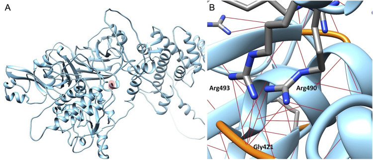
Recessively inherited limb-girdle muscular dystrophy type 1, caused by mutations in the calpain 3 gene, is the most common limb-girdle muscular dystrophy worldwide. Recently, cases of autosomal dominant calpainopathy have been described. A man was referred to our neurological outpatient clinic at the age of 54 for persistent hyperCKemia (>1000 U/l) associated with muscle fatigue and myalgia. Clinical examination revealed mild proximal weakness in the lower limbs. His brother exhibited a moderate increase in serum creatine kinase levels (up to 2000 U/l) without other signs of myopathy. Their father experienced slowly progressive lower limb weakness after the age of 50. The calpain 3 variant c.1478G>A (p.Arg493Gln) in the heterozygous state was identified in both brothers. In silico modeling studies predict that this substitution may disrupt protein folding. This represents the first description of the heterozygous p.Arg493Gln calpain 3 variant as a potential cause of mild calpainopathy.

Keywords: LGMD; LGMDR1; calpain; calpainopathy; case report.


Conflict of interest statement

The authors declare that the research was conducted in the absence of any commercial or financial relationships that could be construed as a potential conflict of interest. The author(s) declared that they were an editorial board member of Frontiers, at the time of submission. This had no impact on the peer review process and the final decision.

Figures

FIGURE 1
FIGURE 1
Muscle Magnetic Resonance Imaging (MRI). In (A), (C), and (E) axial, T1-weighted images of the lower limbs are shown, whilst in (B), (D), and (F) axial T2-weighted images with fat-saturation are displayed. Images (A) and (B) were acquired at the level of the first sacral vertebra, images (C) and (D) at the midportion of the thigh, and images (E) and (F) at the proximal third of the leg. Increased T1-signal related to fibrous and fat infiltration was observed in the gluteus maximus (GM), gluteus medius (Gm), gluteus minimus [white asterisk in (A), (B)], semimebranosus (sm), semitendinosus (st), adductor magnus [white asterisk in (C), (D)], vastus intermedius (vI), medial gastrocnemius [black asterisk in (E), white asterisk in (F)], and soleus (so) muscles. A mild signal intensity increase in the T2-weighted sequence was found in the short head of the biceps femoris (sbf), long head of the biceps femoris (lbf), semimembranosus (sm), and semitendinosus (st) muscles, corresponding to muscle necrosis and consequent inflammatory response. The sartorius (s), gracilis (g), and lateral gastrocnemius (LG) muscles did not present any morphological changes. Muscle alteration pattern is consistent with the findings previously described in autosomal dominant calpainopathies. Vl = vastus lateralis; Vm = vastus medialis; r = rectus femoris.
FIGURE 2
FIGURE 2
Muscle biopsy. Hematoxylin and eosin staining (A), Modified Gomori trichrome staining (B), Nicotinamide adenine dinucleotide staining (NADH) (C, D). Muscle changes are milder than in limb-girdle muscular dystrophy recessive type 1 (LGMDR1). Observations include a mild fiber caliber variation, rare fibers with internalized nuclei, and fiber splitting. No necrosis or interstitial cellular infiltrates were detected, and vessels and connective tissue appeared normal. Oxidative enzymatic activity was unevenly distributed in some muscle fibers, often displaying moth eaten and core-like features. Bar: (A, C) 100 μm, (B, D) 50 µm.
FIGURE 3
FIGURE 3
Calpain-3 3D model. (A) Residue Arg493 is highlighted in light red. (B) Focus on the Arg493 subregion in Calpain-3 (ribbon). Arginine residues are represented as capped sticks, Gly421 residue is depicted in orange, and hydrogen bonds are shown as red dashed lines.

References

    1. Aguti S., Gallus G. N., Bianchi S., Salvatore S., Rubegni A., Berti G., et al. (2024). Novel biomarkers for limb girdle muscular dystrophy (LGMD). Cells 13 (13), 329. 10.3390/CELLS13040329 - DOI - PMC - PubMed
    1. Banerjee S., Radotra B. D., Luthra-Guptasarma M., Goyal M. K. (2024). Identification of novel pathogenic variants of Calpain-3 gene in limb girdle muscular dystrophy R1. Orphanet J. Rare Dis. 19 (1), 140. 10.1186/s13023-024-03158-1 - DOI - PMC - PubMed
    1. Barp A., Laforet P., Bello L., Tasca G., Vissing J., Monforte M., et al. (2020). European muscle MRI study in limb girdle muscular dystrophy type R1/2A (LGMDR1/LGMD2A). J. Neurol. 267, 45–56. 10.1007/S00415-019-09539-Y - DOI - PubMed
    1. Berman H. M., Westbrook J., Feng Z., Gilliland G., Bhat T. N., Weissig H., et al. (2000). The protein Data Bank. Nucleic Acids Res. 28, 235–242. 10.1093/NAR/28.1.235 - DOI - PMC - PubMed
    1. Cerino M., Bartoli M., Riccardi F., Le Goanvic B., Blanck V., Salvi A., et al. (2020b). Autosomal dominant segregation of CAPN3 c.598_612del15 associated with a mild form of calpainopathy. Ann. Clin. Transl. Neurol. 7, 2538–2540. 10.1002/ACN3.51193 - DOI - PMC - PubMed

Publication types

LinkOut - more resources