Review
. 2024 Dec 5:15:1430825.
doi: 10.3389/fimmu.2024.1430825. eCollection 2024.

Macrophages in inflammatory skin diseases and skin tumors

Affiliations
Review

Macrophages in inflammatory skin diseases and skin tumors

Si-Han Liu et al. Front Immunol. .

Abstract

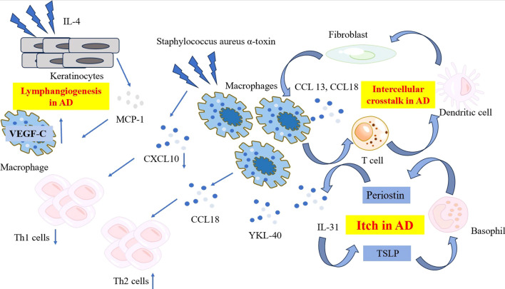
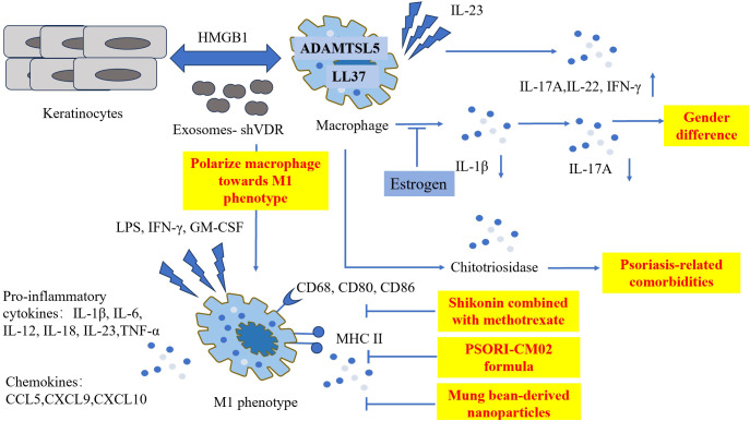
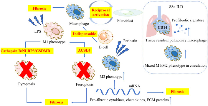
Macrophages, as specialized, long-lasting phagocytic cells of the innate immune system, have garnered increasing attention due to their wide distribution and various functions. The skin, being the largest immune organ in the human body, presents an intriguing landscape for macrophage research, particularly regarding their roles in inflammatory skin diseases and skin tumors. In this review, we compile the latest research on macrophages in conditions such as atopic dermatitis, psoriasis, systemic sclerosis, systemic lupus erythematosus, rosacea, bullous pemphigoid, melanoma and cutaneous T-cell lymphoma. We aim to contribute to illustrating the pathogenesis and potential new therapies for inflammatory skin diseases and skin tumors from the perspective of macrophages.

Keywords: inflammatory skin diseases; macrophage; pathogenesis; skin tumors; treatment.


Conflict of interest statement

The authors declare that the research was conducted in the absence of any commercial or financial relationships that could be construed as a potential conflict of interest.

Figures

Figure 1
Figure 1
The characteristic and pathogenetic roles of the macrophages in AD. Compared to psoriasis, macrophages in AD produce lower level of CXCL10 when exposed to Staphylococcus aureus α-toxin, resulting in reduced Th1 polarization. Instead, macrophages in AD produce high levels of CCL18, recruiting more Th2 cells to affected skin and release YKL-40, an important Th2 marker. A network comprising periostin, TSLP, basophils and macrophage-derived IL-31 contribute to the mechanism of itch in AD. Ligand-receptor interactions data revealed the intracellular crosstalk between CCL13, CCL18-macrohages, T cells, DCs and fibroblasts. Macrophages also get involved in the lymphangiogenesis in AD by expressing significant level of VEGF-C. AD, atopic dermatitis; CCL, C-C Motif Chemokine Ligand; CXCL, C-X-C Motif Chemokine Ligand; DC, dendritic cell; IL, interleukin; MCP-1, monocyte chemoattractant protein-1; TSLP, thymic stromal lymphopoietin; VEGF-C, vascular endothelial growth factor-C; YKL-40, Chitinase 3-like 1.
Figure 2
Figure 2
The pathogenetic roles of macrophages in psoriasis and treatments involving M1 phenotype. Macrophages co-express with two important psoriasis autoantigens LL-37 and ADAMTSL5. Macrophages triggered by IL-23 produce significant quantities of IL-7A, IL-22 and IFN-γ. Chitotriosidase secreted by activated macrophages is related to psoriasis-related comorbidities. Estrogen suppresses the production of IL-1β, furthermore reducing the level of IL-17A. Keratinocytes interact with macrophages via HMGB1, and exosomes derived from VDR-deficient keratinocytes polarize macrophages toward M1 phenotype, exaggerating the inflammation condition. Shikonin combined with methotrexate, PSORI-CM02 formula, and Mung bean-derived nanoparticles exhibit anti-psoriatic properties by hindering M1 polarization. ADAMTSL5, ADAMTS-Like Protein 5; CD, cluster of differentiation; CCL, C-C Motif Chemokine Ligand; CXCL, C-X-C Motif Chemokine Ligand; GM-CSF, granulocyte-macrophage colony-stimulating factor; HMGB1, High Mobility Group Box-1; IFN, interferon; IL, interleukin; LL-37, Cathelicidin; LPS, lipopolysaccharide; MHC, major histocompatibility complex; TNF, tumor necrosis factor; VDR, vitamin D receptor.
Figure 3
Figure 3
The characteristic and pathogenetic roles of the macrophages in SSc. Macrophages and fibroblasts mutually activate each other and contribute to the pathology in SSc. B cells promote the differentiation of profibrotic macrophages, and is indispensable for the progression of SSc. Periostin induces higher ratio of M2 macrophage and upregulates the mRNA level of pro-fibrotic cytokines, chemokines, and ECM proteins. M1 macrophage facilitates fibrosis by pyroptosis and ferroptosis. CD14+ tissue resident pulmonary macrophages in SSc-ILD patients’ lungs show an active profibrotic signature. Elevated levels of mixed M1/M2 phenotype macrophages are observed in the circulation of SSc-ILD patients. ACSL4, Acyl-CoA synthetase long chain family member4; CD, cluster of differentiation; ECM, extracellular matrix; GSDMD, Gasdermin D; LPS, lipopolysaccharide; NLRP3, NOD-like receptor thermal protein domain associated protein 3; SSc-ILD, systemic sclerosis-interstitial lung disease.

References

    1. Gordon S, Taylor PR. Monocyte and macrophage heterogeneity. Nat Rev Immunol. (2005) 5:953–64. doi: 10.1038/nri1733 - DOI - PubMed
    1. Atri C, Guerfali FZ, Laouini D. Role of human macrophage polarization in inflammation during infectious diseases. Int J Mol Sci. (2018) 19:1801. doi: 10.3390/ijms19061801 - DOI - PMC - PubMed
    1. Kloc M, Ghobrial RM, Wosik J, Lewicka A, Lewicki S, Kubiak JZ. Macrophage functions in wound healing. J Tissue Eng regenerative Med. (2019) 13:99–109. doi: 10.1002/term.2772 - DOI - PubMed
    1. Shapouri-Moghaddam A, Mohammadian S, Vazini H, Taghadosi M, Esmaeili SA, Mardani F, et al. . Macrophage plasticity, polarization, and function in health and disease. J Cell Physiol. (2018) 233:6425–40. doi: 10.1002/jcp.v233.9 - DOI - PubMed
    1. Netea MG, Balkwill F, Chonchol M, Cominelli F, Donath MY, Giamarellos-Bourboulis EJ, et al. . A guiding map for inflammation. Nat Immunol. (2017) 18:826–31. doi: 10.1038/ni.3790 - DOI - PMC - PubMed

LinkOut - more resources