. 2024 Dec 19:28:0098.
doi: 10.34133/bmr.0098. eCollection 2024.

Polygonum multiflorum Extracellular Vesicle-Like Nanovesicle for Skin Photoaging Therapy

Affiliations

Polygonum multiflorum Extracellular Vesicle-Like Nanovesicle for Skin Photoaging Therapy

Junjia He et al. Biomater Res. .

Abstract

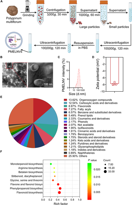
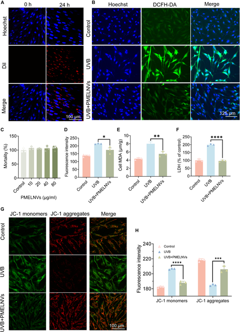
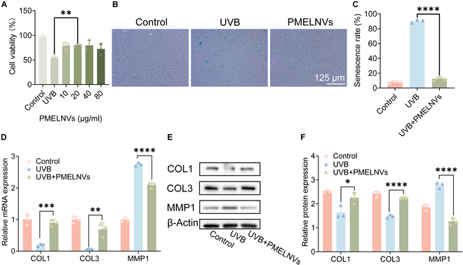
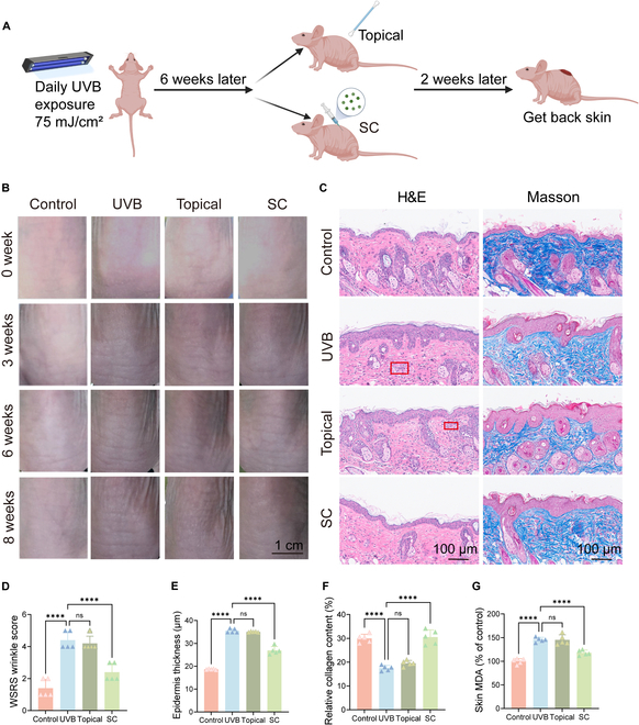
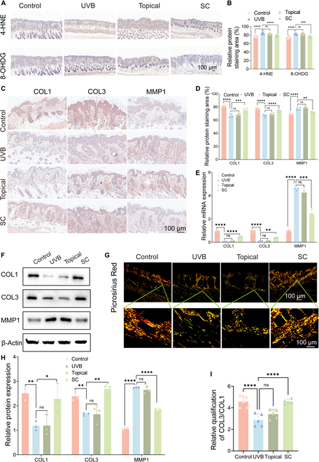
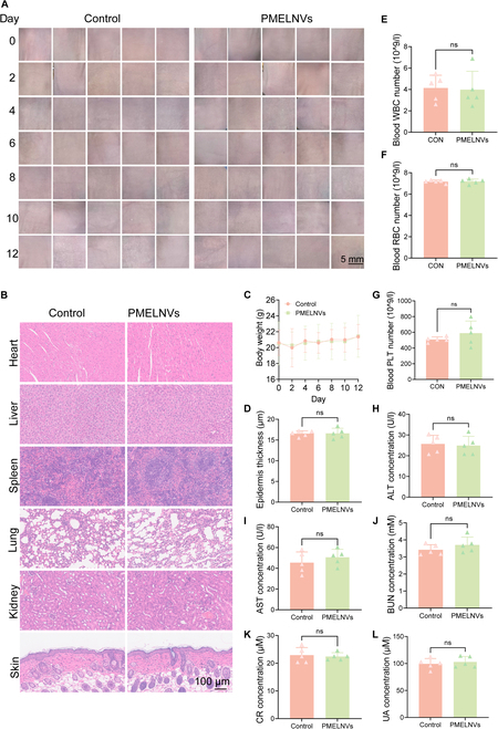
Ultraviolet (UV) irradiation leads to the degradation of the extracellular matrix and collagen, thereby accelerating skin aging and imposing substantial psychological burden on patients. Current anti-aging strategies are limited and often associated with high costs or strong side effects. Plant-derived extracellular vesicle-like nanovesicles, with advantages such as natural availability and cost-effectiveness, show potential in anti-aging interventions. This study extracted extracellular vesicle-like nanovesicle from Polygonum multiflorum (PMELNVs) and systematically investigated their composition and metabolic pathways, further examining their efficacy and underlying mechanisms in combating photoaging. Results revealed the excellent antioxidative properties of PMELNVs, alleviating UV-induced oxidative stress, inhibiting matrix metalloproteinase production, reducing extracellular matrix degradation, promoting collagen synthesis, and ultimately exerting anti-photoaging effects. Additionally, safety assessments demonstrated favorable biocompatibility of PMELNVs. This study provides novel evidence supporting PMELNVs' ability to resist photoaging by reducing oxidative stress and enhancing collagen expression, thereby offering potential as a new natural therapeutic agent against skin photoaging and promising a safer and more effective local anti-aging strategy.


Conflict of interest statement

Competing interests: The authors declare that they have no competing interests.

Figures

Fig. 1.
Fig. 1.
Detection and analysis of PMELNVs. (A) Schematic diagram illustrating the process of isolating and purifying PMELNVs. (B) Morphological characteristics and dimensions of PMELNVs following centrifugation using a sucrose gradient density characterized by TEM. Scale bar, 200 nm. (C) Dynamic light scattering analysis of the size distribution of PMELNVs. (D) Zeta potential of PMELNVs. (E) Identification of all compounds present in PMELNVs. (F) Enrichment analysis of KEGG pathways for all compounds present in PMELNVs.
Fig. 2.
Fig. 2.
Cellular uptake of PMELNVs by HDFs and the antioxidant effect of PMELNVs in HDFs. (A) Uptake of PMELNVs by HDF cells. (B and D) PMELNVs reduce ROS production in UVB-damaged HDF cells and their quantification of fluorescence intensity. (C) Cytotoxic effect of PMELNVs on HDF cells. (E and F) PMELNVs reduce the production of MDA and LDH in UVB-damaged HDF cells. (G and H) JC-1 staining semiquantification of fluorescence images.
Fig. 3.
Fig. 3.
PMELNVs suppress β-gal and matrix metalloproteinase production, enhance HDF viability, and up-regulate collagen expression to counteract aging. (A) PMELNVs enhance the viability of UVB-damaged HDF cells. (B and C) β-Gal staining and its quantification data. (D) PCR examination of COL1, COL3, and MMP1 expression in HDF cells. (E and F) Analysis using the Western blot technique for COL1, COL3, and MMP1 expression in HDF cells and its quantification data.
Fig. 4.
Fig. 4.
Effects of PMELNVs on the nude mouse photoaging model. (A) Schematic diagram of photoaging modeling in mice. (B) Changes in the dorsal skin of nude mice. (C) H&E and Masson staining. (D) Skin wrinkle scores. (E and F) Quantitative analysis of epidermal thickness using H&E staining and collagen deposition using Masson staining. (G) Skin MDA levels.
Fig. 5.
Fig. 5.
Evaluation of the anti-photoaging properties of PMELNVs in vivo. (A) Immunohistochemical detection of 4-HNE and 8-OHDG in the skin through staining. (B) Quantitative analysis of immunohistochemical detection of 4-HNE and 8-OHDG. (C) Immunohistochemical staining of COL1, COL3, and MMP1 in the skin. (E) mRNA expression (COL1, COL3, and MMP1). (F) Western blot analysis of COL1, COL3, and MMP1 in skin tissue. (D and H) Quantitative analysis of immunohistochemistry and Western blot. (G) Sirius Red staining with polarized light analysis. (I) Statistical data of Sirius Red staining for COL3/COL1.
Fig. 6.
Fig. 6.
Safety evaluation of PMELNVs in vivo. (A) Skin condition before dosing every other day. (B and D) H&E-stained sections of major organs and skin tissue. (C) Changes in body weight. (E to G) Levels of RBC, WBC, and PLT. (H to L) Blood biochemical levels of ALT, AST, BUN, CR, and UA.

References

    1. Fisher GJ, Kang S, Varani J, Bata-Csorgo Z, Wan Y, Datta S, Voorhees JJ. Mechanisms of photoaging and chronological skin aging. Arch Dermatol. 2002;138(11):1462–1470. - PubMed
    1. Kammeyer A, Luiten RM. Oxidation events and skin aging. Ageing Res Rev. 2015;21:16–29. - PubMed
    1. Cocetta V, Cadau J, Saponaro M, Giacomini I, Dall’Acqua S, Sut S, Catanzaro D, Orso G, Miolo G, Menilli L, et al. . Further assessment of Salvia haenkei as an innovative strategy to counteract skin photo-aging and restore the barrier integrity. Aging. 2021;13(1):89–103. - PMC - PubMed
    1. Silva S, Michniak-Kohn B, Leonardi GR. An overview about oxidation in clinical practice of skin aging. An Bras Dermatol. 2017;92(3):367–374. - PMC - PubMed
    1. Spierings NMK. Evidence for the efficacy of over-the-counter vitamin A cosmetic products in the improvement of facial skin aging: A systematic review. J Clin Aesthet Dermatol. 2021;14(9):33–40. - PMC - PubMed

LinkOut - more resources