. 2024 Nov 12;32(4):101378.
doi: 10.1016/j.omtm.2024.101378. eCollection 2024 Dec 12.

αFAP-specific nanobodies mediate a highly precise retargeting of modified AAV2 capsids thereby enabling specific transduction of tumor tissues

Affiliations

αFAP-specific nanobodies mediate a highly precise retargeting of modified AAV2 capsids thereby enabling specific transduction of tumor tissues

Olaniyi Olarewaju et al. Mol Ther Methods Clin Dev. .

Abstract

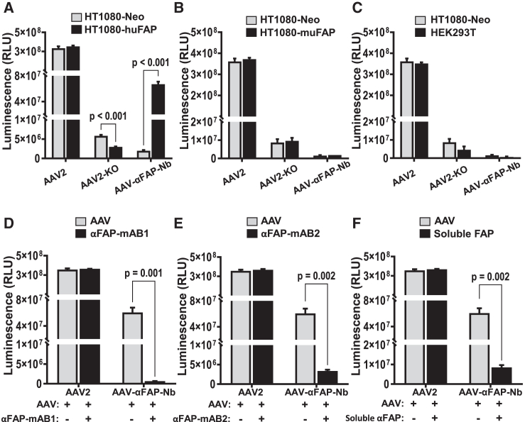
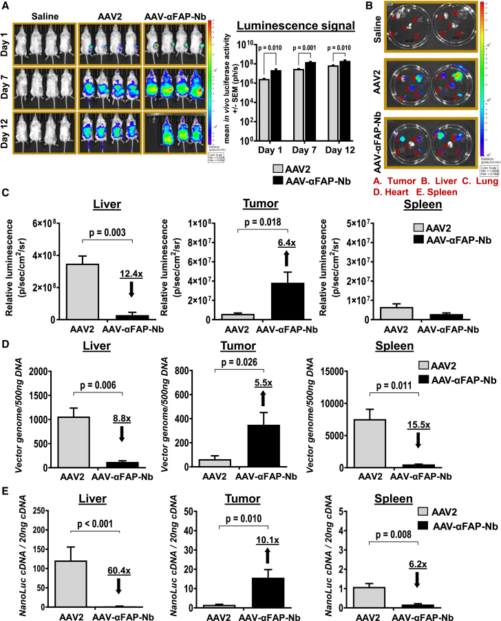
Due to the refractiveness of tumor tissues to adeno-associated virus (AAV) transduction, AAV vectors are poorly explored for cancer therapy delivery. Here, we aimed to engineer AAVs to target tumors by enabling the specific engagement of fibroblast activation protein (FAP). FAP is a cell surface receptor distinctly upregulated in the reactive tumor stroma, but rarely expressed in healthy tissues. Thus, targeting FAP presents an opportunity to selectively transduce tumor tissues. To achieve this, we modified the capsid surface of AAV2 with an αFAP nanobody to retarget the capsid to engage FAP receptor. Following transduction, we observed a 23- to 80-fold increase in the selective transduction of FAP+ tumor cells in vitro, and greater than 5-fold transduction of FAP+ tumor tissues in vivo. Subsequent optimization of the VP1-nanobody expression cassette further enhanced the transduction efficiency of the modified capsids. Due to the limited αFAP nanobodies repertoires, we broadened the versatility of this high-fidelity platform by screening a naive VHH yeast display library, leading to the identification of several novel αFAP nanobody candidates (KD = 0.1 to >100 nM). Hence, our study offers new opportunity for the application of AAV vectors for highly selective delivery of therapeutics to the tumor stroma.

Keywords: AAV; FAP; adeno-associated virus vectors; capsid engineering; fibroblast activation protein; targeted AAV delivery; tumor microenvironment; tumor targeting.


Conflict of interest statement

O.O., F.H., P.M., U.M., and F.d.P. are employees of Boehringer Ingelheim Pharma GmbH & Co. KG, Biberach, Germany. P.C., C.K., and T.P. are employees of Boehringer Ingelheim Pharmaceuticals, Inc., Ridgefield, CT, USA.

Figures

None
Graphical abstract
Figure 1
Figure 1
Genetic incorporation of human αFAP_Nb in AAV2 VP1 and biophysical characterization of AAV-αFAP_Nb (A) Overview of the capsid engineering strategies showing modifications to the viral capsid proteins. The human αFAP_VHH was integrated in the GH2/GH3 loop of VP1 under the control of a CMV promoter and native HSPG receptor-binding motifs were ablated. Recombinant AAVs were packaged using four plasmid transfection and the modified VP1 cassette was supplied in trans. (B) Evaluation of the productivity of the modified AAVs in comparison with AAV2 vector with wild-type capsid and the HSPG-ablated capsid variant. (C) Western blot analyses of the VP1, VP2, and VP3 capsid proteins composition using the monoclonal anti-VP1/VP2/VP3 B1 monoclonal antibody. (D) VP1, VP2 capsid protein analysis using anti-VP1/VP2 A69 monoclonal antibody. (E) Comparative characterization of the particle sizes of AAV2 vector and the modified capsid variants.
Figure 2
Figure 2
Display of αFAP_Nb on AAV capsid surface mediates retargeting of the capsids to human FAP-expressing cells (A) Human HT1080 cells expressing either the human FAP receptor (HT1080-huFAP) or the negative control (HT1080-Neo) were incubated with 1 × 104 VG/cells of the indicated AAV vectors expressing NanoLuc transgene. NanoLuc protein expression was determined and quantified 72 h after incubation. Off-target analyses of the modified AAV-αFAP_Nb in (B) human HT1080 cells expressing the mouse FAP receptor (HT1080-muFAP) and (C) in non-FAP receptor expressing HEK293T cells. Competitive inhibition of AAV-αFAP_Nb by in-house-generated αFAP-mAB1 (D), αFAP-mAB2 (E), and soluble FAP protein (F). The values shown represent three independent experiments. Error bars are presented as ± SEM of the three independent experiments. p value was determined by two-tailed Student’s t test. ∗p < 0.05, ∗∗p < 0.01, ∗∗∗p < 0.001.
Figure 3
Figure 3
αFAP_Nb mediates successful retargeting of AAVs to HT1080-huFAP tumors in vivo (A) HT1080-huFAP tumors were engrafted in the mammary fat pad of immunodeficient NXG mouse strain. After tumor volume of 30–60 mm3 was attained, 1 × 1011 VG of either AAV2 or the retargeted AAV-αFAP_Nb vector was administered intravenously per mouse. In vivo bioluminescence imaging was performed to detect nanoluciferase activity at day 1, day 7, and day 12 post-AAV administration and the corresponding plot of the average luminescence signal. (B) Ex vivo bioluminescence imaging of the nanoluciferase activity in the liver, tumor, lung, heart, and spleen tissues collected after termination of the experiment. (C) Quantification and comparison of the nanoluciferase activity in tissues collected from AAV2 mice and AAV-αFAP_Nb mice. (D) Analysis and comparison of the biodistribution of AAV2 and AAV-αFAP_Nb vector genomes in tissues retrieved from the HT1080-huFAP cell-derived xenograft mice. (E) Quantification of NanoLuc mRNA expression levels in the collected tissues. Data are presented as ± SEM for n = 3–4 mice. p value was determined by two-tailed Student’s t test. ∗p < 0.05, ∗∗p < 0.01, ∗∗∗p < 0.001.
Figure 4
Figure 4
Incorporation of modified VP1-αFAP_Nb during AAV packaging is promoter dependent (A) Schematics of the substitution of CMV promoter with an EF1α promoter for driving the expression of the modified VP1-αFAP_Nb expression cassette during AAV packaging and generation of AAV-αFAP_Nb2 vector. (B) Western blot analyses of the VP1, VP2, and VP3 capsid proteins composition using the monoclonal anti-VP1/VP2/VP3 B1 monoclonal antibody. (C) VP1, VP2 capsid protein analysis using anti-VP1/VP2 A69 monoclonal antibody. (D) Determination of the transduction efficiency AAV-αFAP_Nb2 in HT1080-huFAP cells and the off-target analyses of the improved vector in (E) human HT1080-muFAP cells, as well as (F) in HEK293T cells. The values shown represent three independent experiments. Error bars are presented as ± SEM of the three independent experiments. p value was determined by two-tailed Student’s t test. ∗p < 0.05, ∗∗p < 0.01, ∗∗∗p < 0.001.
Figure 5
Figure 5
Further optimization of the VP1-αFAP_Nb expression cassette with an NLS enhances transduction efficiency (A) Schematics of the capsid engineering strategy showing the introduction of an NLS sequence at the 3′ end of the αFAP_Nb on VP1 and generation of AAV-αFAP_NLS vector. (B) In vitro validation of the transduction efficiency of the optimized AAV-αFAP_NLS in HT1080-huFAP cells and the subsequent off-target analyses in HT1080-muFAP cells (C), as well as in HEK293T cells (D). Competitive inhibition of AAV-αFAP_NLS by in-house-generated αFAP-mAB1 (E), αFAP-mAB2 (F), and soluble FAP protein (G). The cells were transduced with MOI of 1 × 104 VG/cell and cells were analyzed for luminescence expression at 72 h post-transduction. The values shown represent three independent experiments. Error bars are presented as ± SEM of the three independent experiments. p value was determined by two-tailed Student’s t test. ∗p < 0.05, ∗∗p < 0.01, ∗∗∗p < 0.001.
Figure 6
Figure 6
Functionalization of the AAV capsid with novel αFAP VHHs identified in a naive VHH yeast display screening (A) Three novel αFAP VHHs Nb-1610, Nb1611, and Nb-1612 identified from our in-house naive VHH yeast display library screen, were integrated in the AAV capsid as previously shown in Figure 4A. The transduction efficiencies of the novel VHHs displaying AAVs were functionally validated in vitro for HT1080-huFAP cells targeting and compared with that of AAV-αFAP_Nb2. (B) Determination of the transduction efficiencies of the indicated AAVs HT1080-muFAP cells. The cells were transduced with MOI of 1 × 104 VG/cell and cells were analyzed for luminescence expression at 72 h post-transduction. The values shown represent three independent experiments. Error bars are presented as ± SEM of the three independent experiments. p value was determined by two-tailed Student’s t test. ∗p < 0.05, ∗∗p < 0.01, ∗∗∗p < 0.001.

References

    1. Srivastava A., Lusby E.W., Berns K.I. Nucleotide sequence and organization of the adeno-associated virus 2 genome. J. Virol. 1983;45:555–564. doi: 10.1128/jvi.45.2.555-564.1983. - DOI - PMC - PubMed
    1. Atchison R.W., Casto B.C., Hammon W.M.D. Adenovirus-Associated Defective Virus Particles. Science. 1965;149:754–756. doi: 10.1126/science.149.3685.754. - DOI - PubMed
    1. Samulski R.J., Berns K.I., Tan M., Muzyczka N. Cloning of adeno-associated virus into pBR322: Rescue of intact virus from the recombinant plasmid in human cells. Proc. Natl. Acad. Sci. USA. 1982;79:2077–2081. doi: 10.1073/pnas.79.6.2077. - DOI - PMC - PubMed
    1. Laughlin C.A., Tratschin J.-D., Coon H., Carter B.J. Cloning of infectious adeno-associated virus genomes in bacterial plasmids. Gene. 1983;23:65–73. doi: 10.1016/0378-1119(83)90217-2. - DOI - PubMed
    1. Wang D., Tai P.W.L., Gao G. Adeno-associated virus vector as a platform for gene therapy delivery. Nat. Rev. Drug Discov. 2019;18:358–378. doi: 10.1038/s41573-019-0012-9. - DOI - PMC - PubMed

LinkOut - more resources