. 2025 Mar;31(3):56.
doi: 10.3892/mmr.2024.13421. Epub 2024 Dec 20.

Epigenetic downregulation of the proapoptotic gene HOXA5 in oral squamous cell carcinoma

Affiliations

Epigenetic downregulation of the proapoptotic gene HOXA5 in oral squamous cell carcinoma

Ying-Ju Chen et al. Mol Med Rep. 2025 Mar.

Abstract

Homeobox A5 (HOXA5) has been identified as a tumor suppressor gene in breast cancers, but its role in oral squamous cell carcinoma (OSCC) has not been confirmed. The Illumina GoldenGate Assay for methylation identified that DNA methylation patterns differ between tumorous and normal tissues in the oral cavity and that HOXA5 is one of the genes that are hypermethylated in oral tumor tissues. The present study obtained more‑complete information on the methylation status of HOXA5 by using the Illumina Infinium MethylationEPIC BeadChip and bisulfite sequencing assays. The results indicated that HOXA5 hypermethylation has great potential as a biomarker for detecting OSCC. Comparing HOXA5 RNA expression between normal oral tissue and OSCC tissue samples indicated that its median level was 2.06‑fold higher in normal tissues that in OSCC tissues. Moreover, treatment using the demethylating agent 5‑aza‑2'‑deoxycytidine can upregulate HOXA5 expression in OSCC cell lines, verifying that the silencing of HOXA5 is primarily regulated by its hypermethylation. It was also found that upregulation of HOXA5 expression can not only increase OSCC cell death but that it can also enhance the therapeutic effect of cisplatin both in vitro and in vivo, suggesting that HOXA5 is an epigenetically downregulated proapoptotic gene in OSCC.

Keywords: DNA methylation array; apoptosis; homeobox A5; oral squamous cell carcinoma; p53.


Conflict of interest statement

The authors declare that they have no competing interests.

Figures

Figure 1.
Figure 1.
Bisulfite sequencing analysis of the promoter regions for HOXA5. The promoter regions (A) from −1423 to −841 and (B) from −505 to −31 upstream of the transcription start site of HOXA5 comprised 72 CpG sites. The CpG sites are indicated by vertical bars. A representative bisulfite sequencing assay was applied to OSCC and normal oral tissues. For each tissue sample, five randomly chosen clones were sequenced and the methylation statuses of the 72 CpG sites are indicated by the circles: Closed and open circles represent methylated and unmethylated CpG sites, respectively.
Figure 2.
Figure 2.
HOXA5 expression levels in paired OSCC and adjacent normal tissues. HOXA5 mRNA levels were compared between the paired normal oral tissue and OSCC tissue specimens, with latter assigned a value of 1. Error bars indicate standard deviations. OSCC, oral squamous cell carcinoma.
Figure 3.
Figure 3.
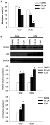
HOXA5 methylation and expression levels following 5-Aza-dC treatment. (A) OC2 and OCSL cells were treated with DMSO, 0.5 µM 5-Aza-dC, or 5 µM 5-Aza-dC for 3 days. The methylation level of HOXA5 was measured using quantitative methylation-specific PCR. (B) The cells were lysed followed by extraction of proteins 3 days after DMSO or 5-Aza-dC treatment and the protein levels of HOXA5, p53 and GAPDH were determined using western blotting (top panel). HOXA5 and p53 proteins were quantified using ImageJ software and normalized relative to GAPDH (middle and bottom panels). Protein levels of cells treated with DMSO were assigned a value of 1. Error bars indicate standard deviations. *P<0.05. 5-Aza-dC, 5-aza-2′-deoxycytidine; OSCC, oral squamous cell carcinoma.
Figure 4.
Figure 4.
HOXA5 upregulates p53 expression in OSCC. (A) OC2 cells were transduced using IG or HIG followed by fluorescence-activated cell sorting and the mRNA levels of HOXA5 and p53 in OC2 were quantified using RT-qPCR. The expression level of each IG-transduced cell was assigned a value of 1. (B) OC2 cells were transduced using lentiviral vector carrying shHOXA5 or shGFP and selected using puromycin. The mRNA expression of HOXA5 and p53 in OC2 were quantified using RT-qPCR. The expression level of each shGFP-transduced cell was assigned a value of 1. (C) The IG- and HIG-transduced OC2 cells were transfected with pGL2b-WTp53-FL or pGL2b-Mutp53-FL and cotransfected with pRL-TK to act as internal control reporters. Bioluminescence intensities of FL and RL were measured using a luminometer and the luciferase activity was calculated from the FL-to-RL ratio. The IG-transduced OC2 cells transfected with pGL2b-Mutp53-FL were assigned a value of 1. Error bars indicate standard deviations. *P<0.05. WT, pGL2b-WTp53-FL; Mut, pGL2b-Mutp53-FL. OSCC, oral squamous cell carcinoma; IG, control lentiviral vector; HIG, HOXA5-expressing lentiviral vector; RT-qPCR, reverse transcription-quantitative PCR; sh, short hairpin; FL, firefly luciferase; RL, Renilla luciferase; Mut, mutant; WT, wild-type.
Figure 5.
Figure 5.
Effect of HOXA5 restoration on OSCC cell viability. (A) The IG- and HIG-transduced OC2 cells were seeded onto culture plates on day 0 and the cells were treated with 2 µg/ml cisplatin on day 1. Cell viability was analyzed on day 4 using MTS assay. (B) The IG- and HIG-transduced OC2 cells were treated with 2 µg/ml cisplatin or PBS for 3 days and stained with propidium iodide and then analyzed using flow cytometry. The proportions of necrotic cells are presented. Error bars indicate standard deviations. *P<0.05. OSCC, oral squamous cell carcinoma; IG, control lentiviral vector; HIG, HOXA5-expressing lentiviral vector; IG/Cis, IG/cisplatin; HIG/Cis, HIG/cisplatin.
Figure 6.
Figure 6.
HOXA5 enhances the therapeutic effect of cisplatin in the animal tumor model. The IG- and HIG-transduced OC2 cells were subcutaneously implanted into the dorsal flanks of nude mice. When the tumor volumes reached ~100 mm3 (at 16 days after tumor implantation), the mice received six weekly intraperitoneal injections of PBS or cisplatin at a dose of 3 mg/kg. Error bars indicate standard deviations. *P<0.05. **P<0.001. IG, control lentiviral vector; HIG, HOXA5-expressing lentiviral vector; IG/Cis, IG/cisplatin; HIG/Cis, HIG/cisplatin.

References

    1. Torre LA, Bray F, Siegel RL, Ferlay J, Lortet-Tieulent J, Jemal A. Global cancer statistics, 2012. CA Cancer J Clin. 2015;65:87–108. doi: 10.3322/caac.21262. - DOI - PubMed
    1. Ferlay J, Soerjomataram I, Dikshit R, Eser S, Mathers C, Rebelo M, Parkin DM, Forman D, Bray F. Cancer incidence and mortality worldwide: Sources, methods and major patterns in GLOBOCAN 2012. Int J Cancer. 2015;136:E359–e386. doi: 10.1002/ijc.29210. - DOI - PubMed
    1. Ministry of Health and Welfare, corp-author. 2021 Cancer Registry Annual Report. https://twcr.tw/wp-content/uploads/2024/02/Top-10-cancers-in-Taiwan-2021.... [ December 18; 2024 ];
    1. Health Promotion Administration and Ministry of Health Welfare, corp-author. 2023 Health Promotion Administration Annual Report. https://www.hpa.gov.tw/EngPages/Detail.aspx?nodeid=1070&pid=18165. [ December 18; 2024 ];
    1. Warnakulasuriya S. Global epidemiology of oral and oropharyngeal cancer. Oral Oncol. 2009;45:309–316. doi: 10.1016/j.oraloncology.2008.06.002. - DOI - PubMed

MeSH terms