. 2025 Jan 10;11(1):238-248.
doi: 10.1021/acsinfecdis.4c00832. Epub 2024 Dec 20.

Identification of Cysteine Metabolism Regulator (CymR)-Derived Pentapeptides as Nanomolar Inhibitors of Staphylococcus aureus O-Acetyl-l-serine Sulfhydrylase (CysK)

Affiliations

Identification of Cysteine Metabolism Regulator (CymR)-Derived Pentapeptides as Nanomolar Inhibitors of Staphylococcus aureus O-Acetyl-l-serine Sulfhydrylase (CysK)

Jordan L Pederick et al. ACS Infect Dis. .

Abstract

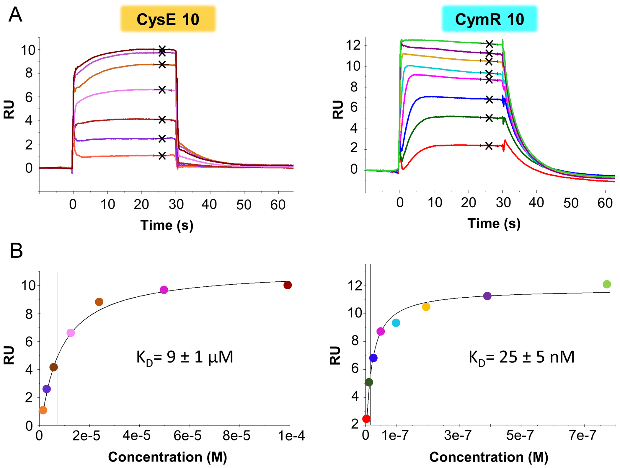
The pathway of bacterial cysteine biosynthesis is gaining traction for the development of antibiotic adjuvants. Bacterial cysteine biosynthesis is generally facilitated by two enzymes possessing O-acetyl-l-serine sulfhydrylases (OASS), CysK and CysM. In Staphylococcus aureus, there exists a single OASS homologue, SaCysK. Knockout of SaCysK was found to increase sensitivity to oxidative stress, making it a relevant target for inhibitor development. SaCysK also forms two functional complexes via interaction with the preceding enzyme in the pathway serine acetyltransferase (CysE) or the transcriptional regulator of cysteine metabolism (CymR). These interactions occur through insertion of a C-terminal peptide of CysE or CymR into the active site of SaCysK, inhibiting OASS activity, and therefore represent an excellent starting point for developing SaCysK inhibitors. Here, we detail the characterization of CysE and CymR-derived C-terminal peptides as inhibitors of SaCysK. Using a combination of X-ray crystallography, surface plasmon resonance, and enzyme inhibition assays, it was determined that the CymR-derived decapeptide forms extensive interactions with SaCysK and acts as a potent inhibitor (KD = 25 nM; IC50 = 180 nM), making it a promising lead for the development of SaCysK inhibitors. To understand the determinants of this high-affinity interaction, the structure-activity relationships of 16 rationally designed peptides were also investigated. This identified that the C-terminal pentapeptide of CymR facilitates the high-affinity interaction with SaCysK and that subtle structural modification of the pentapeptide is possible without impacting potency. Ultimately, this work identified CymR pentapeptides as a promising scaffold for the development of antibiotic adjuvants targeting SaCysK.

Keywords: cysteine biosynthesis; cysteine synthase; enzyme inhibition; peptide inhibitor; sulfur assimilation.


Figures

Fig 1.
Fig 1.. CysK enzymes possess multiple functions.
A) Reaction catalyzed by the O-acetyl-l-serine sulfhydrylase activity possessed by both CysK and CysM holo enzymes. B) The C-terminal regions of CysE and CymR enzymes that form functional complexes with CysK. The strictly conserved C-terminal isoleucine is conserved in all cases, indicated by the green shading. The reported binding affinities or inhibition constants correspond to the underlined sequence.
Fig 2.
Fig 2.. Isolation and functional characterization of SaCysK.
A) SDS-PAGE analysis of SaCysK. Lane 1: NEB unstained protein ladder broad range (10 – 200 KDa), Lane 2: 5 μg purified SaCysK.. B) Appearance of purified SaCysK. The intense yellow color indicated that SaCysK was purified as a PLP-adduct. C) Kinetic characterization of SaCysK activity. Data represents the mean ± standard deviation of three experiments.
Fig 3.
Fig 3.. The overall fold and active site architecture of holo SaCysK.
A) The dimeric assembly of holo SaCysK. The individual monomers of SaCysK are shown in green and dark green. The PLP cofactor shown in yellow sticks. The active site cavity is indicated by the yellow stars. B) Sequence alignment of active site regions in SaCysK, StCysK and StCysM. Positions conserved between 2 or more sequences are indicated by colored shading. C) Structural superposition of the corresponding regions forming the active site in SaCysK, StCysK (PDB: 1OAS) and StCysM (PDB: 2JC3). Loops 1 – 4 are shown as red, orange, pink and blue cartoons respectively, with Helix 4 shown in cyan.
Fig 4.
Fig 4.. Characterizing the binding of CysE 10 and CymR 10 to SaCysK by surface plasmon resonance (SPR).
A) Representative sensorgrams of CysE 10 (left) and CymR 10 (right) binding to SaCysK. Black crosses indicate the steady-state responses used to derive the KD. B) Representative steady-state binding responses for CysE 10 (left) and CymR 10 (right). the vertical line represents the KD value. The reported KD values are the mean ± standard deviation of at least two experiments.
Fig 5.
Fig 5.. Overall binding mode of CysE 10 and CymR 10 within the active site of SaCysK.
A) Simulated annealing composite omit electron density maps (2 Fo-Fc, 1σ; blue mesh) for CysE 10 (left; orange sticks) and CymR 10 (right; cyan sticks). A portion of each 10 amino acid peptide was ordered, indicated by the bold, underlined text. B) Binding of CysE 10 (left; orange sticks) and CymR 10 (right; cyan sticks) within the active site region of SaCysK. The transparent surface of SaCysK is shown in gray, with interacting sidechains shown as green sticks. PLP is shown as yellow sticks.
Fig 6.
Fig 6.. Structural comparison of CysE 10 and CymR 10 binding to SaCysK.
Comparison of binding at A) position 10, B) positions 7 and 9, and C) Positions 5, 6 and 8. CysE 10 and CymR 10 are shown as orange and cyan sticks, respectively. SaCysK is represented by the green cartoon, with interacting residues shown as green sticks. PLP is shown as yellow, transparent sticks. Yellow dashes represent hydrogen bonds, with distances reported in angstroms.
Fig 7.
Fig 7.. Structural characterization of CymR 5 and CymR 5-8b binding to SaCysK.
A) Simulated annealing composite omit electron density maps (2Fo-Fc, 1σ; blue mesh) for CymR 5 (left; pink sticks) and CymR 5-8b (right; purple sticks). B) Structural comparison of CymR 5 (pink sticks) and CymR 10 (cyan sticks) binding to the active site of SaCysK. C) Structural comparison of CymR 5-8b (purple sticks) and CymR 10 (cyan sticks) binding at position 8. D) A cavity lies beyond the hydrophobic channel occupied by Nal8. The SaCysK:CymR 5 and SaCysK:CymR 5-8b complexes are represented by green cartoon, with the SaCysK:CymR 10 complex shown as yellow cartoon. Yellow dashes indicate hydrogen bonds, with distances labelled in angstroms.
Fig 8.
Fig 8.. Proposed avenues for the optimisation of the CymR 5 scaffold.
Chemical modifications at the N-terminus, position 8, and dual modifications at positions 7 and 9 represent feasible routes to maintain potency and improve cell permeability and metabolic stability.

Update of

References

    1. Wright GD Antibiotic Adjuvants: Rescuing Antibiotics from Resistance. Trends Microbiol. 2016, 24 (11), 862–871. 10.1016/j.tim.2016.06.009. - DOI - PubMed
    1. Melander RJ; Melander C The Challenge of Overcoming Antibiotic Resistance: An Adjuvant Approach? ACS Infect. Dis 2017, 3 (8), 559–563. 10.1021/acsinfecdis.7b00071. - DOI - PMC - PubMed
    1. Douafer H; Andrieu V; Phanstiel OI; Brunel JM Antibiotic Adjuvants: Make Antibiotics Great Again! J. Med. Chem 2019, 62 (19), 8665–8681. 10.1021/acs.jmedchem.8b01781. - DOI - PubMed
    1. Campanini B; Pieroni M; Raboni S; Bettati S; Benoni R; Pecchini C; Costantino G; Mozzarelli A Inhibitors of the Sulfur Assimilation Pathway in Bacterial Pathogens as Enhancers of Antibiotic Therapy. Curr. Med. Chem 2015, 22 (2), 187–213. 10.2174/0929867321666141112122553. - DOI - PubMed
    1. Hicks JL; Oldham KEA; McGarvie J; Walker EJ Combatting Antimicrobial Resistance via the Cysteine Biosynthesis Pathway in Bacterial Pathogens. Biosci. Rep 2022, 42 (10), BSR20220368. 10.1042/BSR20220368. - DOI - PMC - PubMed

MeSH terms

LinkOut - more resources