. 2025 Jan 1;32(Pt 1):29-45.
doi: 10.1107/S1600577524010890. Epub 2025 Jan 1.

The Heisenberg-RIXS instrument at the European XFEL

Affiliations

The Heisenberg-RIXS instrument at the European XFEL

Justine Schlappa et al. J Synchrotron Radiat. .

Abstract

Resonant inelastic X-ray scattering (RIXS) is an ideal X-ray spectroscopy method to push the combination of energy and time resolutions to the Fourier transform ultimate limit, because it is unaffected by the core-hole lifetime energy broadening. Also, in pump-probe experiments the interaction time is made very short by the same core-hole lifetime. RIXS is very photon hungry so it takes great advantage from high-repetition-rate pulsed X-ray sources like the European XFEL. The Heisenberg RIXS instrument is designed for RIXS experiments in the soft X-ray range with energy resolution approaching the Fourier and the Heisenberg limits. It is based on a spherical grating with variable line spacing and a position-sensitive 2D detector. Initially, two gratings were installed to adequately cover the whole photon energy range. With optimized spot size on the sample and small pixel detector the energy resolution can be better than 40 meV (90 meV) at any photon energy below 1000 eV with the high-resolution (high-transmission) grating. At the SCS instrument of the European XFEL the spectrometer can be easily positioned thanks to air pads on a high-quality floor, allowing the scattering angle to be continuously adjusted over the 65-145° range. It can be coupled to two different sample interaction chambers, one for liquid jets and one for solids, each state-of-the-art equipped and compatible for optical laser pumping in collinear geometry. The measured performances, in terms of energy resolution and count rate on the detector, closely match design expectations. The Heisenberg RIXS instrument has been open to public users since the summer of 2022.

Keywords: European XFEL; Heisenberg RIXS; RIXS; X-ray Raman scattering; X-ray resonant diffraction; charge order; free-electron lasers; hRIXS; photochemistry; resonant X-ray diffraction; resonant inelastic X-ray scattering; soft X-rays; spin dynamics; spin order; time-resolved spectroscopy.


Figures

Figure 1
Figure 1
(a) The limits set by the Heisenberg uncertainty for time and energy during a soft X-ray spectroscopy experiment. Lower limits for energy and time resolution at given energy resolving power. We notice that a resolution of 30 meV (the present resolution limit for RIXS at synchrotrons at 1000 eV photon energy) sets the time resolution limit to about 138 fs, which is within the scope of SCS (Gerasimova et al., 2022 ▸) and other European XFEL instruments. (b) The influence of instrumental energy resolution on the appearance of a hypothetical RIXS spectrum of a correlated copper oxide material (Ament et al., 2011 ▸).
Figure 2
Figure 2
hRIXS spectrometer grating efficiency. Lines represent the computed values at four incidence angles with Au coating, aspect ratios c/d = 0.60 and 0.65, and groove depths h = 5 nm and 9 nm for the high-resolution grating (HRG) and high-transmission grating (HTG), respectively (Schäfers & Krumrey, 1996 ▸). The measured values for the HTG are shown by symbols (measurement performed at PM1 at BESSY II).
Figure 3
Figure 3
Contributions to the spectrometer energy bandwidth for the two gratings. The total resolution is given by the quadratic combination of the partial values. Calculations made for the nominal values of source size and detector spatial resolution used for the optimization of the VLS parameters (S1 = 5 µm, S2 = 10 µm) and slope error s′ = 0.1 µrad r.m.s.
Figure 4
Figure 4
Representation of the hRIXS spectrometer working points for the HRG (left) and the HTG (right). The graphs at the top show the accessible photon energies for a fixed incident angle α and the corresponding detector position. The bottom graphs show entrance and exit arm, r1 and r2, for a given photon energy and incidence angle α.
Figure 5
Figure 5
Spectrometer energy bandwidth for the two gratings as a function of spot size S1 and of detector spatial resolution S2. The total resolution is given by the quadratic combination of the partial values. Calculations made for the nominal value of the slope error s′ = 0.1 µrad r.m.s.
Figure 6
Figure 6
hRIXS spectrometer overview: diffraction scheme and degrees of motion for operation (a), model of the spectrometer (b), top view of the spectrometer when placed at the interaction point at the two extreme scattering angles: back-scattering at 2Θ = 145° (left) and forward-scattering at tΘ = 65° (right) (c).
Figure 7
Figure 7
Photograph of the hRIXS spectrometer installed in the working point inside the SCS hutch (the high-quality floor is partially covered for protection).
Figure 8
Figure 8
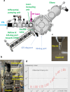
CHEM setup. Model with top view (a), photograph of the CHEM chamber (b), photograph of the liquid jet (c), and performance of the differential pumping unit (d).
Figure 9
Figure 9
Overview of the XRD setup for a solid sample environment: model (a) and photograph (b).
Figure 10
Figure 10
Inner mechanics of the XRD chamber: model revealing degrees of motion for the sample (a), photograph showing the diffractometer and the sample stage (b), and model with overview of the inner mechanics (c).
Figure 11
Figure 11
(a) XAS spectra of NiO and La2CuO4. (b) RIXS spectra of thin-film La2CuO4 at the Cu L3 edge measured at two different incident angles. The surface normal of the sample is parallel to the crystallographic c-direction. (c) Elastic line near specular conditions to estimate the combined resolution taken on La2CuO4. (d) RIXS spectrum of bulk-crystal NiO at the Ni L3 edge. The surface normal is the crystallographic a-direction. The elastic, magnetic and dd excitations are indicated in the spectra. (e) Schematic of the experimental geometry.
Figure 12
Figure 12
RIXS spectrum of liquid water obtained during commissioning at room temperature with the 1000 lines mm−1 grating. The inset shows a close-up of the vibrational progression.

References

    1. Aharonov, Y. & Bohm, D. (1961). Phys. Rev.122, 1649–1658.
    1. Ament, L. J., Ghiringhelli, G., Sala, M. M., Braicovich, L. & van den Brink, J. (2009). Phys. Rev. Lett.103, 117003. - PubMed
    1. Ament, L. J., van Veenendaal, M., Devereaux, T. P., Hill, J. P. & van den Brink, J. (2011). Rev. Mod. Phys.83, 705–767.
    1. Amorese, A., Langini, C., Dellea, G., Kummer, K., Brookes, N., Braicovich, L. & Ghiringhelli, G. (2019). Nucl. Instrum. Methods Phys. Res. A, 935, 222–226.
    1. Arpaia, R., Caprara, S., Fumagalli, R., De Vecchi, G., Peng, Y., Andersson, E., Betto, D., De Luca, G., Brookes, N., Lombardi, F., Salluzzo, M., Braicovich, L., Di Castro, C., Grilli, M. & Ghiringhelli, G. (2019). Science, 365, 906–910. - PubMed

LinkOut - more resources