. 2024 Dec 20;10(51):eadr9139.
doi: 10.1126/sciadv.adr9139. Epub 2024 Dec 20.

A wearable triboelectric impedance tomography system for noninvasive and dynamic imaging of biological tissues

Affiliations

A wearable triboelectric impedance tomography system for noninvasive and dynamic imaging of biological tissues

Peng Yang et al. Sci Adv. .

Abstract

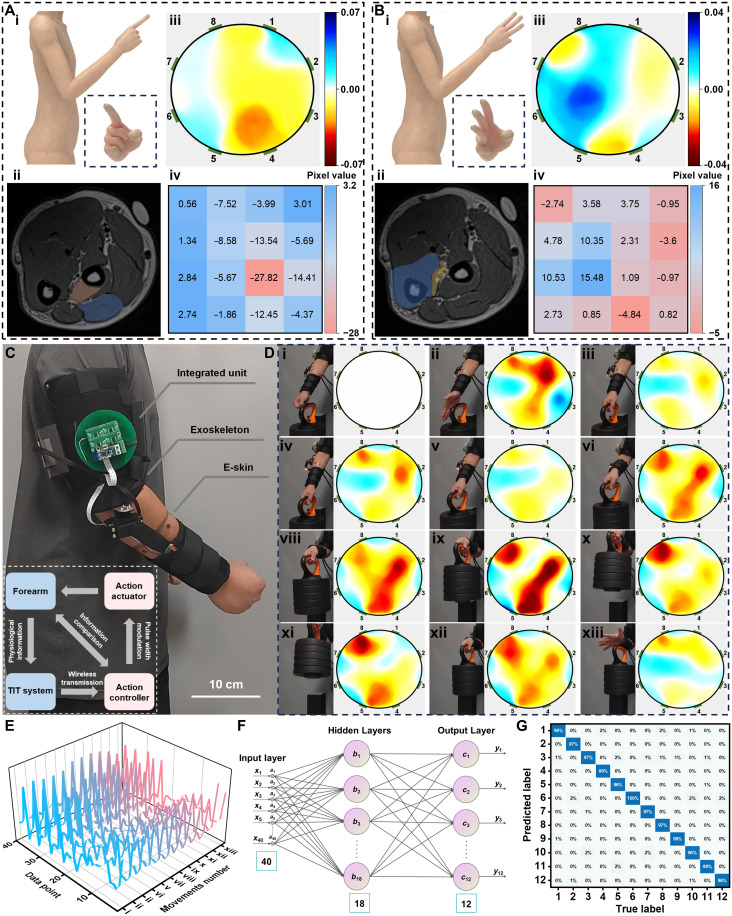
Tissue imaging is usually captured by hospital-based nuclear magnetic resonance. Here, we present a wearable triboelectric impedance tomography (TIT) system for noninvasive imaging of various biological tissues. The imaging mechanism relies on the obtained impedance information from the different soft human tissues. A high-precision signal source is designed on the basis of a composite triboelectric nanogenerator, which exhibits a minimal total harmonic distortion of 0.03% and a peak output signal-to-noise ratio up to 120 decibels. The current density injected into human skin is around 79.58 milliamperes per square meter, far below the safety threshold for medical devices. The TIT system achieves time-resolved tomography of human limbs' soft tissues, and many appealing functions can be realized by using this wearable system, including the observation of muscle movement, the motion intention recognition, and the identification of pathological changes of soft tissue. Hence, this TIT system with excellent biocompatibility can be integrated with various devices, such as medical-assistive exoskeletons and smart protective suit.


Figures

Fig. 1.
Fig. 1.. Overview of the design scheme and system components for the TIT system for soft tissue imaging of limbs.
(A) The design philosophy operational principles, and comparative analysis with imaging results of the TIT system. (B) Operational mode of TIT and the derivation of impedance models pertaining of human tissue, where “I” and ”V” represent the controlled current source and the measuring voltmeter, respectively. (C) The comprehensive framework delineation and data processing strategies used in the TIT system.
Fig. 2.
Fig. 2.. Design scheme of e-skin and electrical parameter characterization of HESS.
(A) The activation time of the adhesion layer at different temperatures. Inset: Activation time of the adhesion layer placed on human skin at different time points. (B) The electrical conductivity (σ) of the PPPA as a function of AgNW solution concentration and the length of AgNWs. The inset illustrates scanning electron microscopy images of the PPPA. (C) Comparison of the contact impedance between the e-skin and commercial AgCl/Ag electrode. (D) Instrumentation photos used for measuring and calibrating the operational imbalance of the HESS. The enlarged image displays physical photographs of the HESS. (E) Full spectrum of the current output from the HESS, ranging from 2 to 200 Hz with amplitudes between 4 and 5 μA. (F) Frequency response characteristics of the THD and OSNR of the ac signal. (G) The derivative curve obtained from the power curve of HESS, whose intersections with 0 correspond to the output resistance. The inset depicts the output resistance corresponding to different thicknesses of polytetrafluoroethylene (PTFE). (H) Human impedance (Z) distribution measured with the TIT principle. (I) The change in current amplitude of the HESS when loaded onto the human body at different operating frequencies. Each set of results is based on 100 data points. kΩ, kilohm; MΩ, megohm.
Fig. 3.
Fig. 3.. Image reconstruction of targets in water tanks and evaluation of imaging results.
(A) Photograph of the tank model. (B) SSNR exhibited by the TIT system with different electrode configurations with each test repeated 200 times. IQR, interquartile range. (C and D) The reconstructed images of single and multiple targets in the water tank using the TIT system, including results obtained with two injection currents of 2 and 200 Hz. In the test, the homogeneous polypropylene rod is selected as the detection target. (E) The ICC comparison for the reconstructed images of single-target and dual-target scenarios at different current injection frequencies. (F) Kernel density map of the imaging accuracy demonstrated by the TIT system, with 200 test trials. (G) Temporal resolution of the TIT device in response to frequency. (H) Evaluation of target position errors and RMSE in 200 tests.
Fig. 4.
Fig. 4.. Image reconstruction of upper limb movements by the TIT system and integration of the TIT system with medical-assistive exoskeleton.
(A) The evaluation results of the TIT system for extending the index finger. (i) Schematic diagram of movement. (ii) MRI result, with color blocks indicating regions where marked changes occur. (iii) The reconstructed images of the TIT system. (iv) Regional change statistics of the reconstructed images. (B) The changes in the internal tissue when the simultaneous extension of the index, middle, and ring fingers. (i) Schematic diagram of movement. (ii) MRI picture. (iii) Reconstructed image. (iv) Regional change statistics. (C) Physical photograph of the integrated exoskeleton, where the integrated unit is the integration of the HESS and microcontroller. Inset: System framework and logical control description of the integrated exoskeleton. (D) The 12 motions (i to xiii) involved in the weight lifting experiment along with their reconstructed images. (E) The boundary data for 12 distinct motions. (F) Schematic diagram of the GA-BP model. (G) Classification results of the GA-BP model on the motion datasets.
Fig. 5.
Fig. 5.. Reconstruction of images for forearm lipomas by the TIT system and evaluation of imaging results.
(A) The MRI images of the patient, along with the images reconstructed by the TIT system. (i) Coronal MRI graph. (ii) Corresponding MRI cross-sectional images. (iii) Reconstructed TIT images, which reveals information such as the presence, size, and location of lipomas. (B) The images at different positions (i to vi), with a longitudinal spacing of 4.5 cm. (C) The 3D model constructed from sectional images. (i) Reconstructed 3D model of the lipoma, where (i) to (iv) correspond to six sectional images. (ii) Localization and morphology of the lipoma within the forearm model. (iii) Cross-sectional images of the forearm model.
Fig. 6.
Fig. 6.. The identification and reconstruction of pathological tissues caused by fractures using the TIT system.
(A) The schematic diagram of TIT forearm testing (i) and the images at the respective locations (ii and iii). (B) Regional variation statistics of the image in A(iii). (C) Comparison of capacitance values at the testing positions on the left and right forearms of the patient, where 1-4, 2-3, 5-8, and 6-7 are the four electrode pairs (i to iv). (D) X-ray imaging of the patient’s left radius and ulna in the anteroposterior and lateral views. (E) Axial extent of the increase region in conductivity measured by the TIT system.

References

    1. Hu H., Huang H., Li M., Gao X., Yin L., Qi R., Wu R. S., Chen X., Ma Y., Shi K., Li C., Maus T. M., Huang B., Lu C., Lin M., Zhou S., Lou Z., Gu Y., Chen Y., Lei Y., Wang X., Wang R., Yue W., Yang X., Bian Y., Mu J., Park G., Xiang S., Cai S., Corey P. W., Wang J., Xu S., A wearable cardiac ultrasound imager. Nature 613, 667–675 (2023). - PMC - PubMed
    1. Wang C., Chen X., Wang L., Makihata M., Liu H.-C., Zhou T., Zhao X., Bioadhesive ultrasound for long-term continuous imaging of diverse organs. Science 377, 517–523 (2022). - PubMed
    1. Boto E., Holmes N., Leggett J., Roberts G., Shah V., Meyer S. S., Muñoz L. D., Mullinger K. J., Tierney T. M., Bestmann S., Barnes G. R., Bowtell R., Brookes M. J., Moving magnetoencephalography towards real-world applications with a wearable system. Nature 555, 657–661 (2018). - PMC - PubMed
    1. Lin M., Hu H., Zhou S., Xu S., Soft wearable devices for deep-tissue sensing. Nat. Rev. Mater. 7, 850–869 (2022).
    1. Frahm J., Voit D., Uecker M., Real-time magnetic resonance imaging: Radial gradient-echo sequences with nonlinear inverse reconstruction. Invest. Radiol. 54, 757–766 (2019). - PubMed

LinkOut - more resources