. 2024 Dec 20;10(51):eadq3632.
doi: 10.1126/sciadv.adq3632. Epub 2024 Dec 20.

Tracking of spaceflight-induced bone remodeling reveals a limited time frame for recovery of resorption sites in humans

Affiliations

Tracking of spaceflight-induced bone remodeling reveals a limited time frame for recovery of resorption sites in humans

Matthias Walle et al. Sci Adv. .

Abstract

Mechanical unloading causes bone loss, but it remains unclear whether disuse-induced changes to bone microstructure are permanent or can be recovered upon reloading. We examined bone loss and recovery in 17 astronauts using time-lapsed high-resolution peripheral quantitative computed tomography and biochemical markers to determine whether disuse-induced changes are permanent. During 6 months in microgravity, resorption was threefold higher than formation. Upon return to Earth, targeted bone formation occurred in high mechanical strain areas, with 31.8% of bone formed in the first 6 months after flight at sites resorbed during spaceflight, significantly higher than the 2.7% observed 6 to 12 months after return. Limited bone recovery at resorption sites after 6 months on Earth indicates a restricted window for reactivating bone remodeling factors in humans. Incomplete skeletal recovery may arise from these arrested remodeling sites, representing potential targets for new interventions, thus providing means to counteract this long-term health risk for astronauts.


Figures
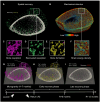
Fig. 1.
Fig. 1.. Spatial recovery and mechanoregulation of bone remodeling in astronauts.
We investigated whether coupling factors or mechanical signals dictate the recovery of resorption sites by bone formation in 17 astronauts during 4- to 7-month missions and 1 year recovery. The outcomes of the study are illustrated using 3D visuals of a distal tibia bone from a representative astronaut, obtained through high-resolution peripheral quantitative computed tomography. (A) Spatial recovery was assessed by analyzing coupled bone formation at previous resorption sites. (B) The mechanical stimulus at the same sites was simulated using finite element analysis to assess the mechanoregulation of bone remodeling. (C to E) Detailed views showing bone resorption sites in purple (C), recovered resorption sites in green (D), and all bone formation sites in orange (E). (F) Detailed view of the mechanical signal stimulus quantified by strain energy density to assess whether bone resorption and formation were more likely to occur in regions of low mechanical strain (shades of blue) and high strain (shades of red), respectively. (G) Our results demonstrated that bone loss due to the absence of gravitational loading was primarily driven by bone resorption. (H) In the first 6 months after return to Earth, increased bone formation occurred and targeted previous resorption sites. (I) In the following 6 months, bone formation decreased and targeted new regions in the microstructure. Throughout the recovery, we identified a relationship between remodeling sites and mechanical signals. This finding, along with identified time constraints for recovery, suggested that bone remodeling was jointly influenced by mechanical stimuli and biological coupling factors.
Fig. 2.
Fig. 2.. Bone remodeling activity visualized in vivo by HR-pQCT at the distal tibia.
(A) Total formation (Tt.F) and resorption (Tt.R) quantified over the mission timeline at the distal tibia reveal imbalanced turnover in microgravity that reverses within the first 6 months (R + 0 days—R + 6 months) after flight and stabilizes in the second 6 months (R + 6—R + 12 months) of recovery. Individual markers represent individual astronauts. Significant differences between Tt.F and Tt.R are indicated (**P < 0.01, ****P < 0.0001). (B) Participant odds ratios quantify the mechanoregulation of formation (ORF) and resorption (ORR) events. ORF and ORR reflect the percent change in formation or resorption likelihood per percent strain change. Notably, there is an increase in both ORF and ORR upon return to Earth, indicating adaptation to gravitational forces during reconditioning. Significant differences between time points are indicated (***P < 0.001). (C and D) Representative images show bone microstructure with resorption (purple) and formation (orange) sites that occurred during microgravity (C) and within the first 6 months of reloading after flight (D). (E) Strain distribution in bone microstructure simulated using micro–finite element (FE) analysis. Micro-FE analysis is a computational technique to estimate local deformations (i.e., strain) by simulating a virtual mechanical compression test of the bone microstructure. Color map represents strain magnitude under simulated gravitational loading.
Fig. 3.
Fig. 3.. Connecting local and systemic bone turnover.
(A and B) Correlations between remodeling measures (Tt.F, Tt.R) represented on log scale (A) and mechanoregulation (ORF, ORR) of the left and right distal tibia (B). (C to F) Correlations between these metrics and absolute bone turnover markers, including osteocalcin (OCN), procollagen 1 N-terminal propeptide (P1NP), sclerostin, C-telopeptide of cross-linked collagen type I (CTX-I), and type I collagen N-telopeptide (NTX-I). Correlations performed using Spearman’s rank correlation, where positive associations are shaded in red, and negative correlations are in blue. This analysis assesses relationships between local bone remodeling outcomes from high-resolution peripheral quantitative computed tomography and systemic bone metabolic activity. Biochemical bone turnover markers were measured in serum and urine during the following periods: in-flight (FD15-FD180), 6 months post-flight (R + 1, R + 6) and 12-months post-flight (R + 12). Original data on bone turnover markers are available in a previously published work (13). Significant correlations are indicated (*P < 0.05, **P < 0.01).
Fig. 4.
Fig. 4.. Bone recovery after spaceflight.
(A) Recovery of resorbed bone during early (R + 0 to R + 6 months) and late (R + 6 to R + 12 months) phases at the exact spatial locations previously resorbed in microgravity. Also shown is subsequent resorption after return to Earth at sites formed in microgravity. Individual markers represent individual astronauts. Significant differences between time points are indicated (*P < 0.05, ****P < 0.0001). (B) Linear regression between recovered resorption (early phase) and increase in CTX-I during spaceflight (measured on flight day 15 and before return on flight day 120 or 180). (C and D) Representative high-resolution peripheral quantitative computed tomography images show bone microstructure with resorption (purple) and formation (orange) sites identified in (C) microgravity and (D) at 6 months after return to Earth. (E) Spatial recovery of the same locations previously resorbed in microgravity during the early recovery phase. Periosteal contraction representing reduced cross-sectional area (i.e., bone size) during spaceflight and subsequent expansion may drive the observed elevated periosteal remodeling, potentially explaining previously reported post-mission changes in bone size (15).

References

    1. Frost H. M., The mechanostat: A proposed pathogenic mechanism of osteoporoses and the bone mass effects of mechanical and nonmechanical agents. Bone Miner. 2, 73–85 (1987). - PubMed
    1. Frost H. M., Bone’s mechanostat: A 2003 update. Anat. Rec. A Discov. Mol. Cell. Evol. Biol. 275, 1081–1101 (2003). - PubMed
    1. Schaffler M. B., Cheung W. Y., Majeska R., Kennedy O., Osteocytes: Master orchestrators of bone. Calcif. Tissue Int. 94, 5–24 (2014). - PMC - PubMed
    1. Qin L., Liu W., Cao H., Xiao G., Molecular mechanosensors in osteocytes. Bone Res. 8, 23 (2020). - PMC - PubMed
    1. Everts V., Delaissé J. M., Korper W., Jansen D. C., Tigchelaar-Gutter W., Saftig P., Beertsen W., The bone lining cell: Its role in cleaning Howship’s lacunae and initiating bone formation. J. Bone Miner. Res. 17, 77–90 (2002). - PubMed

LinkOut - more resources