. 2025 Jan;301(1):108110.
doi: 10.1016/j.jbc.2024.108110. Epub 2024 Dec 18.

The physicochemical properties of lipopolysaccharide chemotypes regulate activation of the contact pathway of blood coagulation

Affiliations

The physicochemical properties of lipopolysaccharide chemotypes regulate activation of the contact pathway of blood coagulation

André L Lira et al. J Biol Chem. 2025 Jan.

Abstract

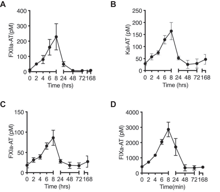
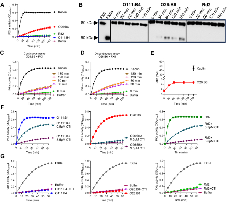
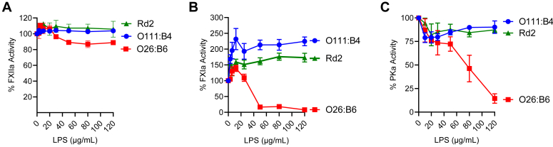
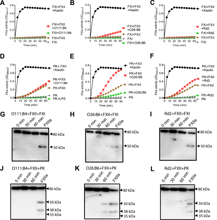
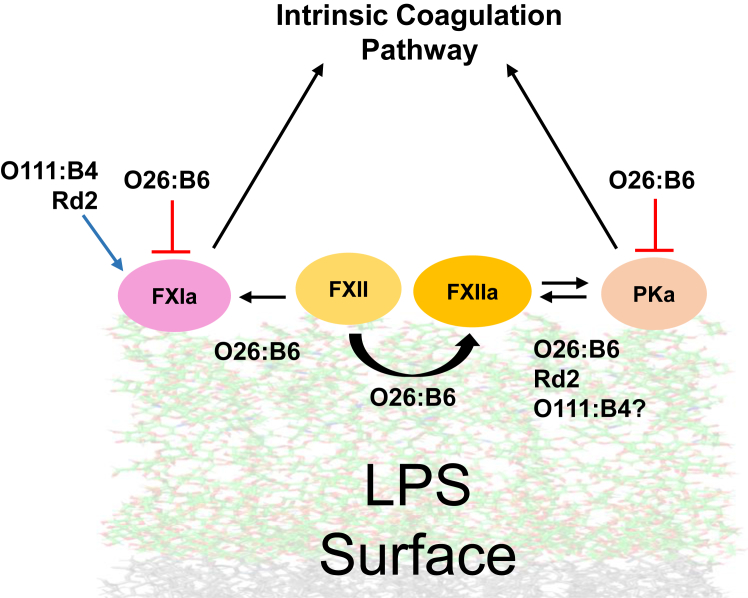
Lipopolysaccharide (LPS) is the primary pathogenic factor in Gram-negative sepsis. While the presence of LPS in the bloodstream during infection is associated with disseminated intravascular coagulation, the mechanistic link between LPS and blood coagulation activation remains ill-defined. The contact pathway of coagulation-a series of biochemical reactions that initiates blood clotting when plasma factors XII (FXII) and XI (FXI), prekallikrein (PK), and high molecular weight kininogen interact with anionic surfaces-has been shown to be activated in Gram-negative septic patients. In this study, using an in vivo baboon model of Gram-negative Escherichia coli sepsis, we observed activation of the contact pathway including FXII, FXI, and PK. We examined whether E.coli LPS molecules could bind and activate contact pathway members by quantifying the interaction and activation of either FXII, FXI, or PK with each of the three chemotypes of LPS: O111:B4, O26:B6, or Rd2. The LPS chemotypes exhibited distinct physicochemical properties as aggregates and formed complexes with FXII, FXI, and PK. The LPS chemotype O26:B6 uniquely promoted the autoactivation of FXII to FXIIa and, in complex with FXIIa, promoted the cleavage of FXI and PK to generate FXIa and plasma kallikrein, respectively. Furthermore, in complex with the active forms of FXI or PK, LPS chemotypes were able to regulate the catalytic activity of FXIa and plasma kallikrein, respectively, despite the inability to promote the autoactivation of either zymogen. These data suggest that the procoagulant phenotype of E.coli is influenced by bacterial strain and the physicochemical properties of the LPS chemotypes.

Keywords: coagulation; contact pathway; factor XII; lipopolysaccharides; sepsis.


Conflict of interest statement

Conflicts of interest C. U. L. and E. I. T. are employees of Aronora, Inc, a company that may have a commercial interest in the results of this research. J. J. S. serves as a medical consultant for Aronora, Inc. This potential conflict of interest has been reviewed and managed by conflict of interest in research committee of the Oregon Health & Science University. The remaining authors declare that they have no conflicts of interest with the contents of this article.

Figures

Figure 1
Figure 1
Escherichia coli infusion induced contact pathway activation in baboons. Activation of contact coagulation cascade in baboons challenged with E. coli. Time course dynamics of complexes of (A) FXIIa- AT, (B) kallikrein-AT, (C) FXIa-AT, and (D) FIXa-AT in bacterial-challenged animals are shown.
Figure 2
Figure 2
LPS chemotypes form aggregates with distinct physicochemical characteristics.A, illustration of LPS molecule. LPS is composed by lipid A, core oligosaccharide (core OS; divided into an inner and an outer region), and a O-polysaccharide chain (O-Antigen; or OAg). The conserved lipid A and inner core region are highly anionic due to multiple negative charges from PO42 (yellow circles) and COO (blue circles). LPS with a long OAg are classified as smooth strains (or S-LPS) while those lacking the OAg are considered rough strains (or R-LPS). Bacteria that express LPS chemotypes which contain only one repeating OAg subunit are classified as semi-rough strains (or SR-LPS). B, a model of the Escherichia coli outer membrane containing LPS chemotype molecules was created using CHARMM (http://charmm-gui.org/) and processed with PyMol. The lipid A moieties are shown as gray chains, while the inner and outer core and O-antigen are shown as green chains. O111:B4 (smooth or S-LPS), O26:B6 (semi-rough or SR-LPS), Rd2 (rough or R-LPS) LPS chemotypes are differentiated by the length of the O-antigen and core region. C, LPS chemotypes O111:B4, O26:B6, or Rd2 (50 μg/ml) were submitted to electrophoresis on a 10 to 20% polyacrylamide gel in the presence of SDS and detected by silver staining. The apparent molecular weights obtained, determined by comparison with molecular weight standards, were 20 to 100 kDa for O111:B4, 13 kDa for O26:B6, and 3 kDa for Rd2. D, statistical analysis and quantifications of size diameters, polydispersity index (PdI), and zeta potential (ZP) of LPS aggregates. The size distribution of LPS aggregates is shown for O111:B4 (blue), O26:B6 (red), and Rd2 (green), which peak with a dh of 95 ± 6 nm, 196.0 ± 7, and 183 ± 11, respectively. The ZP values confirmed the expected negative surface charges of O26:B6 and Rd2, as well as the overall neutral charge of O111:B4. The aggregates were analyzed in 20 mM Hepes buffer, pH 7.4 in the absence or presence of 150 mM NaCl. The PdI of the aggregates were in general <0.3, which indicates a good stability and homogeneity.
Figure 3
Figure 3
Illustration of electrostatic potential surfaces of contact pathway proteins, calculated by the APBS module as implemented in PyMol. The following PDB structures were used in the calculations: 5EOD (FXI) and 2ANW (PK). The PDB structure for HK and FXII was downloaded from the AlphaFold Database (entry P01042 and P00748, respectively). Additionally, the figure provides information on molecular weights and hydrodynamic diameters (HD).
Figure 4
Figure 4
Analysis of LPS–zymogens interactions using fluorescence spectroscopy. The normalized tryptophan intensities at an emission of 336 nm of (A) FXII (0.5 μM), (B) PK (0.5 μM), or (C) FXI (0.5 μM) were plotted as a function of O111:B4, O26:B6, or Rd2 (3–120 μg/ml) in Hepes buffer solution supplemented with 150 mM NaCl. Lines are guides to the eye. Tryptophan emission spectra of (D) FXII (0.5 μM), (E) PK (0.5 μM), or (F) FXI (0.5 μM) in the absence or presence of LPS (120 μg/ml). Spectra were normalized from 0 to 1 for ease of comparison. The data points represent the averages of triplicate measurements, with error bars indicating ±1 SD.
Figure 5
Figure 5
Effects of LPS aggregates on zymogens activation.A, FXII (100 nM) was incubated with O111:B4, O26:B6, and Rd2 (50 μg/ml) for 2 h. Continuous formation of amidolytic activity was measured using the substrate S-2302 (300 μM) and monitored at 405 nm. Kaolin (5 μg/ml) was used as a positive control and buffer as a vehicle control. B, O26:B6-induced generation of FXIIa analyzed by immunblotting. FXII, 100 nM; LPS, 25 μg/ml. FXII (80 kDa) and FXIIa (50 kDa heavy chain) were used as control. C, assessment of FXII autoactivation in a continuous assay with O26:B6. FXII (100 nM) and O26:B6 (25 μg/ml) were incubated from 0 to 180 min, followed by the measurement of FXII amidolytic activities in a continuous reaction with S-2302 (300 μM). D, assessment of FXII autoactivation in a discontinuous assay with O26:B6. Timed aliquots from an FXII and O26:B6 mixture were quenched with 0.1 mg/ml polybrene, and FXII amidolytic activities were subsequently measured. E, apparent concentration of FXIIa generated at each quenched time point in the discontinuous assay. Concentrations were calculated from the initial velocities in (D) with reference to a standard curve. Kaolin promotes the formation of ∼50 nM FXIIa (black sphere). Data points are the averages of triplicate measurements. F, autoactivation of PK in the presence of LPS and HK appears to be due to contamination of HK with traces of FXII(a). The apparent autoactivation was inhibited by CTI. PKa levels were measured as a function of time after incubating PK (100 nM) with HK (120 nM) and LPS aggregates (25 μg/ml) in the presence of 0, 0.5, or 2.5 μM CTI, and PK amidolytic activities were subsequently measured using the substrate S-2302 (300 μM) at 405 nm. G, assessment of autoactivation of FXI in the presence of LPS. FXI (50 nM) with HK (120 nM) and LPS aggregates (25 μg/ml) were mixed, and FXI amidolytic activities were subsequently measured using the substrate S-2366 (300 μM) at 405 nm. Data are expressed as mean ± SD of three independent experiments.
Figure 6
Figure 6
Effects of LPS on FXIIa, PKa, and FXIa amidolytic activities. (A) FXIIa (50 nM), (B) PKa (50 nM), or (C) FXIa (50 nM) was incubated with LPS (0–120 μg/ml), and amidolytic activity was measured using the substrate S-2302 (300 μM) for FXIIa or PKa and S-2366 (300 μM) for FXIa. The LPS showed distinct abilities to regulate the catalytic function of the proteases. Data are expressed as mean ± SD of three independent experiments.
Figure 7
Figure 7
The complex FXIIa–LPS presents catalytic activity towards FXI and PK. FXI (30 nM) was incubated with FXII (5 nM), with (A) O111:B4, (B) O26:B6, and (C) Rd2 (25 μg/ml) or with both in the presence of S2302 (300 μM) and the reaction was subsequently monitored at 405 nm. Samples from 0, 30, and 60 min time point were separated under reducing conditions and immunoblotted with an antibody against FXI in the presence of (G) O111:B4, (H) O26:B6, or (I) Rd2. PK (50 nM) was incubated with FXII (5 nM), with (D) O111:B4, (E) O26:B6, and (F) Rd2 (25 μg/ml) or with both in the presence of S2302 (300 μM) and the reaction was subsequently monitored at 405 nm. Samples from 0, 30, and 60 min time point were separated under reducing conditions and immunoblotted with an antibody against PK in the presence of (J) O111:B4, (K) O26:B6, or (L) Rd2. Data are expressed as mean ± SD of three independent experiments.
Figure 8
Figure 8
Effects of “masked” monomeric LPS on contact pathway proteases.A, schematic representation of LPS masking. In aqueous solution, LPS naturally forms aggregates in the form of micelles or vesicles. Addition of citrate (chelator) weakens the aggregate structure by removing divalent cations. Surfactants like Triton X-100 then intercalate into the aggregates. Eventually, the aggregates completely disperse into monomers. B, FXIIa generation in the presence of LPS aggregates or monomers. LPS (50 μg/ml) in the form of aggregates or monomers were incubated with FXII (100 nM) for 2 h followed by the addition of S-2302 (300 μM). LPS in their aggregate form are more potent surfaces to stimulate FXII autoactivation than monomers. The effects on the proteases (C) FXIIa, (D) PKa, and (E) FXIa in the presence of aggregates or monomers were evaluated. The dashed line indicates the point of maximum enzyme activity (100%). Data are expressed as mean ± SD of three independent experiments.
Figure 9
Figure 9
Detection of FXII/PK activation during contact activation in plasma by LPS. Contact activation was triggered by the incubation of (A) O111:B4, (B) O26:B6, or (C) Rd2 (25, 50, or 100 μg/ml) in citrated plasma for 30 min at 37 °C, followed by the measurement of FXII/PK amidolytic activity using the substrate S-2302 (300 μM) and monitored at 405 nm. Clot times of human normal plasma by (D) O111:B4, (E) O26:B6, or (F) Rd2 (25, 50, or 100 μg/ml) after incubation for 5 min. After recalcification, time to clot formation was assayed by turbidity measurements at 405 nm. Kaolin (5 μg/ml) was used as a positive control and buffer as a vehicle control. Error bars are the SD of three independent experiments performed in technical triplicate. Results were analyzed by one-way ANOVA; ∗p < 0.05, ∗∗p < 0.001, ∗∗∗p < 0.0001 with respect to buffer control.
Figure 10
Figure 10
Simplified schematic illustration of the contact pathway activation by LPS. Although the zymogens FXII, FXI, and PK bind to the LPS surface, the nature of their interactions and influence differ according to the chemotype. Only FXII autoactivates upon O26:B6 chemotype LPS surface contact, generating FXIIa. The FXII–O26:B6 complex activates both PK and FXI, whereas the FXII–Rd2 activates only PK. The LPS chemotypes also uniquely regulate protease catalytic activities. O26:B6 acts as a regulator for PKa and FXIa showing a dose-dependent reduction of amidolytic activity (red arrows), while Rd2 and O111:B4 boost the catalytic activity of FXIa (blue arrow).

Similar articles

Cited by

References

    1. Kellum J.A., Formeck C.L., Kernan K.F., Gómez H., Carcillo J.A. Subtypes and mimics of sepsis. Crit. Care Clin. 2022;38:195–211. - PubMed
    1. Remick D.G. Pathophysiology of sepsis. Am. J. Pathol. 2007;170:1435–1444. - PMC - PubMed
    1. Popescu N.I., Lupu C., Lupu F. Disseminated intravascular coagulation and its immune mechanisms. Blood. 2022;139:1973–1986. - PMC - PubMed
    1. Czempik P.F., Wiórek A. Management strategies in septic coagulopathy: a review of the current literature. Healthcare (Basel) 2023;11:227. - PMC - PubMed
    1. Iba T., Levy J.H., Raj A., Warkentin T.E. Advance in the management of sepsis-induced coagulopathy and disseminated intravascular coagulation. J. Clin. Med. 2019;8:728. - PMC - PubMed

Publication types

LinkOut - more resources