. 2024 Dec 20;8(1):285.
doi: 10.1038/s41698-024-00771-y.

Feature-interactive Siamese graph encoder-based image analysis to predict STAS from histopathology images in lung cancer

Affiliations

Feature-interactive Siamese graph encoder-based image analysis to predict STAS from histopathology images in lung cancer

Liangrui Pan et al. NPJ Precis Oncol. .

Abstract

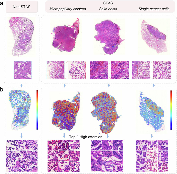
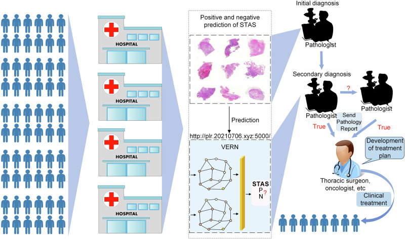
Spread through air spaces (STAS) is a distinct invasion pattern in lung cancer, crucial for prognosis assessment and guiding surgical decisions. Histopathology is the gold standard for STAS detection, yet traditional methods are subjective, time-consuming, and prone to misdiagnosis, limiting large-scale applications. We present VERN, an image analysis model utilizing a feature-interactive Siamese graph encoder to predict STAS from lung cancer histopathological images. VERN captures spatial topological features with feature sharing and skip connections to enhance model training. Using 1,546 histopathology slides, we built a large single-cohort STAS lung cancer dataset. VERN achieved an AUC of 0.9215 in internal validation and AUCs of 0.8275 and 0.8829 in frozen and paraffin-embedded test sections, respectively, demonstrating clinical-grade performance. Validated on a single-cohort and three external datasets, VERN showed robust predictive performance and generalizability, providing an open platform ( http://plr.20210706.xyz:5000/ ) to enhance STAS diagnosis efficiency and accuracy.


Conflict of interest statement

Competing interests: The authors declare that they have no known competing financial interests or personal relationships that could have appeared to influence the work reported in this paper.

Figures

Fig. 1
Fig. 1. Workflow including data preprocessing, model training and inference, interpretability analysis, and multicenter validation.
a Data preprocessing involves digitization of WSI, extraction of regions of interest, segmentation of tissue into patches, and patch data augmentation. b The model architecture includes feature extraction from pretrained models (KimiaNet, CTransPath), construction of WSI spatial topological maps, feature-interactive Siamese graph encoder (VERN) module, and diagnostic results for STAS. c Detailed architecture of the VERN. d Interpretability analysis of WSI. e The STAS dataset includes internal training and validation sets, a test set, and an external validation set.
Fig. 2
Fig. 2. Experimental results of STAS prediction using the VERN trained with five-fold cross-validation on internal and external datasets.
a ROC curves of the VERN from five-fold cross-validation. The five curves represent the testing performance of the VERN after each fold of training, with the diagonal line showing the in-domain test results. b PRC of the VERN from five-fold cross-validation. c Accuracy, precision, recall, F1 score, specificity, and AUC values of the VERN from five-fold cross-validation testing. d Confusion matrix of the VERN from five-fold cross-validation testing. e ROC curves of the VERN tested on digital FSs and PSs. f PRC curves of the VERN tested on digital FSs and PSs. g Confusion matrix results of the VERN tested on digital FSs and PSs.
Fig. 3
Fig. 3. Interpretability analysis of the VERN.
a WSIs with STAS-negative and STAS-positive annotations, where pathologists outlined the main tumor body and STAS subtypes, including micropapillary, solid nests, and single cells. b Attention scores for each patch based on the VERN. High values (red) indicate higher model-predicted contributions, while low values (purple) indicate lower contributions. Additionally, the nine patches with the highest contributions were identified.
Fig. 4
Fig. 4. Evaluation of VERN performance in predicting STAS in single-cohort and multicenter experiments.
a ROC and PRC curves validating the VERN’s effectiveness based on 356 histopathological images from the Second Xiangya Hospital of Central South University. b ROC and PRC curves validating the VERN’s effectiveness based on 91 histopathological images from the Cancer Hospital of Zhengzhou University and Henan Cancer Hospital. c ROC and PRC curves validating the VERN’s effectiveness based on 101 histopathological images from TCGA. d ROC and PRC curves validating the VERN’s effectiveness based on 100 histopathological images from CPTAC. e Accuracy, precision, recall, F1 score, specificity, and AUC values of the VERN predicting the presence or absence of STAS across the four datasets.
Fig. 5
Fig. 5. Workflow diagram of the application of the VERN in clinical practice.
This system can be used as a supplement to the interpretation of STAS by pathologists or pathology departments in hospitals in areas with insufficient medical resources. The process involves patients undergoing surgery in the hospital, pathology sections being prepared and scanned by the pathology department, and testing through the STAS testing website. The three-level diagnosis and treatment system is combined with the test results of the VERN. The pathologist provides a detailed pathological diagnosis report, and the thoracic surgeon determines the surgical method suitable for the patient (such as lobectomy, sublobar resection, etc.), and the oncologist develops a follow-up treatment plan to provide patients with personalized and precise treatment. We welcome you to visit our test platform http://plr.20210706.xyz:5000/, or download the VERN from https://github.com/pengsl-lab/STAS/tree/main to use.

Similar articles

References

    1. Onozato, M. L. et al. Tumor islands in resected early-stage lung adenocarcinomas are associated with unique clinicopathologic and molecular characteristics and worse prognosis. Am. J. Surgical Pathol.37, 287–294 (2013). - PMC - PubMed
    1. Kadota, K. et al. Tumor spread through air spaces is an important pattern of invasion and impacts the frequency and location of recurrences after limited resection for small stage I lung adenocarcinomas. J. Thorac. Oncol.10, 806–814 (2015). - PMC - PubMed
    1. Warth, A. et al. Prognostic impact of intra-alveolar tumor spread in pulmonary adenocarcinoma. Am. J. Surgical Pathol.39, 793–801 (2015). - PubMed
    1. Chae, M. et al. Prognostic significance of tumor spread through air spaces in patients with stage IA part-solid lung adenocarcinoma after sublobar resection. Lung Cancer152, 21–26 (2021). - PubMed
    1. Eguchi, T. et al. Lobectomy is associated with better outcomes than sublobar resection in spread through air spaces (STAS)-positive T1 lung adenocarcinoma: a propensity score–matched analysis. J. Thorac. Oncol.14, 87–98 (2019). - PMC - PubMed

LinkOut - more resources