. 2025 Aug;48(4):2503-2515.
doi: 10.1007/s10753-024-02206-z. Epub 2024 Dec 21.

CircIRAK3 Promotes Neutrophil Extracellular Trap Formation by Improving the Stability of ELANE mRNA in Sepsis

Affiliations

CircIRAK3 Promotes Neutrophil Extracellular Trap Formation by Improving the Stability of ELANE mRNA in Sepsis

Yao Lu et al. Inflammation. 2025 Aug.

Abstract

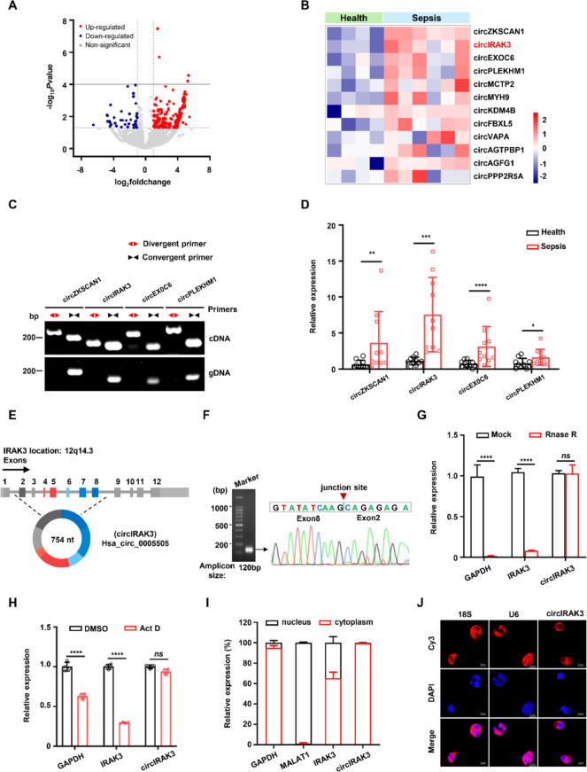
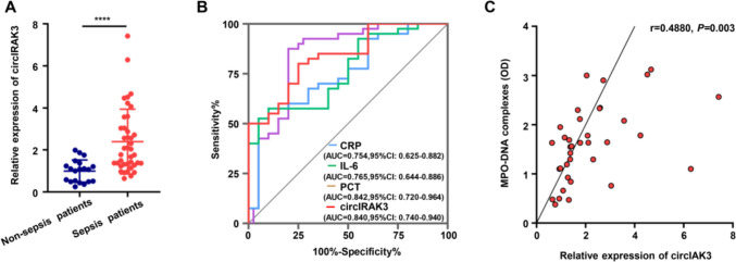
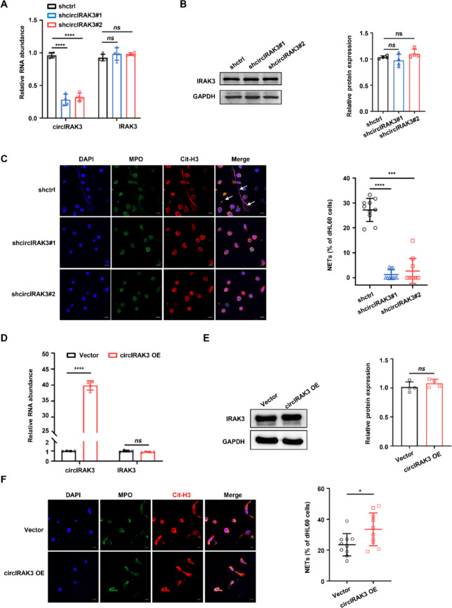
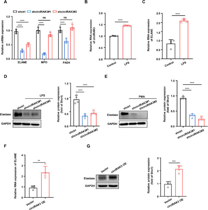
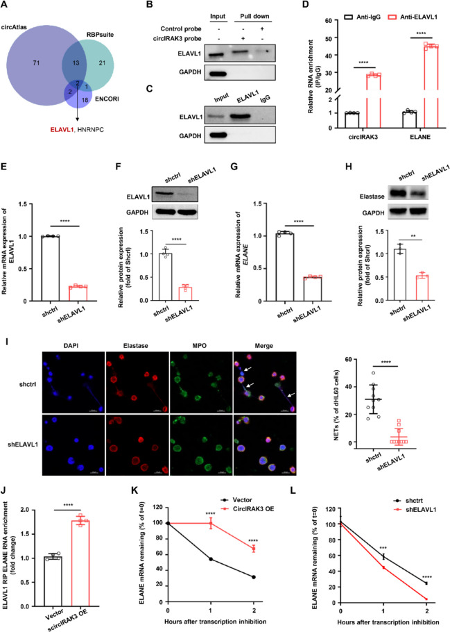
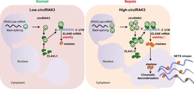
Excessive formation of neutrophil extracellular traps (NETs) has been shown to exacerbate inflammatory injury and organ damage in patients with sepsis. Circular RNAs (circRNAs) abnormally expressed in immune cells of sepsis patients, and play an important role in the pathogenesis of dysregulated immune responses. However, the functions of circRNAs in NET formation during sepsis remain unknown. Here, we identified circIRAK3, a novel circRNA that was upregulated in peripheral blood neutrophils of sepsis patients. Combining clinical data, we revealed that elevated circIRAK3 was positively correlated with blood NET levels. Furthermore, knockdown and overexpression in differentiated HL-60 (dHL-60) neutrophil-like cells demonstrated that circIRAK3 promoted NET formation. In addition, we found that circIRAK3 promoted NET formation via positively regulating elastase expression in dHL-60 cells when treated with inflammatory stimuli. Mechanistically, circIRAK3 directly interacted with ELAVL1 to improve ELANE mRNA stability and consequently promote elastase protein expression. In summary, our study reveals that circIRAK3 promotes NET formation in sepsis by increasing ELANE mRNA levels.

Keywords: Circular RNA; ELANE; ELAVL1; Neutrophil extracellular trap; Sepsis; mRNA stability.


Conflict of interest statement

Declarations. Ethics: The present study was approved by the Ethics Committee at Daping Hospital of Army Medical University (No. 66, 2020). All study participants provided written informed consent. Conflict of Interest: The authors declare no competing interests.

Figures

Fig. 1
Fig. 1
Identification and characterization of circIRAK3 in neutrophils of septic patients. A Volcano plot of differentially expressed circRNAs in neutrophils of septic patients relative to healthy volunteers. B Heatmap for top 12 up-regulated circRNAs in neutrophils from patients with sepsis. C PCR amplification using divergent or convergent primers against complementary DNA (cDNA) or genomic DNA (gDNA) from human neutrophils. D RT-qPCR to check for the expression of up-regulated circRNAs in neutrophils of sepsis patients (n = 10) and healthy donors (n = 10). Data shown are mean ± SD. *P < 0.05, **P < 0.01, ***P < 0.0001, ****P < 0.0001. E Schematic diagram illustrating the genomic location and back-splicing of circIRAK3. F The back-splicing junction site of circIRAK3 was confirmed by PCR amplification and Sanger sequencing. G RNase R treatment followed by RT-qPCR of circIRAK3 and it’s host gene IRAK3. GAPDH represents mRNA. ****P < 0.0001, ns, no significance. H Actinomycin D (Act D) treatment of neutrophils followed by RT-qPCR. Linear mRNA controls: GAPDH and IRAK3. ****P < 0.0001, ns, no significance. I Cytoplasm/nucleus fraction isolation of neutrophils followed by RT-qPCR. GAPDH represents cytoplasmic RNA and MALAT1 represents nuclear RNA. J Images of FISH for circIRAK3. 18S, cytoplasmic RNA control; U6, nuclear RNA control
Fig. 2
Fig. 2
High expression of circIRAK3 in neutrophils is positively correlated with blood NET levels in patients with sepsis. A Expression of circIRAK3 in neutrophils isolated from ICU septic patients (n = 40) compared to ICU non-sepsis patient controls (n = 20), ****P < 0.0001. B ROC curve showing biomarker value of circIRAK3 for sepsis with AUC = 0.840. C Correlation of circIRAK3 and blood NET levels identified by plasma MPO-DNA complexes of sepsis patients (n = 36), P = 0.003, r = 0.4880
Fig. 3
Fig. 3
CircIRAK3 contributed to NET formation in dHL-60 neutrophil-like cells. A The expression level of circIRAK3 and IRAK3 mRNA in shcircIRAK3- transfected HL-60 cells, ****P < 0.0001, ns, no significance. B Western blot analysis of IRAK3 in circIRAK3- knockdown HL-60 cells, ns, no significance. C Reduced NET formation in shcircIRAK3- transfected dHL-60 stimulated with 100 nM PMA for 4 h. Left: Representative immunofluorescence images of NETs. The cells stained with DAPI (blue), MPO (green) and Cit-H3 (red). Right: Quantification of NETs release from dHL-60 cells. The percentage of NETs was calculated as the average of ten fields normalized to the total number of dHL-60 cells, ***P < 0.001, ****P < 0.0001. D The expression level of circIRAK3 and IRAK3 mRNA in circIRAK3- overexpressed dHL-60 cells, ****P < 0.0001, ns, no significance. E Western blot analysis of IRAK3 in circIRAK3-overexpressed dHL-60 cells, ns, no significance. F Increased NET formation in circIRAK3-overexpressed dHL-60 stimulated with 100 nM PMA for 4 h. Left: Representative immunofluorescence images of NETs. The cells stained with DAPI (blue), MPO (green) and Cit-H3 (red). Right: Quantification of NETs release from dHL-60 cells, *P < 0.05
Fig. 4
Fig. 4
CircIRAK3 positively regulated ELANE expression in human neutrophils. A RT-qPCR to detect the mRNA expression of NET formation related genes, ie. ELANE, MPO and PAD4. B The expression level of circIRAK3 in dHL60 cells stimulated by 1 µg/ml LPS for 4 h, ****P < 0.0001. C The mRNA level of ELANE in dHL60 cells stimulated by 1 µg/ml LPS for 4 h, ****P < 0.0001. D Elastase protein levels in shcircIRAK3- transfected dHL-60 stimulated by LPS for 4 h, ****P < 0.0001. E Elastase protein levels in shcircIRAK3- transfected dHL-60 stimulated by PMA for 4 h, ****P < 0.0001. F RT-qPCR to detect the mRNA expression of ELANE in circIRAK3- overexpressed dHL60 cells, **P < 0.0001. G Elastase protein levels in circIRAK3- overexpressed dHL-60 cells, ***P < 0.001
Fig. 5
Fig. 5
CircIRAK3 interacted with ELAVL1 to promote ELANE mRNA stability and expression. A Venn diagram showed the intersection of proteins that binding with circIRAK3 predicated by three databases. B RNA-pulldown followed by western blot analysis showing that circIRAK3 interacts with ELAVL1. C ELAVL1-RIP followed by western blot to confirm the interaction between ELAVL1 antibody and ELAVL1. D The binding of ELAVL1 with circRIAK3 and ELANE were detected by ELAVL1-RIP followed by RT-qPCR. E The mRNA level of ELAVL1 in shELAVL1-transfected dHL-60 cells, ****P < 0.0001. F ELAVL1 protein levels in shELAVL1-transfected dHL-60 cells, ****P < 0.0001. G The mRNA level of ELANE in shELAVL1-transfected dHL-60 cells, ****P < 0.0001. H Elastase protein levels in shELAVL1-transfected dHL-60 cells, **P < 0.01. I Knockdown of ELAVL1 resulted in the reduction of NETs formation in dHL-60 cells stimulated with 100 nM PMA for 4 h. Left: Representative immunofluorescence images of NETs. The cells stained with DAPI (blue), MPO (green) and elastase (red). Right: Quantification of NETs release from dHL-60 cells. The percentage of NETs was calculated as the average of ten fields normalized to the total number of dHL-60 cells, ****P < 0.0001. J ELAVL1-RIP followed by RT-qPCR in circIRAK3 overexpressed dHL-60 cells. K The stability of ELANE mRNA in circIRAK3 overexpressed dHL-60 cells, ****P < 0.0001. L The stability of ELANE mRNA in shELAVL1-transfected dHL-60 cells, ***P < 0.001, ****P < 0.0001
Fig. 6
Fig. 6
Schematic illustration of the mechanism whereby circIRAK3 regulates ELANE mRNA stability via ELAVL1. In nuclear, circIRAK3 is generated by backsplicing of the IRAK3 gene and exported to the cytoplasm. During sepsis, more circIRAK3 molecules are expressed to stabilize ELANE mRNA through their interactions with ELAVL1, thus facilitating chromatin decondensation and NET formation under inflammatory stimulation

Similar articles

References

    1. Rudd, K.E., et al. 2020. Global, regional, and national sepsis incidence and mortality, 1990–2017: analysis for the Global Burden of Disease Study. The Lancet 395: 200–211. 10.1016/S0140-6736(19)32989-7. - PMC - PubMed
    1. Giamarellos-Bourboulis, E.J., et al. 2024. The pathophysiology of sepsis and precision-medicine- based immunotherapy. Nature Immunology 25: 19–28. 10.1038/s41590-023-01660-5. - PubMed
    1. Zhang, H., et al. 2023. Neutrophil, neutrophil extracellular traps and endothelial cell dysfunction in sepsis. Clinical and Translational Medicine 13: e1170. 10.1002/ctm2.1170. - PMC - PubMed
    1. Zhang, H., et al. 2024. Ferritin-mediated neutrophil extracellular traps formation and cytokine storm via macrophage scavenger receptor in sepsis-associated lung injury. Cell Communication and Signaling 22: 97. 10.1186/s12964-023-01440-6. - PMC - PubMed
    1. Hawez, A., et al. 2022. c-Abl kinase regulates neutrophil extracellular trap formation and lung injury in abdominal sepsis. Laboratory Investigation 102: 263–271. 10.1038/s41374-021-00683-6. - PMC - PubMed

LinkOut - more resources