. 2024 Dec 20;41(1):3.
doi: 10.1007/s10565-024-09946-6.

miR-21-loaded bone marrow mesenchymal stem cell-derived exosomes inhibit pyroptosis by targeting MALT1 to repair chemotherapy-induced premature ovarian insufficiency

Affiliations

miR-21-loaded bone marrow mesenchymal stem cell-derived exosomes inhibit pyroptosis by targeting MALT1 to repair chemotherapy-induced premature ovarian insufficiency

Lichao Tang et al. Cell Biol Toxicol. .

Abstract

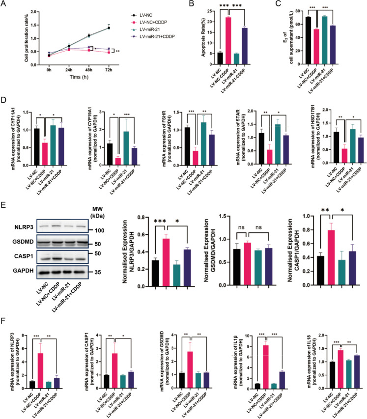
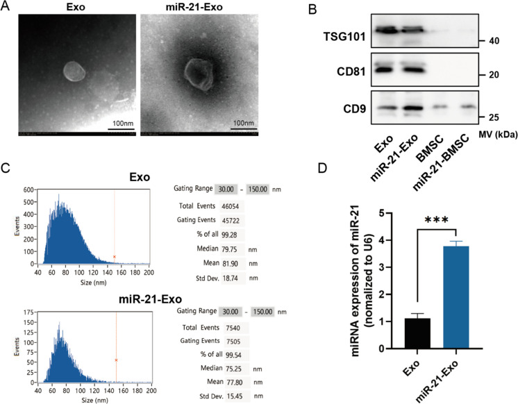
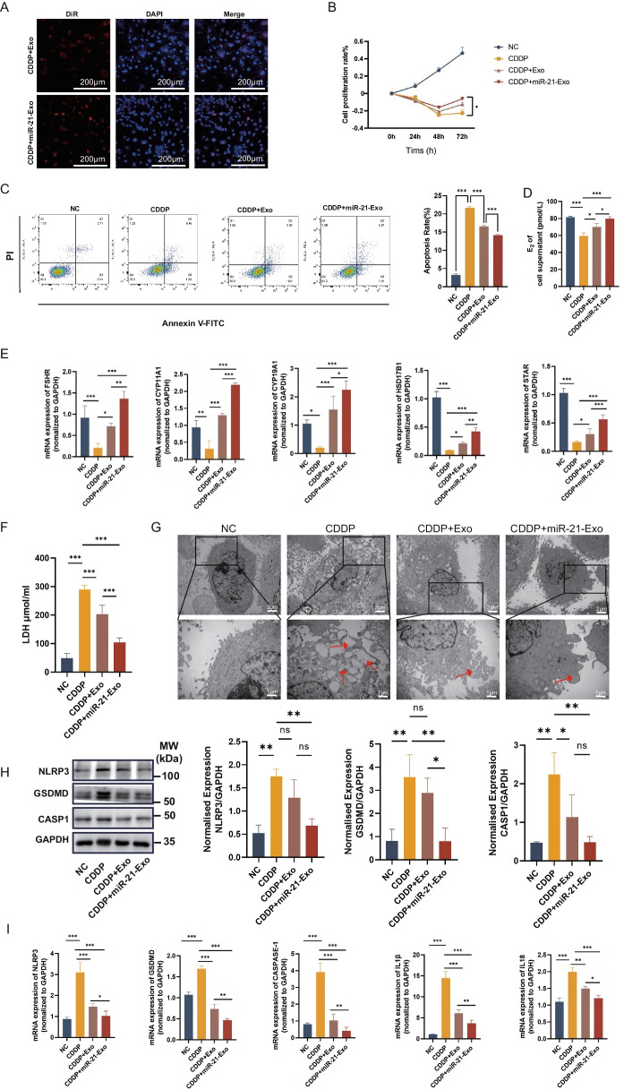
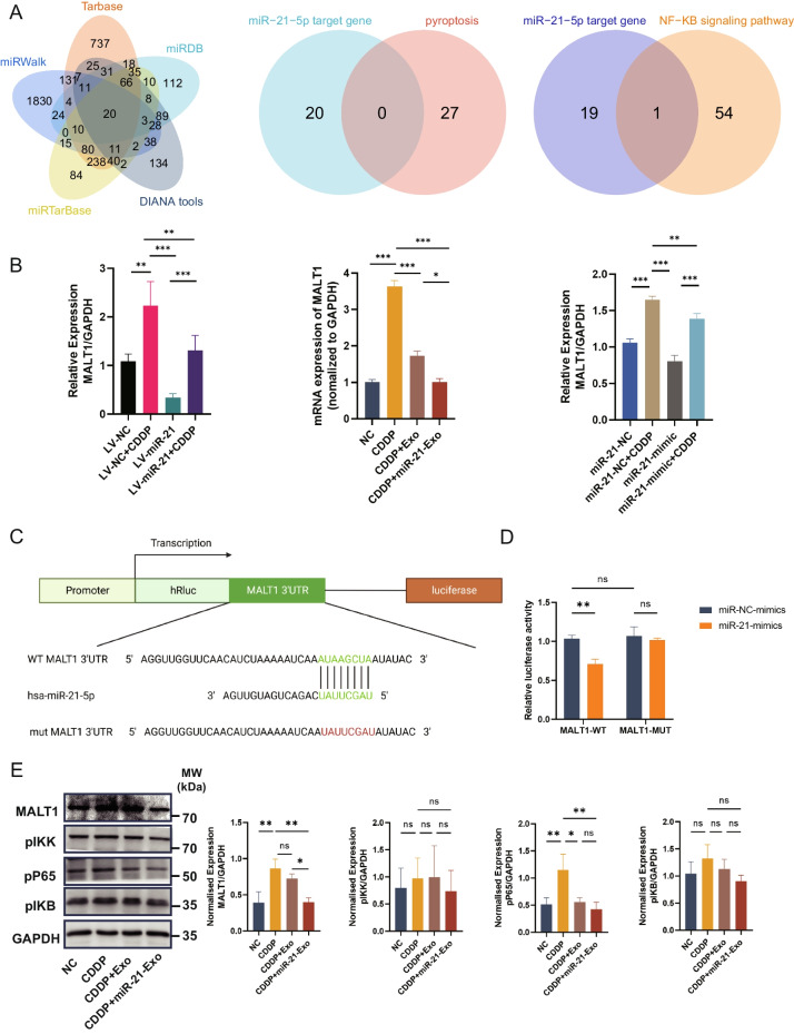
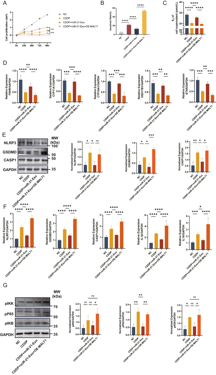
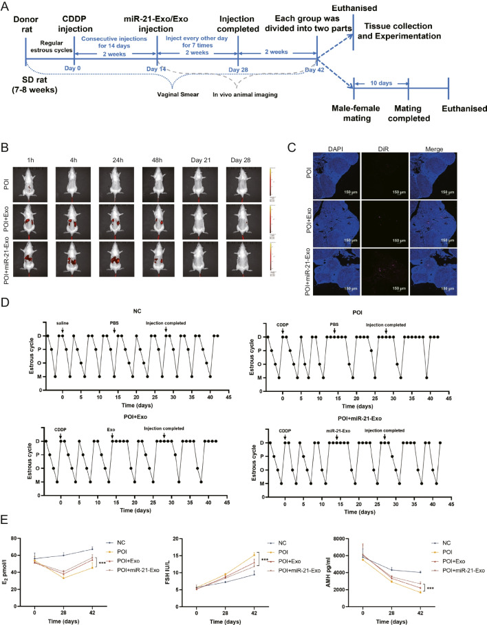
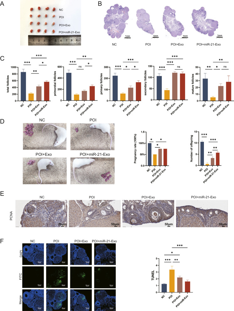
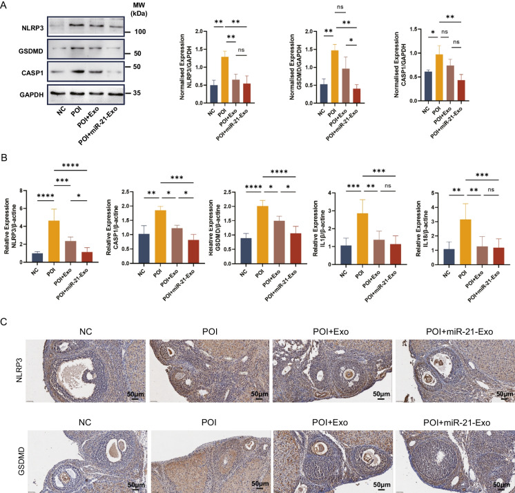
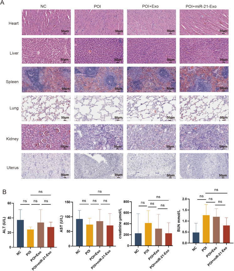
Chemotherapy is essential for treating malignant tumors, but it can cause premature ovarian insufficiency (POI). Recent studies suggest that exosomes enriched with miR-21 (miR-21-Exo) may help mitigate POI, though the underlying mechanisms remain largely unexplored. This research investigates how miR-21-Exo influences chemotherapy-induced POI using an experimental model where KGN cells are exposed to cisplatin. We assessed the impact of miR-21 on cellular activity and generated miR-21 overexpressing bone marrow mesenchymal stem cells (miR-21-BMSC) via lentiviral modification. Isolated miR-21-Exo was analyzed for its effects on cellular function. Bioinformatics identified Mucosa-Associated Lymphoid Tissue Lymphoma Translocation Protein 1 (MALT1) as a target of miR-21. We confirmed that miR-21-Exo regulates MALT1 and the NF-κB signaling pathway to prevent cell pyroptosis. Further studies in a rat model demonstrated the therapeutic potential and safety of miR-21-Exo. Overall, our findings highlight a novel strategy for addressing chemotherapy-induced POI by modulating MALT1 and the NF-κB pathway, offering significant therapeutic implications.

Keywords: Bone mesenchymal stem cell; Exosome; MALT1; MiR-21; Premature ovarian insufficiency; Pyroptosis.


Conflict of interest statement

Declarations. Ethics approval and informed consent: All animal experiments conducted was compliant with the Ethics Committee of Southern Medical University Zhujiang Hospital (Nọ: LAEC-2022–208). Consent for publication: Not applicable. Competing interests: The authors declare no competing interests.

Figures

Fig. 1
Fig. 1
The restorative effect of miR-21 on cisplatin-induced granulosa cells. (A) The CCK8 test (n = 3) was used to determine the proliferation rate. (B) The level of apoptosis was assessed using fluorescence-activated cell sorting (n = 3). (C) An enzyme-linked immunosorbent assay was used to quantify the levels of E2 in the cell supernatants (n = 3). (D) The mRNA levels of FSHR, CYP11A1, CYP19A1, HSD17B1, and STAR were measured using qRT-PCR and standardized against GAPDH (n = 3). (E) The protein levels of pyroptosis-related genes NLRP3, CASP1, and GSDMD were measured using Western Blotting and analyzed using ImageJ software (n = 3). (F) The mRNA levels of NLRP3, CASP1, GSDMD, IL1β, and IL18 were measured using qRT-PCR and standardized against GAPDH (n = 3)
Fig. 2
Fig. 2
Extraction and identification of miR-21-Exo. (A) Transmission electron microscopy (n = 3) was utilized to investigate the structure of Exo and miR-21-Exo. (B) Exo and miR-21-Exo (n = 3) were analyzed with a Flow NanoAnalyzer to assess their particle sizes. (C) TSG101, CD81, and CD9 membrane protein levels in Exo and miR-21-Exo samples (n = 3) were measured. (D) The expression level of the miR-21 gene was assessed in BMSC and in Exo generated from BMSC transfected with miR-21 (n = 3). Standardization of the expression levels to U6 was done
Fig. 3
Fig. 3
miR-21-Exo enhanced the activity of CDDP-treated KGN cells. (A) CDDP-induced KGN cells (n = 3) were employed to observe the absorption of DiR-labeled Exo or miR-21-Exo using confocal microscopy. (B) Using the CCK8 test to calculate the proliferation rate (n = 3). (C) Using fluorescence-activated cell sorting to determine the rate of apoptosis (n = 3). (D) Measurement of estradiol concentration in cell supernatants using ELISA (n = 3). (E) qRT-PCR analysis (n = 3) of the mRNA levels of FSHR, CYP11A1, CYP19A1, HSD17B1, and STAR1, and normalized to GAPDH. (F) Quantification of LDH content in cell supernatants (n = 3). (G) Observation of cell morphology under electron microscopy. (H) Using Western Blotting and ImageJ software, the protein expressions of the pyroptosis-related genes NLRP3, CASP1, and GSDMD were analyzed (n = 3). (I) Using qRT-PCR (n = 3) and normalization to GAPDH, the mRNA levels of NLRP3, CASP1, GSDMD, IL1β, and IL18 were determined
Fig. 4
Fig. 4
miR-21-Exo regulated the MALT1 and NF-κB signaling pathways. (A) P The identification of pyroptosis-related MALT1 as a putative target gene of miR-21 via intersection (Data Sources: Tarbase, miRDB, DIANA tools, miRTarBase, miRWalk, rectome and GeneCards). (B) The expression of MALT1 mRNA is regulated by miR-21 (n = 3). (C) miRDB database predicts the specific binding site of miR-21 with MALT1 (n = 3). (D) Results from dual-luciferase reporter assays demonstrate the specific targeting of MALT1 by miR-21 (n = 3). The sequence's altered portion is denoted as MALT1-MUT. MALT1-WT denotes the wild-type MALT1 (n = 3). (E) Validation of the impact of miR-21 on the protein expression levels of the NF-κB signaling pathway through Western Blotting (n = 3)
Fig. 5
Fig. 5
The increase of MALT1 suppressed the activity of miR-21-Exo. (A) Determination of proliferation rate using CCK8 test (n = 3). (B) Flow cytometry was used to determine the rate of apoptosis (n = 3). (C) Estradiol concentration in cell supernatants was measured using ELISA (n = 3). (D) Using qRT-PCR (n = 3), the mRNA levels of FSHR, CYP11A1, CYP19A1, HSD17B1, and STAR1 were determined and normalized against GAPDH. (E) Using Western Blotting and ImageJ software, the protein expressions of the pyroptosis-related genes NLRP3, CASP1, and GSDMD were analyzed (n = 3). (F) Using qRT-PCR (n = 3) and normalization to GAPDH, the mRNA levels of NLRP3, CASP1, GSDMD, IL1β, and IL18 were determined. (G) Using ImageJ software and Western Blotting, the NF-κB signaling pathway's protein expressions were analyzed (n = 3)
Fig. 6
Fig. 6
miR-21-Exo restored the estrous cycle and hormones in chemically-induced ovarian insufficiency rats. (A) Schematic schematic illustrating the animal experimental design. (B) Intraperitoneal injection of DiR-labeled Exo or miR-21-Exo resulted in in vivo metabolism observed through live animal imaging. (C) Ovarian frozen sections showed uptake of DiR fluorescently labeled exosomes by the ovaries. (D) Representative estrous cycles of rats exhibiting either regular or interrupted estrous cycles within each group. (E) The levels of serum sex hormones E2, FSH, and AMH were measured using ELISA (n = 4)
Fig. 7
Fig. 7
miR-21-Exo repaired the structure of the rats ovaries, improved reproductive function, promoted proliferation, and inhibited apoptosis. (A) Macroscopic observation of ovaries in each group (B) HE staining of ovarian tissues. (C) The number of follicles per ovary was determined in rats (n = 4). (D) Litter position diagrams, pregnancy rate, and offspring count in each group (n = 4). (E) Immunohistochemistry was used to evaluate the expression of PCNA protein (n = 4). (F) The apoptosis of granulosa cells was evaluated using TUNEL labeling (n = 4)
Fig. 8
Fig. 8
miR-21-Exo inhibited ovarian pyroptosis in chemotherapy-induced POI rats. (A) Western blot analysis was used to determine the expression levels of NLRP3, CASP1, and GSDMD proteins. (B) mRNA levels of NLRP3, CASP1, GSDMD, IL1β, and IL18 in the miR-21-Exo group were analyzed by qRT-PCR. (C) Immunohistochemical staining was performed for NLRP3 and GSDMD in the ovaries
Fig. 9
Fig. 9
The application of exosomes did not affect the structure and function of major organs in rats. (A) HE diagram of paraffin sections of each organ in each group. (B) Detection of liver and kidney function in each group

References

    1. Cai JH, Sun YT, Bao S. Hucmscs-exosomes containing mir-21 promoted estrogen production in ovarian granulosa cells via lats1-mediated phosphorylation of loxl2 and yap. Gen Comp Endocrinol. 2022;321–322:114015. 10.1016/j.ygcen.2022.114015. - PubMed
    1. Chen D, Hu N, Xing S, Yang L, Zhang F, GuoMa SH. Placental mesenchymal stem cells ameliorate nlrp3 inflammasome-induced ovarian insufficiency by modulating macrophage m2 polarization. J Ovarian Res. 2023;16(1):58. 10.1186/s13048-023-01136-y. - PMC - PubMed
    1. Chen Y, Zhao Y, Miao C, Yang L, Wang R, Chen B, Zhang Q. Quercetin alleviates cyclophosphamide-induced premature ovarian insufficiency in mice by reducing mitochondrial oxidative stress and pyroptosis in granulosa cells. J Ovarian Res. 2022;15(1):138. 10.1186/s13048-022-01080-3. - PMC - PubMed
    1. Ding C, Qian C, Hou S, Lu J, Zou Q, Li H, Huang B. Exosomal mirna-320a is released from hamscs and regulates sirt4 to prevent reactive oxygen species generation in poi. Mol Ther Nucleic Acids. 2020a;21:37–50. 10.1016/j.omtn.2020.05.013. - PMC - PubMed
    1. Ding C, Zhu L, Shen H, Lu J, Zou Q, HuangHuang CB. Exosomal mirna-17-5p derived from human umbilical cord mesenchymal stem cells improves ovarian function in premature ovarian insufficiency by regulating sirt7. Stem Cells. 2020b;38(9):1137–48. 10.1002/stem.3204. - PubMed

Publication types

MeSH terms