. 2024 Dec 21;41(1):12.
doi: 10.1007/s10565-024-09960-8.

Identification of gastric cancer stem cells with CD44 and Lgr5 double labelling and their initial roles on gastric cancer malignancy and chemotherapy resistance

Affiliations

Identification of gastric cancer stem cells with CD44 and Lgr5 double labelling and their initial roles on gastric cancer malignancy and chemotherapy resistance

Zhida Chen et al. Cell Biol Toxicol. .

Abstract

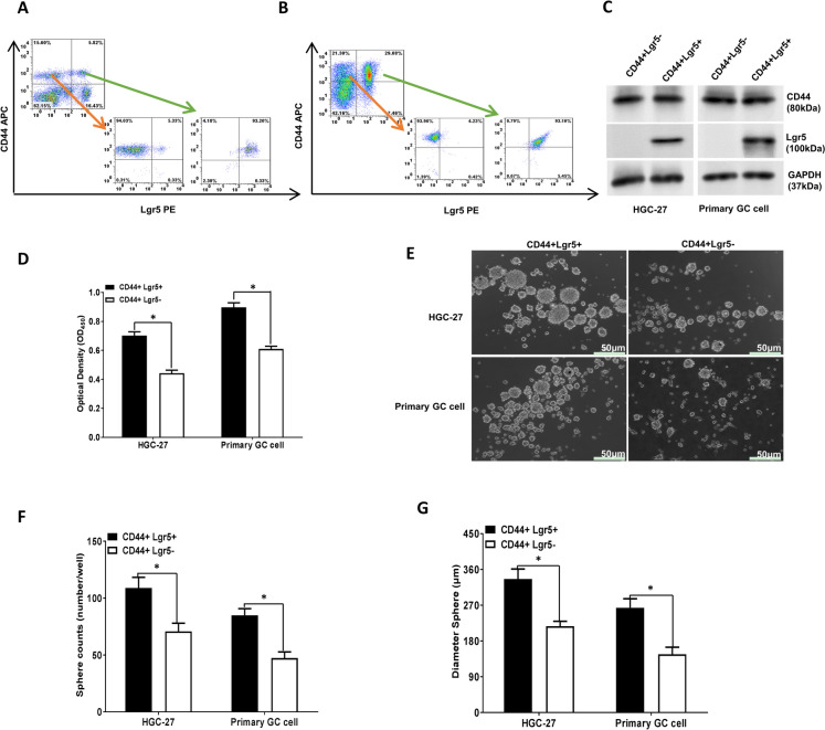
Accumulating evidences have indicated that cancer stem cells (CSCs) can initiate tumor progression and cause recurrence after therapy. However, specific markers of gastric CSCs (GCSCs) from different origins have not been comprehensively revealed. Here, we further detected whether cell populations labelled with CD44 and Lgr5, well-recognized stem markers for gastric cancer (GC), can better emphasize cancer initiation, therapeutic resistance and recurrence. Flow cytometry was utilized to sort the CD44 + Lgr5 + and CD44 + Lgr5- cells from GC cell line HGC-27 and primary GC cells. The influences of CD44 and Lgr5 GCSCs on the malignant behaviors and their potential mechanisms was investigated, respectively. In our study, we reported the identification and validation of CD44 + Lgr5 + cells that presented stronger stemness characteristics, as evidenced by increase of sphere forming ability, elevation of stem cell transcriptional activity. Additionally, CD44 + Lgr5 + double positive cells have lower apoptosis, greater chemotherapy resistance, and higher EMT capacity and LC3 density compared with CD44 + Lgr5- cells. Tumor xenograft experiments also verified the faster carcinogenesis of CD44 + Lgr5 + GCSCs. Furthermore, a series of key proteins in the Wnt, Hedgehog, Notch, and TGF-β pathways were elevated in the CD44 + Lgr5 + double positive subpopulation, except for Notch 1 and Smad 1. In conclusion, the binding of CD44 and Lgr5 can serve as a precise GCSCs marker that initiate malignant progression and chemotherapy resistance in GC by activating Wnt, Hedgehog, Notch, TGF-β pathways. Those evidences raise the needs to target both markers simultaneously as a potential approach for the GC treatment.

Keywords: CD44; Cancer stem cell; Chemotherapy resistance; Epithelial-mesenchymal transformation; Gastric cancer; Lgr5.


Conflict of interest statement

Declarations. Ethical approval: All animal experiments were approved by the Animal Ethics Committee of Beijing Viewsolid Biotechnology Co. LTD (VS2126A23528). Conflicts of interest: The authors declare no competing interests.

Figures

Fig. 1
Fig. 1
CD44 + Lgr5 + GC cell populations displayed greater stemness-like characteristics. Sorting and enrichment of CD44 + Lgr5- and CD44 + Lgr5 + cell subsets of HGC-27 GC cells (A) and (B) primary GC cells were tested by flow cytometry; (C) Western blot was employed to detect the levels of CD44 and Lgr5 in sorted populations; (D) CCK-8 results displayed the OD value of proliferation experiment of gastric cancer stem cells of sorting subgroup; (EG) Sphere formation assay was employed to detect the spheroidization effect of sorted CD44 + Lgr5- and CD44 + Lgr5 + cells (scale bar = 50 μm); GCSCs, gastric cancer stem cells. *P < 0.05; All data were displayed from at least three independent experiments
Fig. 2
Fig. 2
CD44 + Lgr5 + GC cell populations displayed greater stemness-related transcriptional activity. (A-D) Western blot assay was utilized to detect the levels of stemness-related transcriptional factors including OCT4, SOX2 and Nanog in sorted subsets of cells; (EG) qRT-PCR assay was used to detect the expressions of stemness-related transcriptional factors including OCT4, SOX2 and Nanog in sorting subgroup cells; (H) The transfection efficacy of siRNA-Lgr5 was displayed; (I-K) CCK-8 was employed to evaluate the chemotherapy drug resistance after treatment with a series concentrations of Oxaliplatin, Taxol and 5-FU in sorting subgroup cells; (L-N) The inhibition rates of sorting subgroup cells after treatment with a series concentrations of Oxaliplatin, Taxol and 5-Fu were shown; *, **P < 0.05, P < 0.01; All data were displayed from at least three independent experiments. 5-FU, 5-fluorouracil
Fig. 3
Fig. 3
CD44 + Lgr5 + GCSCs had stronger tumorigenesis, and proliferation abilities in vivo and in vitro. (A-C) Subcutaneous tumor xenograft assays of CD44 + Lgr5 + GCSCs and CD44 + Lgr5- cell subsets were established and showed the tumor volume (n = 4 per group); (D-E) Body weight and tumor weight in different groups were shown (n = 4 per group); Immunohistochemistry assay was utilized to detect the Ki67 (F, H) and Cyclin D1 (G, I) levels in xenograft tumors (Scar bar = 50 μm). (J, L) The migratory abilities of CD44 + Lgr5 + and CD44 + Lgr5- GCSCs from HGC-27 and primary GC cells were determined by Transwell assay; (K, M) The cell cycle distribution was detected by flow cytometry with propidium iodide staining; GCSCs, gastric cancer stem cells; *P < 0.05; All in vitro data were displayed from at least three independent experiments
Fig. 4
Fig. 4
CD44 + Lgr5 + GCSCs displayed lower apoptosis, higher autophagy and greater chemotherapy resistance. (A, B) The apoptosis of CD44 + Lgr5 + and CD44 + Lgr5- GCSCs was tested by flow cytometry assay; (C-F) The levels of Caspase 3, Caspase 9 and Bcl-2 in CD44 + Lgr5 + and CD44 + Lgr5- GCSCs were examined by western blot; (G) The autophagy of sorted subsets was detected with immunofluorescence assay; (H-M) The expressions of ERCC1 and MDR1 in GCSCs after chemotherapy drugs such as oxaliplatin and 5-Fu treatment were determined by qRT-PCR and western blot assays; GCSCs, gastric cancer stem cells; *, **P < 0.05, P < 0.01; All data were displayed from at least three independent experiments
Fig. 5
Fig. 5
The schematic mechanism of signaling pathways involved in GCSCs stemness. (A-C) Expressions of key proteins, β-catenin and TCF4 in the Wnt signaling pathway were determined by western blot; (D-F) Expressions of key proteins, Gli1 and Smo in the Hedgehog signaling pathway were determined by western blot; (G-L) Western blot showed the expressions of key proteins including Notch 1–4 and NICD in Notch signaling pathway; (M-R) Western blot showed the expressions of key proteins, TGF-β1 and Smad 1–4 in the TGF-β signaling pathway; *, **P < 0.05, P < 0.01; All data were displayed from at least three independent experiments

Similar articles

Cited by

References

    1. Akyala AI, Peppelenbosch MP. Gastric cancer and Hedgehog signaling pathway: emerging new paradigms. Genes Cancer. 2018;9:1–10. - PMC - PubMed
    1. Bekaii-Saab T, El-Rayes B. Identifying and targeting cancer stem cells in the treatment of gastric cancer. Cancer. 2017;123:1303–12. - PMC - PubMed
    1. Bigagli E, Cinci L, D’Ambrosio M, Luceri C. Transcriptomic characterization, chemosensitivity and regulatory effects of exosomes in spontaneous EMT/MET transitions of breast cancer cells. Cancer Genomics Proteomics. 2019;16:163–73. - PMC - PubMed
    1. Cao HZ, Liu XF, Yang WT, Chen Q, Zheng PS. LGR5 promotes cancer stem cell traits and chemoresistance in cervical cancer. Cell Death Dis. 2017;8:e3039. - PMC - PubMed
    1. Cao W, Li M, Liu J, Zhang S, Noordam L, Verstegen MMA, Wang L, Ma B, Li S, Wang W, Bolkestein M, Doukas M, Chen K, Ma Z, Bruno M, Sprengers D, Kwekkeboom J, van der Laan LJW, Smits R, Peppelenbosch MP, Pan Q. LGR5 marks targetable tumor-initiating cells in mouse liver cancer. Nat Commun. 2020;11:1961. - PMC - PubMed

Publication types

MeSH terms

LinkOut - more resources