. 2024 Dec 20;12(1):119.
doi: 10.1186/s40635-024-00707-7.

Lung immune incompetency after mild peritoneal sepsis and its partial restoration by type 1 interferon: a mouse model study

Affiliations

Lung immune incompetency after mild peritoneal sepsis and its partial restoration by type 1 interferon: a mouse model study

Qiuming Meng et al. Intensive Care Med Exp. .

Abstract

Background: Sepsis is commonly associated with acute respiratory distress syndrome (ARDS). Although the exaggerated inflammation may damage intact lung tissues, a percentage of patients with ARDS are reportedly immunocompromised, with worse outcomes. Herein, using a murine sepsis model, time-course immune reprogramming after sepsis was evaluated to explore whether the host is immunocompromised. Leukocyte kinetics in the lung tissue were evaluated in a male C57/BL6 mouse model of mild peritoneal sepsis induced by cecal ligation and puncture, with the survival rate exceeds 90%. Lung immune reactivity was evaluated by intratracheal instillation of lipopolysaccharide (LPS; 30 µg). Furthermore, the effect of interferon (IFN)-β in vivo and ex vivo was evaluated.

Results: Four days after sepsis, the lung water content remained high, even among mice in clinical recovery. While monocytes and neutrophils gradually accumulated in the lung interstitium, the inflammatory cytokine/chemokine expression levels in the lungs continued to decline. Intratracheal LPS instillation induced more leukocyte trafficking and protein leakage into the alveoli in the septic lung, indicating more severe lung injury. However, LPS stimulation-associated mRNA expression of tnf, il6, ccl2, and cxcl1 was suppressed. Intra-alveolar expression of tumor necrosis factor (TNF)-α, interleukin (IL)-6, monocyte chemoattractant protein (MCP)-1, and keratinocyte-derived cytokine (KC) was also suppressed. Monocytes isolated from the lung tissue showed an impaired response in il6, ccl2, and cxcl1 to LPS. Systemic IFN-β restored the above impaired regulator function of monocytes, as did coculturing these cells from lung tissue with IFN-β.

Conclusions: Histologically accelerated inflammation and paradoxically suppressed immunological regulator signaling were observed in the early recovery phase of sepsis. This observation may provide a model for the immunologically irresponsive state that occurs in some patients with sepsis. Systemic IFN-β partly restored the post-septic immunocompromised state, indicating its therapeutic potential for the immunosuppressive state seen in some patients with sepsis/ARDS.

Keywords: ARDS; Acute lung injury; Immune reprogramming; Immune suppression; Innate immunity; Interferon-β; Sepsis.


Conflict of interest statement

Declarations. Ethics approval and consent to participate: The experimental protocols used for all animal experiments were approved by the University of Tokyo Graduate School of Medicine Institutional Review Board (#Med-P20-106), conformed to the NIH Guide for the Care and Use of Laboratory Animals (1985 revision), and were conducted with strict compliance with ethical guidelines issued by the University of Tokyo Graduate School of Medicine. Male C57/BL6 mice (Jackson Laboratories, Clea Japan, Tokyo, Japan), 8–10 weeks of age and 20–25 g in body weight, were purchased for use in animal experiments. Consent for publication: Not applicable. Competing interests: Uchida K is in collaboration with Nihon Kohden Corporation and Nipro Corporation. Neither of these collaborations have any relationship with the present study. No conflicts of interest are declared by any other authors.

Figures
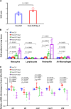
Fig. 1
Fig. 1
Sepsis-related lung immune reprogramming. A Lung wet/dry weight ratios showing a significant increase from pre-cecal ligation and puncture (CLP) to day 4 post-CLP in the CLP group, indicating that the post-CLP lung remained edematous despite clinical recovery. B White blood cell populations over time after sepsis. White blood cells in the lung tissue were isolated by enzymatic digestion, counted with a hemocytometer, and differentiated by flow cytometry with immune staining (Figure E3 in the data supplement). C Time-course changes in mRNA expression after sepsis. All evaluated cytokines/chemokines except for il10 exhibited decreased expression over time and significant reductions on day 4 compared with expression pre-CLP. Data are expressed as the mean + SD, accompanied by individual data points (A, B). Data are expressed as the median ± [25th, 75th percentile], accompanied by individual data points (C). Exact P values are shown for each comparison
Fig. 2
Fig. 2
Sepsis aggravates lung injury histologically and biochemically. A Representative hematoxylin and eosin-stained images of the lung at 4 day post-cecal ligation and puncture (CLP) with or without intratracheal (i.t.) lipopolysaccharide (LPS) instillation. Compared with the nonintervention control (upper left panel), the 4-day CLP-treated lung showed mild thickening of the interstitium and capillary congestion (upper right panel). At 1 day post i.t. LPS challenge, the lung without CLP showed mild infiltration of inflammatory cells into the alveoli and mild accumulation of edema fluid (bottom left panel). Lungs after 4 days of CLP and 1 day after i.t. LPS exhibited increased migration of inflammatory cells into the alveoli and protein-rich edema fluid in the alveoli observed macroscopically (bottom right panel). B i.t. LPS-induced lung fluid protein concentration in the bronchoalveolar lavage fluid (BALF) from mice with sepsis was significantly higher than that in the noCLP group. CE I.t. LPS-induced intra-alveolar influx of white blood cells. C Sepsis alone did not increase the total number of recovered cells above that of the nonintervention group, whereas LPS administration induced a significant increase in the number of cells migrating into the alveoli. Prior sepsis further significantly (P = 0.0212) increased the number of recovered cells. D, E Sepsis induced a significant increase in migrated monocytes (D), whereas neutrophils were not significantly increased (E). Data are expressed as the median ± [25th, 75th percentile], accompanied by individual data points. Exact P values are shown for each comparison
Fig. 3
Fig. 3
Changes in the monocyte populations in lung tissue after sepsis. A Proportions of Ly6Clo and Ly6Chi monocytes in the control group were nearly equivalent (47% vs 53%), whereas these proportions changed to Ly6Clo dominant (67% vs 33%) at 4 days post-cecal ligation and puncture (CLP). The monocyte population returned to pre-CLP levels (50:50 for Ly6Chi and Ly6Clo) with systemic interferon (IFN)-β administration. B Following intratracheal (i.t.) instillation of lipopolysaccharide (LPS), 95% of monocytes migrating into the alveolar space (BALF) were Ly6Chi. Data are expressed as the median ± [25th, 75th percentile], accompanied by individual data points (A). Data are expressed as the mean + SD, accompanied by individual data points (B). Exact P values are shown for each comparison
Fig. 4
Fig. 4
Effect of sepsis on the intra-alveolar cytokine response to intratracheal (i.t.) lipopolysaccharide (LPS) instillation. AE I.t. LPS instillation (30 µg in PBS) induced significant increases in the indicated cytokines and chemokines except for IL-10 in both the cecal ligation and puncture (CLP) and noCLP groups compared with their respective controls (i.e., noLPS). However, the magnitude of the increases was lower in the CLP group than in the noCLP group. Data are expressed as the median ± [25th, 75th percentile], accompanied by individual data points. Exact P values are shown for each comparison
Fig. 5
Fig. 5
Effect of sepsis on lung cytokine mRNA expression in response to intratracheal (i.t.) lipopolysaccharide (LPS). AE mRNA expression of the indicated cytokines and chemokines in the lung tissue of mice with or without cecal ligation and puncture (CLP)-induced sepsis and at 1 day post i.t. LPS instillation (30 µg). In the noCLP group, all five cytokines and chemokines evaluated showed significant increases after LPS stimulation compared with levels in the noCLP without LPS group. Although all cytokines/chemokines except for IL-10 were also significantly increased with i.t. LPS in the CLP group compared with the group without LPS, the magnitude of the increases was significantly lower compared with those in the noCLP + i.t. LPS group. FJ Real-time PCR evaluation of the mRNA expression of the indicated cytokines and chemokines in monocytes isolated from lung tissues of mice with or without CLP-induced sepsis and stimulated with LPS (10 ng/mL) or PBS in vitro. Similar to the response patterns in lung tissue, the in vitro LPS-induced expression of il6, ccl2, and cxcl1 were lower in monocytes isolated from the lungs of the CLP group compared with those from the noCLP group, with the exception of tnf, which had higher levels than those in the noCLP group. Data are expressed as the median ± [25th, 75th percentile], accompanied by individual data points. Exact P values are shown for each comparison
Fig. 6
Fig. 6
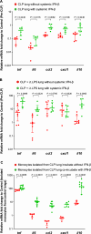
Effect of interferon (IFN)-β administration on lung immune function. A mRNA expression levels in lung tissue 4 days after cecal ligation and puncture (CLP) with systemic IFN-β administration 1 h after CLP were significantly higher than those in the CLP without IFN-β group. B Expression levels of tnf and il6 in lung tissue, with systemic IFN-β administration 1 h after CLP and 4 days later upon stimulation with i.t. LPS, were significantly higher than those in the CLP without IFN-β group. C LPS-stimulated expression of il6, ccl2, and cxcl1 in isolated monocytes from the CLP group was restored by coculture with IFN-β. Data are expressed as the median ± [25th, 75th percentile], accompanied by individual data points. Exact P values are shown for each comparison

References

    1. Skrupky LP, Kerby PW, Hotchkiss RS (2011) Advances in the management of sepsis and the understanding of key immunologic defects. Anesthesiology 115:1349–1362 - PMC - PubMed
    1. Evans L, Rhodes A, Alhazzani W, Antonelli M, Coopersmith CM, French C et al (2022) Surviving sepsis campaign: international guidelines for management of sepsis and septic shock in adults 2021. Acta Anaesthesiol Scand 66:634–635 - PMC - PubMed
    1. Cohen J, Vincent JL, Adhikari NK, Machado FR, Angus DC, Calandra T et al (2015) Sepsis: a roadmap for future research. Lancet Infect Dis 15:581–614 - PubMed
    1. Rubenfeld GD, Caldwell E, Peabody E et al (2005) Incidence and outcomes of acute lung injury. N Engl J Med 353:1685–1693 - PubMed
    1. Iskander KN, Osuchowski MF, Stearns-Kurosawa DJ, Kurosawa S, Stepien D, Valentine C et al (2013) Sepsis: multiple abnormalities, heteroge-neous responses, and evolving understanding. Physiol Rev 93:1247–1288 - PMC - PubMed

LinkOut - more resources