. 2024 Dec 20;30(1):261.
doi: 10.1186/s10020-024-01011-6.

Endothelial-specific deletion of connexin 43 improves renal function and structure after acute kidney injury

Affiliations

Endothelial-specific deletion of connexin 43 improves renal function and structure after acute kidney injury

Magali Genest et al. Mol Med. .

Abstract

Background: We have previously reported that the gap junction protein connexin 43 (Cx43) was upregulated in chronic renal disease in humans and rodents and plays a crucial role in the progression of experimental nephropathy. In this study, we investigated its role after renal ischemia/reperfusion (rIR), which is a major mechanism of injury in acute renal injury (AKI) and renal transplant graft dysfunction.

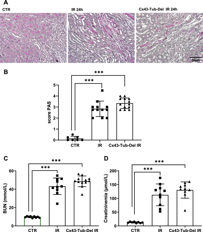
Methods: Wild-type mice (WT) and mice in which Cx43 expression was genetically reduced by half (Cx43 ±) were unilaterally nephrectomized. The left renal artery was subsequently clamped, with reperfusion of varying duration. Mice with tubular- or endothelial-specific deletion of Cx43 were also used to assess the effect of this connexin in each cell type after rIR. Kidneys were assessed for histological evaluation, immunohistochemistry, and RT-PCR.

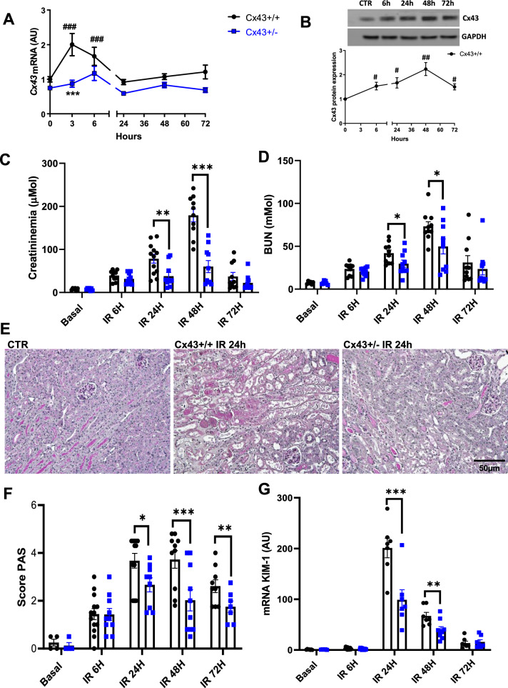
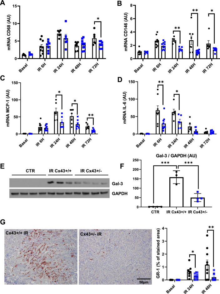
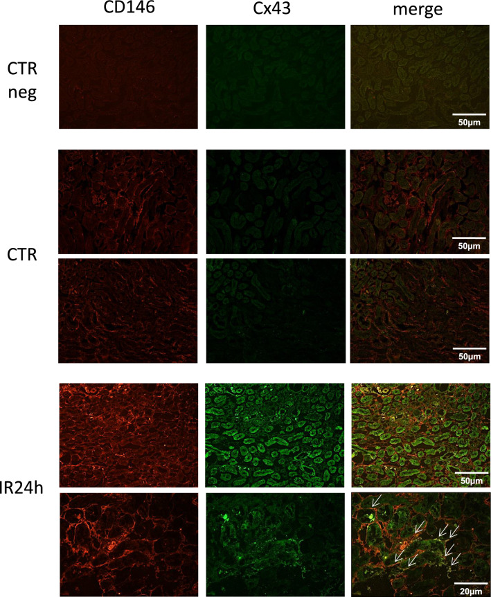
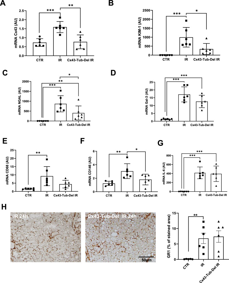
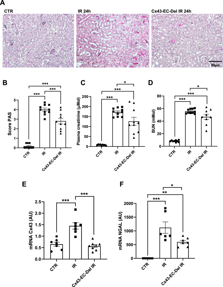
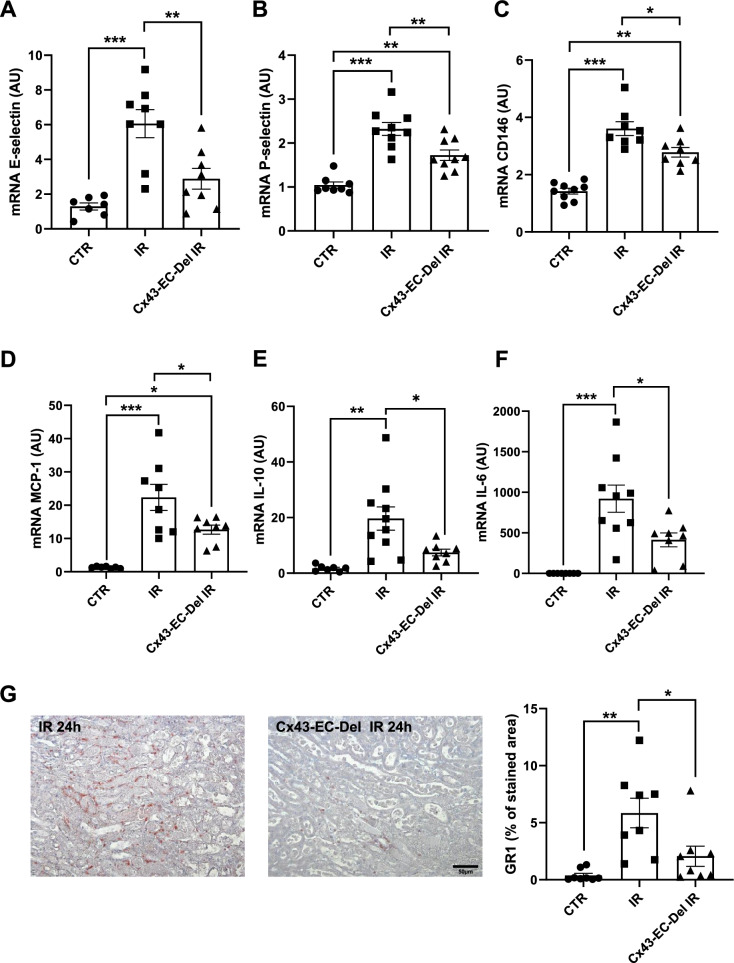
Results: Blood urea nitrogen and creatininemia were progressively elevated in WT mice and picked up 48 h after rIR. At the same time point, severe tubular necrosis and dilation occurred in the cortico-medullary junction of the injured kidneys with accompanying massive neutrophil infiltration. Interestingly, Cx43 expression was progressively increased within the tubulointerstitial compartment during kidney damage progression and was paralleled closely by that of markers of renal dysfunction. Cx43 ± mice showed fewer tubular lesions, less inflammation, and further improved renal function. Similar results were observed in mice where Cx43 was specifically deleted within the vascular endothelium. In contrast, Cx43 deletion in renal tubules did not significantly improve renal structure and function after rIR.

Conclusion: Our findings suggest that endothelial Cx43 plays a crucial role in AKI.

Keywords: Acute kidney injury; Connexin 43; Inflammation.


Conflict of interest statement

Declarations. Ethics approval and consent to participate: Written informed consent was provided by all participants. All animal protocols were approved by the appropriate committee of the National Institute for Health and Medical Research (Inserm) and the Sorbonne University (Paris, France). Consent for publication: Not applicable. Competing interests: The authors have declared that no conflict of interest exists.

Figures

Fig. 1
Fig. 1
Cx43 ± mice showed improved renal function and structure after IR. Cx43 expression is progressively increased at the mRNA (A) and protein levels (B) in injured kidneys after renal IR. Renal function was evaluated by plasma creatinine (C) and BUN (D) measurements. All parameters reveal a slower progress of the disease in Cx43 ± animals. Representative images of PAS staining (E) show that renal structure was preserved in Cx43 ± vs Cx43 + / + since they developed less tubular necrosis and dilation at 24, 48 and 72 h after IR (F). qPCR for KIM-1 confirmed tubular protection in Cx43 ± mice (G). Values are presented as mean ± SEM; n = 7–10 mice from each group; *P < 0.05, **P < 0.01, ***P < 0.001. * compares Cx43 + / + versus Cx43 ± , # fold induction initial versus different time points. Scale bar, 50 μm
Fig. 2
Fig. 2
Cx43 ± mice develop less renal inflammation after AKI. qPCR for CD68 (A), CD146 (B), MCP-1 (C) and IL-6 (D) show that increased expression of mRNAs of all these inflammatory markers was blunted in Cx43 ± kidneys at distinct time points after AKI. Representative western blots for Gal-3 and GAPDH were performed by using renal cortex of sham controls and kidneys after 24 h of IR (E). Graphs show quantification of western blots expressed as the ratio of Gal3 versus GAPDH signal for each sample (F). Gal-3 increased expression was moderated in Cx43 ± mice 48 h after renal IR. At the same time point, quantification of immunohistochemistry for GR1 indicate that Cx43 ± develop less inflammation compared to Cx43 + / + mice (G). Values are presented as mean ± SEM; n = 7–10 mice from each group except for WB (n = 3–4 mice per group); *P < 0.05, **P < 0.01, ***P < 0.001. ● represents Cx43 + / + , and ■ Cx43 ± . Scale bar, 50 μm
Fig. 3
Fig. 3
Cx43 expression was mainly increased within damaged tubules and renal peritubular capillaries after IR. Immunofluorescence showed increased expression of Cx43 in damaged tubules after 24 h of renal IR. Double immunofluorescense for Cx43 (green) and the endothelial marker CD146 (red) in damaged kidneys indicate localization of Cx43 in renal microcirculation (white arrows) after 24 h of renal IR. Scale bars 50 μm and 20 μm. Negative controls (CTR neg) included the omission of primary antibodies
Fig. 4
Fig. 4
Cx43-specific deletion in renal tubules does not affect renal structure and function after AKI. Representative images of PAS staining (A) show that renal structure was not preserved in Cx43-Tub-Del mice (A) since tubular damage was similar in mice with and without tubule-specific deletion of Cx43 after 24 h of renal IR (B). Both BUN (C) and creatininemia measurements showed similar data regarding renal function (D). Values are presented as mean ± SEM; n = 7–12 mice from each group; *P < 0.05, **P < 0.01, ***P < 0.001. Scale bar 50 μm
Fig. 5
Fig. 5
mRNA expression for tubular and inflammatory markers in kidneys after renal IR. qPCR for Cx43 (A), KIM-1 (B), NGAL (C), Gal3 (D), CD68 (E), CD146 (F) and IL-6 (G), using kidneys from mice with and without Cx43 tubular-specific deletion after 24 h of renal IR. Data are expressed in graphs as arbitrary units (AU) that represents the ratio of the target gene/internal control gene (RPL32). Representative immunostainings for GR1 and its quantification (H). n = 6 mice per group. *, P < 0.05; **, P < 0.01; ***, P < 0.001
Fig. 6
Fig. 6
Cx43-specific deletion within vascular endothelium improves renal structure and function after AKI. Representative images of PAS staining (A) show that kidney structure was preserved in Cx43-EC-Del mice, since we observed less tubular necrosis and dilation after 24 h of renal IR (B). Renal function was evaluated by creatininemia (C) and BUN (D) measurements. qPCR for Cx43 (E) and NGAL (F) are presented in graphs as arbitrary units (AU), that represent the ratio of the target gene/internal control gene (RPL32). Values are presented as mean ± SEM; n = 7–10 mice from each group; * *P < 0.05, **P < 0.01, ***P < 0.001. Scale bar, 50 μm
Fig. 7
Fig. 7
Cx43-specific deletion within vascular endothelium protects against renal inflammation after AKI. mRNA expressions for E-selectin (A), P-selectin (B), CD146 (C), MCP-1 (D), IL-10 (E) and IL-6 (F), in kidneys from mice with and without Cx43 endothelial-specific deletion after 24 h of renal IR. At the same time point, quantification of GR1 immunostaining (G) indicates that Cx43-CE-Del developed less inflammation compared to mice in which Cx43 was overexpressed. qPCR data are illustrated in graphs as arbitrary units (AU), representing the ratio of the target gene/internal control gene (RPL32). Values are presented as mean ± SEM; n = 7–10 mice from each group; * *P < 0.05, **P < 0.01, ***P < 0.001. Scale bar, 50 μm

References

    1. Abed A, Toubas J, Kavvadas P, Authier F, Cathelin D, Alfieri C, Boffa JJ, Dussaule JC, Chatziantoniou C, Chadjichristos CE. Targeting connexin 43 protects against the progression of experimental chronic kidney disease in mice. Kidney Int. 2014;86:768–79. - PubMed
    1. Abed A, Leroyer AS, Kavvadas P, Authier F, Bachelier R, Foucault-Bertaud A, Bardin N, Cohen CD, Lindenmeyer MT, Genest M, Joshkon A, Jourde-Chiche N, Burtey S, Blot-Chabaud M, Dignat-George F, Chadjichristos CE. Endothelial-specific deletion of CD146 protects against experimental glomerulonephritis in mice. Hypertension. 2021;77(4):1260–72. - PubMed
    1. Bardin N, Reumaux D, Geboes K, Colombel JF, Blot-Chabaud M, Sampol J, Duthilleul P, Dignat-George F. Increased expression of CD146, a new marker of the endothelial junction in active inflammatory bowel disease. Inflamm Bowel Dis. 2006;12(1):16–21. - PubMed
    1. Bardin N, Blot-Chabaud M, Despoix N, Kebir A, Harhouri K, Arsanto JP, Espinosa L, Perrin P, Robert S, Vely F, Sabatier F, Le Bivic A, Kaplanski G, Sampol J, Dignat-George F. CD146 and its soluble form regulate monocyte transendothelial migration. Arterioscler Thromb Vasc Biol. 2009;29(5):746–53. - PubMed
    1. Basile DP, Anderson MD, Sutton TA. Pathophysiology of acute kidney injury. Compr Physiol. 2012;2:1303–53. - PMC - PubMed

LinkOut - more resources