. 2024 Dec 20;23(1):450.
doi: 10.1186/s12933-024-02490-x.

L-Arginine supplementation as mitochondrial therapy in diabetic cardiomyopathy

Affiliations

L-Arginine supplementation as mitochondrial therapy in diabetic cardiomyopathy

Antonella Fiordelisi et al. Cardiovasc Diabetol. .

Abstract

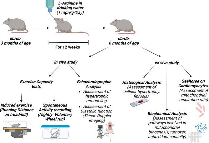
In patients with type II diabetes, the development of diabetic cardiomyopathy (DC) is associated with a high risk of mortality. Left ventricular hypertrophy, diastolic dysfunction, and exercise intolerance are the first signs of DC. The underlying mechanisms are not fully elucidated, and there is an urgent need for specific biomarkers and molecular targets for early diagnosis and treatment. Mitochondrial alterations play a key role in the development of DC, and microRNAs regulating mitochondrial function are emerging as potential biomarkers of metabolic stress in DC. L-Arginine (Arg) supplementation has been shown to be an effective strategy for improving mitochondrial function and energetics, with a significant impact on physical performance. The aim of the current study was to evaluate the effects of Arg supplementation on cardiac mitochondrial function, DC development, and relative phenotypes including exercise intolerance. We used db/db mice as a model of type II diabetes, chronically treated with Arg (1 mg/kg/day) for 12 weeks. Arg-treated db/db mice showed preserved diastolic function and left ventricular morphology compared with untreated diabetic mice. Arg supplementation also improved exercise tolerance and the propensity to physical activity. Mitochondrial respiration was significantly increased in cardiomyocytes isolated from treated db/db mice, as well as in diabetic cardiomyocytes treated with Arg in vitro. The improvement of cardiac mitochondrial function in db/db + Arg mice was associated with an increase in PGC-1-alpha levels, mitochondrial biogenesis, recycling, and antioxidant capacity. Arg treatment prevented the accumulation of circulating and cardiac miR-143 in db/db mice, which is an index of metabolic stress and activation of mitochondrial damage mechanisms. In conclusion, Arg supplementation is effective in preventing the development of DC, preserving diastolic function and exercise tolerance by improving mitochondrial fitness and homeostasis. Additionally, miR-143 could potentially be employed to monitor cardiac metabolic stress and the effects of Arg treatment in diabetes.


Conflict of interest statement

Declarations. Competing interests: Gaetano Santulli declares that he is Associate Editor of Cardiovascular Diabetology and that the article was assigned to another Editor to assume responsibility for overseeing peer review. This submissions was subject to the exact same review process as any other manuscript submitted to the journal. Licenses: Figure 1 was created in BioRender. License was granted to Iaccarino, G. (2024- BioRender.com/c38b746).

Figures

Fig. 1
Fig. 1
Study Protocol. Starting from 3 months of age db/db mice were treated with Arg in drinking water at the indicated dose. After 12 weeks of treatment, cardiac function and remodeling and exercise capacity were assessed in vivo. Rodents were sacrificed and ex vivo experiments performed: histological and biochemical analysis on cardiac tissue, as well as mitostress test on isolated cardiomyocytes by Seahorse.
Fig. 2
Fig. 2
Echocardiographic analysis of wt, db/db, and Arg-treated db/db mice showing: M-mode representative images for short axis (A), Left Ventricular Mass automatically determined by echocardiography machine (LVmass (normalized by body weight) (B), Left Ventricular end Diastolic Volume (LVEDV) (C). Tissue doppler imaging was performed to evaluate diastolic function. Representative images of e’ and a’ waves (D). The automatically calculated e′/a′ (E) and E/e′ (F) ratio were displayed for each group. WGA staining was performed to determine cellular hypertrophy. Representative images for WGA staining on cardiac histologic sections (G), and cumulative quantification of cardiomyocytes cross sectional area (H). ANOVA followed by Bonferroni correction was used to assess the significative differences among groups. (WT, db/db, db/db + Arg n = 8) *p < 0.05 vs WT, # p < 0.05 vs db/db.
Fig. 3
Fig. 3
Exercise tolerance was assessed by induced running on treadmill, with increasing speed to exhaustion. The total distance covered by running was calculated for each mouse (WT n = 5, db/db n = 5, db/db + Arg n = 7) (A). The propensity to physical activity was determined by spontaneous overnight wheel run. The automatically calculated count of wheel runs is displayed (WT n = 4, db/db n = 5, db/db + Arg n = 5) (B). ANOVA followed by Bonferroni correction was used to assess the differences among groups. *p < 0.05 vs. WT, # p < 0.05 vs. db/db.
Fig. 4
Fig. 4
Adult Ventricular cardiomyocytes were isolated from WT, db/db, and fArg-treated db/db mice. The day after isolation, the Oxygen Consumption Rate was determined by Seahorse, in basal conditions and in response to Oligomycin, FCCP and Rotenone (A). Basal (B) and maximal respiration rate (C), alongside with ATP- coupled respiration (D) were determined. ANOVA followed by Bonferroni correction was used to assess the significative differences among groups (WT, db/db, ddb/db + Arg n = 4). *p < 0.05 vs. WT, # p < 0.05 vs. db/db.
Fig. 5
Fig. 5
Western blot analysis of PGC1 alpha in cardiac lysate, and relative quantification using Actin as loading control (A). Real-time PCR for TfaM mRNA levels in cardiac extract (B). Mitochondrial biogenesis determined by assessing the copy number of mitochondrial genes NADH and Cytochrome B by Real-time PCR, using a nuclear gene (GAPDH) as control (C). Active expression of mitochondrial genes was determined by evaluating the levels of a mitochondrial proteins encoded by mitochondrial DNA, mtCOX-1 and mtCOX-3, compared with the levels of a mitochondrial protein encoded by nuclear genome SDH-A; GAPDH was used as loading control (D). The images are representative of 3 independent experiments (WT, db/db and db/db + Arg n = 4) ANOVA followed by Bonferroni correction was used to assess the significative differences among groups. *p < 0.05 vs. WT, # p < 0.05 vs. db/db.
Fig. 6
Fig. 6
Mitochondria were isolated from cardiac tissue of wt, db/db and treated db/db mice and western blot analyses on mitochondrial extract was conducted to evaluate the levels of LC3II, DRP1 (A) and SOD (B). VDAC1 was used as loading control for mitochondrial extracts. To assess redox status GSH/ GSSG ratio was determined by immunoenzymatic based assay conducted on cardiac tissue from WT, db/db and Arg treated db/db mice (C). The images are representative of 3 independent experiments. (WT, db/db, db/db + Arg n = 4). ANOVA followed by Bonferroni correction was used to assess the significative differences among groups. *p < 0.05 vs. WT, # p < 0.05 vs. db/db.
Fig. 7
Fig. 7
Measurement of microRNA-143 (miR-143) levels in plasma (A) and in cardiac tissue (B) isolated from wt, db/db, and treated db/db mice. (WT, db/db, db/db + Arg n = 6) ANOVA followed by Bonferroni correction was used to assess the significative differences among groups. *p < 0.05 vs. WT, # p < 0.05 vs. db/db.

References

    1. Huo JL, Feng Q, Pan S, Fu WJ, Liu Z, Liu Z. Diabetic cardiomyopathy: early diagnostic biomarkers, pathogenetic mechanisms, and therapeutic interventions. Cell Death Discov. 2023;9(1):256. - PMC - PubMed
    1. Grubic Rotkvic P, Planinic Z, Liberati Prso AM, Sikic J, Galic E, Rotkvic L. The Mystery of Diabetic Cardiomyopathy: from early concepts and underlying mechanisms to novel therapeutic possibilities. Int J Mol Sci 2021;22(11). - PMC - PubMed
    1. Yin L, Sun Z, Ren Q, Su X, Zhang D. Long non-coding RNA BANCR is overexpressed in patients with diabetic retinopathy and promotes apoptosis of retinal pigment epithelial cells. Med Sci Monit. 2019;25:2845–51. - PMC - PubMed
    1. Duncan JG, Fong JL, Medeiros DM, Finck BN, Kelly DP. Insulin-resistant heart exhibits a mitochondrial biogenic response driven by the peroxisome proliferator-activated receptor-alpha/PGC-1alpha gene regulatory pathway. Circulation. 2007;115(7):909–17. - PMC - PubMed
    1. Flarsheim CE, Grupp IL, Matlib MA. Mitochondrial dysfunction accompanies diastolic dysfunction in diabetic rat heart. Am J Physiol. 1996;271(1 Pt 2):H192–202. - PubMed

Publication types

MeSH terms

LinkOut - more resources