. 2024 Dec 20;22(1):786.
doi: 10.1186/s12951-024-02965-9.

Targeted lipid nanoparticles distributed in hydrogel treat osteoarthritis by modulating cholesterol metabolism and promoting endogenous cartilage regeneration

Affiliations

Targeted lipid nanoparticles distributed in hydrogel treat osteoarthritis by modulating cholesterol metabolism and promoting endogenous cartilage regeneration

Zhibo Zhao et al. J Nanobiotechnology. .

Abstract

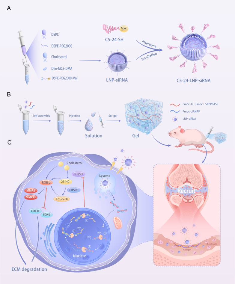
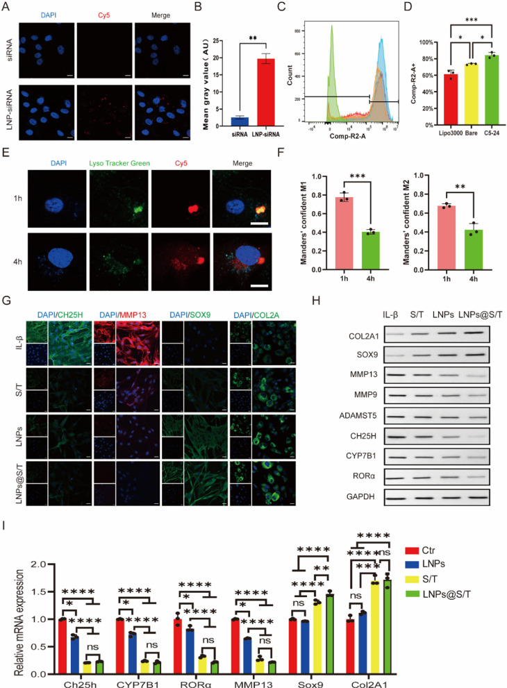
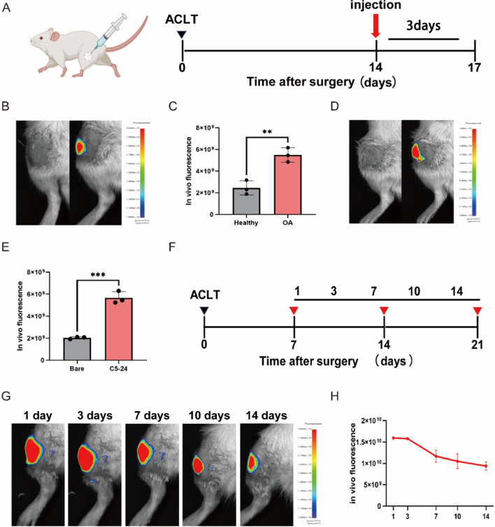
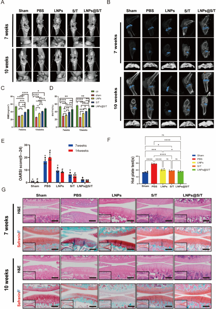
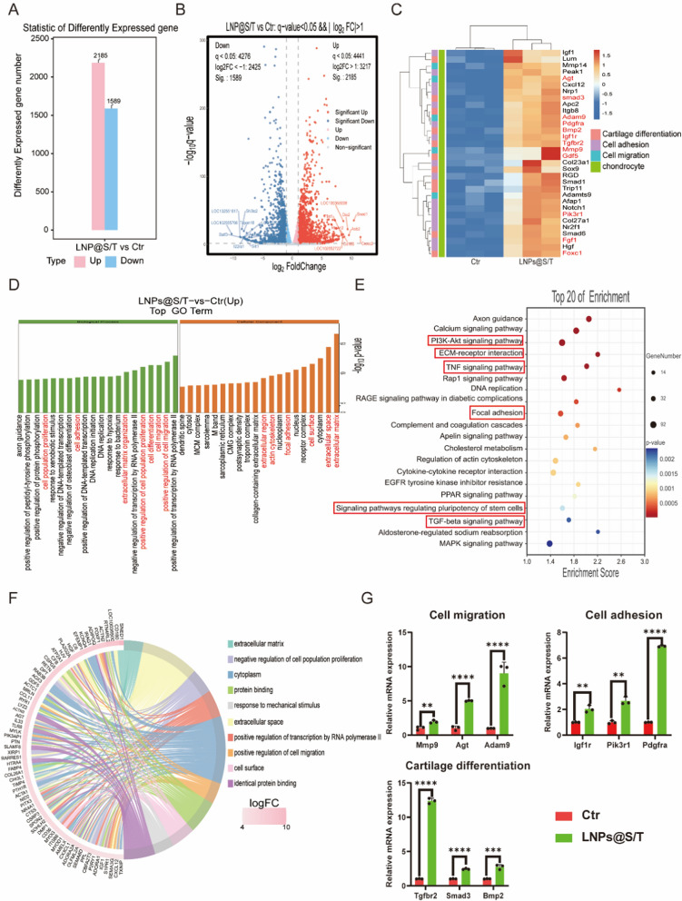
Osteoarthritis (OA) is the most common disease in aging joints and has characteristics of cartilage destruction and inflammation. It is currently considered a metabolic disease, and the CH25H-CYP7B1-RORα axis of cholesterol metabolism in chondrocytes plays a crucial catabolic regulatory role in its pathogenesis. Targeting of this axis in chondrocytes may provide a therapeutic approach for OA treatment. Here, in this study, we propose to use a combination of stem cell-recruiting hydrogels and lipid nanoparticles (LNPs) that modulate cholesterol metabolism to jointly promote a regenerative microenvironment. Specifically, we first developed an injectable, bioactive hydrogel composed of self-assembling peptide nanofibers that recruits endogenous synovial stem cells (SMSCs) and promotes their chondrogenic differentiation. At the same time, LNPs that regulate cholesterol metabolism are incorporated into the hydrogel and slowly released, thereby improving the inflammatory environment of OA. Enhancements were noted in the inflammatory conditions associated with OA, alongside the successful attraction of mesenchymal stem cells (MSCs) from the synovial membrane. These cells were then observed to differentiate into chondrocytes, contributing to effective cartilage restoration and chondrocyte regeneration, thereby offering a promising approach for OA treatment. In summary, this approach provides a feasible siRNA-based therapeutic option, offering a potential nonsurgical solution for treatment of OA.

Keywords: Cartilage rejuvenation; Cholesterol metabolism; Hydrogel; Lipid nanoparticles; Osteoarthritis.


Conflict of interest statement

Declarations. Ethics approval and consent to participate: All animal experiments were conducted in compliance with the Guidelines for the Care and Use of Laboratory Animals and were approved by the Animal Ethics Committee of Shandong Provincial Hospital (No.2023–004). Consent for publication: All the authors have approved the manuscript to publish. Competing interests: The authors declare no competing interests.

Figures

Fig. 1
Fig. 1
Schematic diagram of the study design. A Schematic illustration of the preparation of C5-24-LNPs-siRNA. B Schematic illustration of the C5-24-LNPs-siRNA coating on the hydrogel. C Schematic diagram of C5-24-LNPs-siRNA regulating chondrocyte cholesterol metabolism to inhibit cartilage degeneration and peptide hydrogel recruiting SMSCs to cartilage defects and promoting cartilage differentiation in the ACLT rat model
Fig. 2
Fig. 2
Design and characterization of the LNPs@S/T coating. A Photographs of the peptide gelator solutions and hydrogel. B The injectable hydrogel was freely molded into diverse geometric shapes using a syringe. C Representative cryogenic transmission electron microscopy (cryo-TEM) image showing the morphology of the C5-24-LNPs. Scale bar: 100 nm. D Efficiency of siRNA entrapment assayed by Ribogreen. Bars show the means of three biologically independent samples; black points show individual samples. Error bars represent ± SD. E, H Hydrodynamic size, polydispersity index (PDI), and zeta potential of the C5-24-LNPs. Data are presented as mean ± SD. F SEM (scale bars, 20 µm) and TEM (scale bar, 200 nm) images of the hydrogels. G Remaining mass of the hydrogel system in vitro. I Cy5-labeled LNPs in hydrogels. J Changes in shear viscosity with increasing shear rates of the hydrogels. K Rheology traces of the hydrogels. L, M CD spectra (L) and FTIR spectra (M) of the hydrogels. Data are presented as mean ± SD
Fig. 3
Fig. 3
siCH25H-mediated regulation of cholesterol metabolism and alleviation of OA cartilage degeneration in rat inflammatory chondrocytes. A Confocal images of inflammatory chondrocytes treated with free siRNA or LNP-siRNA. The nuclei were counterstained with DAPI (blue). Scale bar, 20 µm. B Quantification of the data shown in (A). C Cellular uptake of LNP-siRNA linked to C5-24 targeting peptide, bare LNP-siRNA, or Lipo3000-siRNA by inflammatory chondrocytes measured by flow cytometry analysis. D Quantification of the data shown in (C). E Typical confocal images of inflammatory chondrocytes incubated with LNP-siRNA for 1 or 4 h at 37 °C. Nuclei were stained with DAPI (blue), endosomes/lysosomes were stained with LysoTracker Green (green), and siRNAs were labeled with Cy5 (red). Scale bars: 20 µm. F Quantitative analysis of the colocalization of Cy5-labeled siRNA with endosomes/lysosomes labeled with LysoTracker Green. G Immunofluorescence staining of CH25H, MMP13, SOX9, and Col2A1 in rat chondrocytes. Scale bar, 40 µm. H Expression of Col2A1, SOX9, MMP13, MMP9, ADAMTS5, CH25H, CYP7B1, and RORα at 2 days post-transfection of rat chondrocytes after different cultures determined by WB (n = 3). I qPCR evaluation of Col2A1, SOX9, MMP13, CH25H, CYP7B1, and RORα expression at 2 days post-transfection of rat chondrocytes after different cultures (n = 3). Data are presented as mean ± SD. Statistical analysis of B was performed using two-tailed Student's t-test. Statistical analysis of D and I was performed using one-way ANOVA. Statistical analysis of F was performed using two-tailed Student's t-test. *P < 0.05, **P < 0.01, ***P < 0.001, ****P < 0.0001. NS, not significant
Fig. 4
Fig. 4
LNPs@S/T-mediated induction of SMSCs recruitment and promotion of chondrogenic differentiation. A Schematic illustration of the recruitment process for SMSCs during cartilage repair. B Representative images of Transwell bottom membranes stained with crystal violet after 24 h of culture and Alcian blue staining of SMSCs pellets after a 7-day culture in the indicated treatment groups. Scale bar, 250 µm. C Quantification of cells observed at the bottom of the membranes (n = 3). D Schematic illustration of the Transwell assay to monitor SMSCs recruitment in vitro. E Immunofluorescence staining of ACAN and Col2A1 in SMSCs cultured on different hydrogels for 7 days. Scale bar, 40 µm. F Quantification of the data shown in (E). G, H WB and qRT-PCR analyses of the protein (G) and mRNA (H) levels of Sox9, ACAN, and Col2A1 in SMSCs pellets in the indicated treatment groups (n = 3). I Quantitative analysis of the data shown in (G) (n = 3). Data are presented as mean ± SD. Statistical analysis was performed using one-way ANOVA. *P < 0.05, **P < 0.01, ***P < 0.001, ****P < 0.0001. NS not significant
Fig. 5
Fig. 5
siCH25H is locally retained relative to bare and C5-24 peptide-functionalized LNPs in the knee joints of a short-term mouse model of OA. A Schematic illustration of the experimental design. B, C Intravital imaging B and quantification C of C5-24-LNPs retained in healthy versus OA knee joints at 3 days (OA vs. Healthy) (n = 4). D, E Representative in vivo imaging D and quantification E of C5-24-LNPs retained compared with bare-LNPs in OA knee joints explanted at 3 days (Bare-LNPs vs. C5-24-LNPs) (n = 4). F Schematic illustration of the experimental design. G, H In vivo biodegradation of Cy5-labeled C5-24-LNPs sustained-released through hydrogel was observed by in vivo fluorescence imaging of the knee joint on day 14 after injection (n = 4). Data are presented as mean ± SD. Statistical analysis was performed using two-tailed Student's t-test. **P < 0.01, ***P < 0.001
Fig. 6
Fig. 6
LNPs@S/T-mediated rescue of OA cartilage degeneration after ACLT surgery in rats. A Representative two-dimensional images in the sagittal and coronal planes and three-dimensional images of the subchondral bone reconstructed by micro-CT. B Three-dimensional images of rat knee joints showing the abnormal growth of osteophytes in the control, Sham, PBS, LNPs, S/T, and LNPs@S/T groups at 7 and 10 weeks post-ACLT. C, D Quantitative statistics of the total BMD C and percentage of BV/TV D on micro-CT (n = 4). E OARSI scores of rat joints at 7 and 10 weeks (n = 4). The OARSI score is calculated as grade * stage. F Hot plate test of rats at 7 and 10 weeks. n = 4 rats per group. G Representative rat knee joint images stained with safranin O/fast green and H&E at 7 and 10 weeks. Scale bar, 200 µm. Data are presented as mean ± SD. Statistical analysis was performed using one-way ANOVA. *P < 0.05 versus LNPs@S/T group at 7 weeks, #P < 0.05 versus LNPs@S/T group at 10 weeks, *P < 0.05, **P < 0.01, ***P < 0.001, ****P < 0.0001. NS not significant
Fig. 7
Fig. 7
Immunofluorescence after LNPs@S/T treatment of OA in vivo and recruitment of SMSCs. A Representative immunohistochemistry images of COL2A1 (scale bar, 200 µm), ACAN (scale bar, 200 µm), and MMP13 (scale bar, 200 µm) in rat knee joints from the Sham, PBS, LNPs, S/T, and LNPs@S/T groups at 7 and 10 weeks. B Quantification of cells with histological positivity for COL2A1, MMP13, and ACAN (n = 4). C Immunofluorescence staining of CD73 (green) and CD90 (red) in rat knee joints at 7 and 10 weeks. Nuclei were stained with DAPI. Scale bar, 500 μm. Data are presented as mean ± SD.Statistical analysis was performed using one-way ANOVA. *P < 0.05 versus LNPs@S/T group at 7 weeks, #P < 0.05 versus LNPs@S/T group at 10 weeks, *P < 0.05, **P < 0.01, ***P < 0.001, ****P < 0.0001. NS not significant
Fig. 8
Fig. 8
Intrinsic mechanisms for LNPs@S/T effect on SMSCs behavior. A Differentially expressed mRNAs in the Ctr and LNPs@S/T groups (*P < 0.05, | log2(fold change) |> 1). B Volcano plot of genes differentially expressed in untreated versus LNPs@S/T-treated SMSCs. C Heatmap of genes related to cell adhesion, cartilage development, and migration in the Ctr and LNPs@S/T groups (n = 3). D GO enrichment bar plots. E KEGG enrichment analysis (PI3K-Akt signaling pathway, ECM-receptor interaction, TNF signaling pathway, focal adhesion, signaling pathways regulating pluripotency of stem cells, and TGF-β signaling pathway). F Expression and crossover of each gene. G qRT-PCR analysis of genes associated with cell migration, cell adhesion, and cartilage development (n = 3). Data are presented as mean ± SD. Statistical analysis was performed using two-tailed Student's t-test. *P < 0.05, **P < 0.01, ***P < 0.001, ****P < 0.0001. NS not significant
Fig. 9
Fig. 9
Intrinsic mechanisms for LNPs@S/T effect on chondrocytes behavior. A Differentially expressed mRNAs in the IL-1β and LNPs@S/T groups (*P < 0.05, | log2(fold change) |> 1). B Volcano plot of genes differentially expressed in IL-1β and LNPs@S/T groups. C Heatmap of genes in the IL-1β and LNPs@S/T groups (n = 3). D GO enrichment bar plots. E KEGG enrichment analysis (ECM-receptor interaction, TGF-β signaling pathway, Wnt signaling pathway, cholesterol metabolism, MAPK signaling pathway, NF-kappa B signaling pathway, and JAK-STAT signaling pathway). F qRT-PCR analysis of genes associated with ECM homeostasis and inflammation (n = 3). Data are presented as mean ± SD. Statistical analysis was performed using two-tailed Student's t-test. *P < 0.05, **P < 0.01, ***P < 0.001, ****P < 0.0001. NS not significant

References

    1. Gelber AC. Knee osteoarthritis. Ann Intern Med. 2024;177:itc129–44. 10.7326/annals-24-01249. - PubMed
    1. Loeser RF, Goldring SR, Scanzello CR, Goldring MB. Osteoarthritis: a disease of the joint as an organ. Arthritis Rheum. 2012;64:1697–707. 10.1002/art.34453. - PMC - PubMed
    1. Nho SJ, Kymes SM, Callaghan JJ, Felson DT. The burden of hip osteoarthritis in the United States: epidemiologic and economic considerations. J Am Acad Orthop Surg. 2013;21(Suppl 1):S1-6. 10.5435/JAAOS-21-07-S1. - PubMed
    1. Duong V, Oo WM, Ding C, Culvenor AG, Hunter DJ. Evaluation and treatment of knee pain: a review. JAMA. 2023;330:1568–80. 10.1001/jama.2023.19675. - PubMed
    1. Mahmoudian A, Lohmander LS, Mobasheri A, Englund M, Luyten FP. Early-stage symptomatic osteoarthritis of the knee—time for action. Nat Rev Rheumatol. 2021;17:621–32. 10.1038/s41584-021-00673-4. - PubMed

MeSH terms

LinkOut - more resources