. 2024 Dec 21;12(1):197.
doi: 10.1186/s40478-024-01904-x.

Selective retinoid X receptor agonism promotes functional recovery and myelin repair in experimental autoimmune encephalomyelitis

Affiliations

Selective retinoid X receptor agonism promotes functional recovery and myelin repair in experimental autoimmune encephalomyelitis

Gracious D S Kasheke et al. Acta Neuropathol Commun. .

Abstract

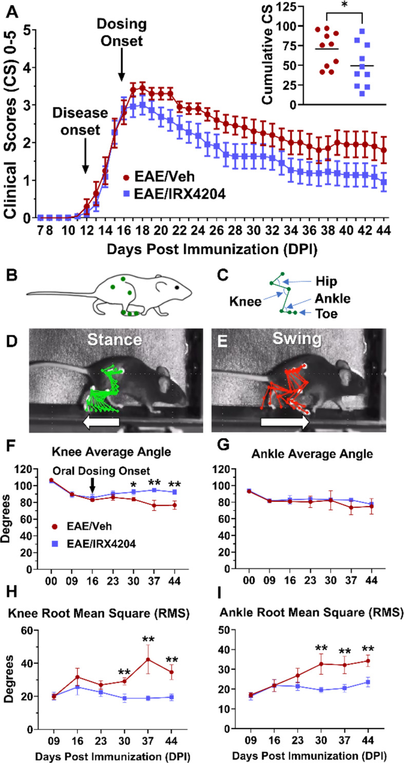
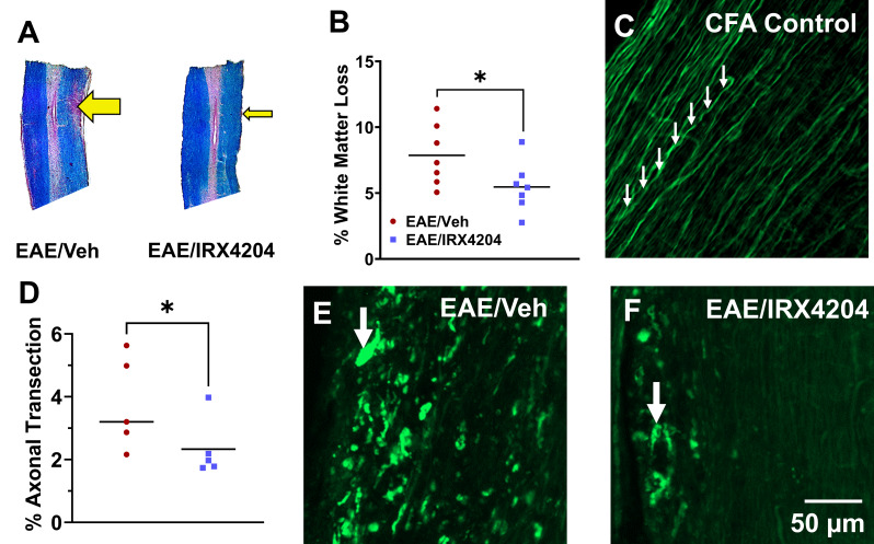
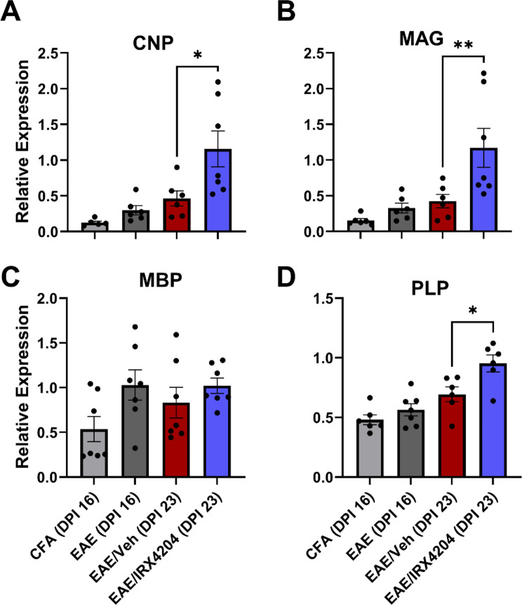
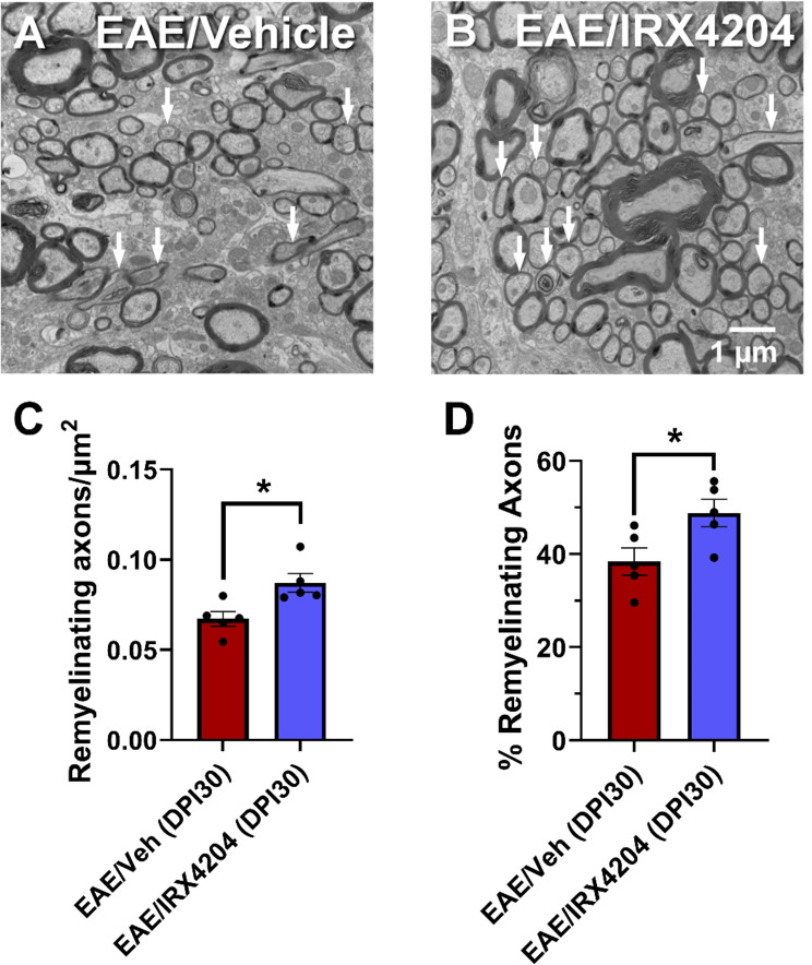
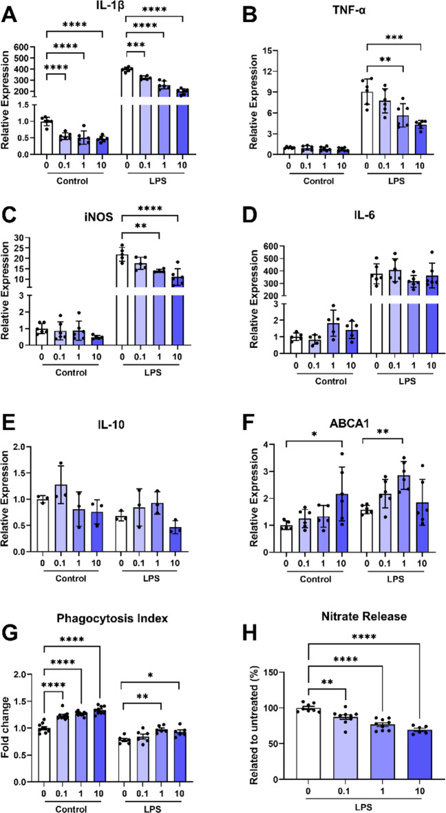
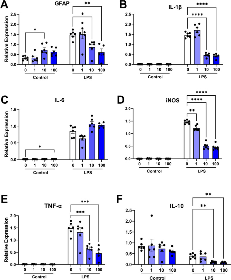
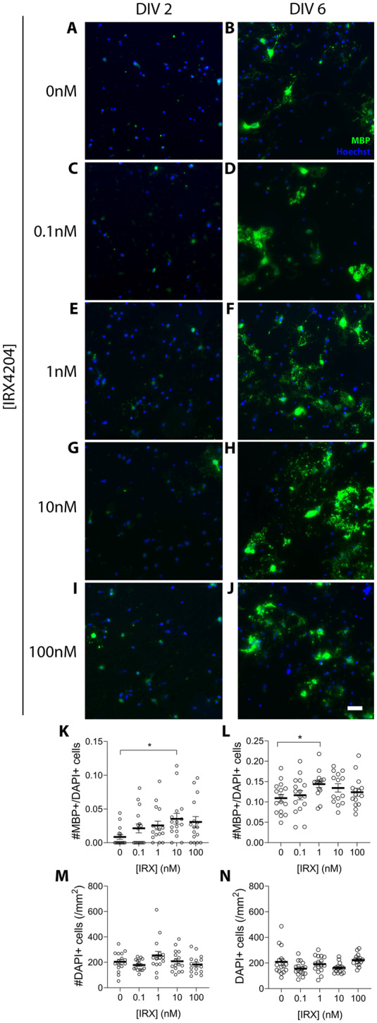
Evidence that myelin repair is crucial for functional recovery in multiple sclerosis (MS) led to the identification of bexarotene (BXT). This clinically promising remyelinating agent activates multiple nuclear hormone receptor subtypes implicated in myelin repair. However, BXT produces unacceptable hyperlipidemia. In contrast, IRX4204 selectively activates the retinoid X receptor (RXR). Given compelling links between RXR activation and increased myelin repair, we employed IRX4204 to investigate the impact of RXR agonism alone on functional recovery in mice subjected to experimental autoimmune encephalomyelitis (EAE). Since gait deficits are common in MS, we used machine learning to obtain highly sensitive and reliable measurements of sagittal hindleg joint movements for mice walking on a treadmill. IRX4204 not only blocked the progressive loss of knee and ankle movements but also reversed joint movement impairments in EAE mice. Our biochemical, transcriptional and histological measurements in spinal cord suggest these gait improvements reflect increased axon survival and remyelination and reduced inflammation. Using microglia, astrocytes and oligodendrocyte progenitor cells, we present additional data suggesting that IRX4204 may act on multiple glial subtypes to orchestrate myelin repair. These results inform the discovery of restorative neural therapeutics for MS by demonstrating that selective RXR agonism is sufficient for effective myelin repair. Moreover, our findings support the therapeutic potential of IRX4204 to promote functional recovery in MS.

Keywords: Multiple sclerosis; Neural repair; Neuroprotection; Oligodendrocyte progenitor cell; Rehabilitation.


Conflict of interest statement

Declarations. Ethics approval and consent to participate: Not applicable. Consent for publication: Not applicable. Competing interests: Drs. Vuligonda and Sanders are equity holders, officers, and members of the Board of Directors of Io Therapeutics, Inc., which is developing IRX4204 for commercialization as a treatment for multiple sclerosis and other neurodegenerative diseases.

Figures

Fig. 1
Fig. 1
Oral administration of IRX4204 beginning at peak disease (DPI 16) reduces clinical scores and reverses gait deficits in EAE mice. (A) Daily clinical scores from DPI 07–44 for EAE mice treated with vehicle (Veh) or IRX4204 (mean ± SEM). Relative to Veh (5 mL/kg/day, po), IRX4204 (12 mg/kg/day, po) administration beginning at peak disease (DPI 16) reduced cumulative clinical scores (insert; n = 10, one-tailed Mann-Whitney U test, P < 0.05, mean ± SEM). (BE) Six points of the hindlimb (B) were tracked to construct a stick model for leg movements (C) during the stance (D) and swing (E) phases of a gait cycle while mice walked on a treadmill. (FI) Gait was measured at baseline (DPI − 02) and DPI 9, 16, 23, 30, 37 and 44. EAE resulted in gait deficits by DPI 16. Relative to Veh, IRX4204 partially reversed the loss of knee average angle (F), completely normalized knee RMS differences (H) and prevented the progressive increase of ankle RMS differences (I) from DPI 30–44 (n = 10, two-way ANOVA, Šidák’s multiple comparisons test, mean ± SEM; *p < 0.05, **p < 0.01)
Fig. 2
Fig. 2
Oral administration of IRX4204 beginning at peak disease (DPI 16) reduces white matter loss and axonal transection in the lumbar spinal cord of EAE mice. (A) Representative spinal cord sections from EAE mice treated orally with vehicle (Veh; 5 mL/kg/day; left) or IRX4204 (12 mg/kg/day; right) stained with Eriochrome Cyanine and counterstained with Neutral Red. Arrows depict regions of white matter loss. (B) Quantification of regions of white matter loss revealed a significant reduction in white matter loss at DPI 44 in IRX4204/EAE compared to EAE/Veh mice (n = 7). (C, E, F) Representative images of eYFP fluorescence in corticospinal axons in white matter regions of spinal cord sections from EAE mice treated orally with Veh (5 mL/kg/day) or IRX4204 (12 mg/kg/day). (D) Axon damage, quantified at DPI 44 by calculating percent area of suprathreshold punctate eYFP labelling, revealed a reduction in axonal transection in EAE/IRX4204 relative to EAE/Veh mice (n = 5, one-tailed Mann-Whitney U test, mean; *p < 0.05)
Fig. 3
Fig. 3
Oral administration with IRX4204 at peak disease (DPI 16) increases pro-myelinating gene expression in the spinal cord of EAE mice. (AD) Expression of 2’,3’-Cyclic nucleotide 3’-phosphodiesterase (CNP; A), myelin-associated glycoprotein (MAG; B), myelin basic protein (MBP; C), and proteolipid protein (PLP; D) normalized relative to reference gene expression in mouse spinal cord. CNP, MAG, and PLP, expression increased in EAE mice treated with IRX4204 (12 mg/kg/day) compared to EAE mice treated with Veh (5 mL/kg/day) beginning at peak disease (n = 7, one-tailed Mann-Whitney U test, mean ± SEM; *p < 0.05, **p < 0.01)
Fig. 4
Fig. 4
Oral administration with IRX4204 at peak disease (DPI 16) reduces expression of IL-17, iNOS, IBA-1, GFAP, and HIF-1α in the spinal cord of EAE mice. (A-I) Expression of interferon-γ (IFN-γ; A), interleukin 1β (IL-1β; B), interleukin 6 (IL-6; C), interleukin 17 (IL-17; D), inducible nitric oxide synthase (iNOS; E), tumour necrosis factor-α (TNF-α; F), ionized calcium-binding adapter molecule 1 (IBA-1; G), glial fibrillary acidic protein (GFAP; H), and hypoxia inducible factor-1α (HIF-1α; I) normalized relative to reference gene expression in mouse spinal cord. Expression of IL-17, iNOS, IBA-1, GFAP, and HIF-1α decreased in EAE mice following treatment with IRX4204 (12 mg/kg/day) compared to EAE mice treated with vehicle (5 mL/kg/day) starting at peak disease (n = 7, one-tailed Mann-Whitney U test, mean ± SEM; *p < 0.05)
Fig. 5
Fig. 5
Oral administration with IRX4204 at peak disease (DPI 16) promotes remyelination in the lumbar spinal cord of EAE mice. (A and B) Representative electron microscopy images of the L3/L4 spinal cord region of EAE mice treated orally with vehicle (5 mL/kg/day; A) or IRX4204 (12 mg/kg/day; B). Arrows depict remyelinating axons. (C and D) Axonal counts revealed that remyelinating axons were more abundant (C) and made up a higher percentage of axons (D) in EAE mice following treatment with IRX4204 compared to EAE mice treated with vehicle starting at peak disease (n = 5, one-tailed Mann-Whitney U test, mean ± SEM; *p < 0.05)
Fig. 6
Fig. 6
IRX4204 attenuates LPS-induced pro-inflammatory gene expression, increases expression of the cellular transport gene ABCA1, and promotes pro-repair actions of C8-B4 microglia in vitro. (AF) IL-1β (A), TNF-α (B), iNOS (C), IL-6 (D), IL-10 (E), and ABCA1 (F), gene expression normalized relative to reference gene expression in microglial cell cultures pre-treated with IRX4204 (24 h) in normal conditions and after treatment with LPS (24 h; n = 3–6 wells). Pre-treatment with IRX4204 produced concentration-dependent attenuation of IL-1β, TNF-α, and iNOS expression in microglial cultures incubated with LPS (24 h) and increased ABCA1 expression in microglial cultures at 10 nM in normal conditions and at 1 nM following incubation with LPS. (G and H) Pre-treatment of these cultures with IRX4204 also increased phagocytic activity in normal conditions at 0.1 nM, 1 nM, and 10 nM (n = 6 wells; G) and following incubation with LPS at 1 nM and 10 nM. Pre-treatment with IRX4204 at 0.1 nM, 1 nM, and 10 nM was also sufficient to produce a concentration-dependent attenuation of nitrate release (n = 6–9 wells; H) in microglial cell cultures after incubation with LPS (ordinary one-way ANOVA, Šidák’s multiple comparisons test, *P < 0.05, **P < 0.01, ***P < 0.001, ****P < 0.0001, mean ± SEM)
Fig. 7
Fig. 7
IRX4204 attenuates LPS-induced pro-inflammatory gene expression in primary mouse astrocyte cultures. (AF) GFAP (A), IL-1β (B), IL-6 (C), iNOS (D), TNF-α (E), and IL-10 (F), gene expression normalized relative to reference gene expression in astrocyte cultures pre-treated with IRX4204 (24 h) at basal conditions or after incubation with LPS (16 h; n = 4–6 wells). IRX4204 produced concentration-dependent attenuation in expression of each of these genes in astrocyte cultures in LPS conditions and increased GFAP expression and IL-6 expression at 10 nM and 100 nM, respectively, in basal conditions (ordinary one-way ANOVA, Šidák’s multiple comparisons test, *P < 0.05, **P < 0.01, ***P < 0.001, ****P < 0.0001, mean ± SEM)
Fig. 8
Fig. 8
IRX4204 stimulates differentiation of primary rat OPCs in vitro. (AJ) Representative images of overlain MBP and Hoechst immunofluorescence in OPC cultures. One day following isolation and plating, OPCs were treated with increasing concentrations of IRX4204 for either 24 h (DIV 2; A, C, E, G, I) or 5 days in vitro (DIV 6; B, D, F, H, J). (KN) Proportion of MBP-positive cells to DAPI-positive cells (K and L) and number of DAPI-positive cells per mm2 (M and N) after 24 h or 5 days of treatment with increasing concentrations of IRX4204. After 24 h treatment, 10 nM of IRX4204 significantly increased the fraction of MBP immunopositive cells, without altering the total number of cells. After 5 days of treatment, the fraction of MBP immunopositive cells increased with IRX4204 treatment, with a statistically significant increase detected for a concentration of 1 nM IRX4204 (n = 14–16 fields/condition; non-parametric Kruskal-Wallis test, Dunn’s multiple comparisons test, *P < 0.05, mean ± SEM; scale bar 50 μm)

References

    1. (2016) 2016 Emerging Science Program Schedule. Neurology 87: e20–e25 Doi 10.1212/WNL.0000000000002784
    1. Akay T, Acharya HJ, Fouad K, Pearson KG (2006) Behavioral and electromyographic characterization of mice lacking EphA4 receptors. J Neurophysiol 96:642–651. 10.1152/jn.00174.2006 - PubMed
    1. Armstrong RC (1998) Isolation and characterization of immature oligodendrocyte lineage cells. Methods 16:282–292. 10.1006/meth.1998.0685 - PubMed
    1. Bauer NM, Moos C, van Horssen J, Witte M, van der Valk P, Altenhein B, Luhmann HJ, White R (2012) Myelin basic protein synthesis is regulated by small non-coding RNA 715. EMBO Rep 13:827–834. 10.1038/embor.2012.97 - PMC - PubMed
    1. Benedetti MG, Piperno R, Simoncini L, Bonato P, Tonini A, Giannini S (1999) Gait abnormalities in minimally impaired multiple sclerosis patients. Mult Scler 5:363–368. 10.1177/135245859900500510 - PubMed

Publication types

MeSH terms

LinkOut - more resources