. 2024 Dec 20;15(1):493.
doi: 10.1186/s13287-024-04115-2.

Small extracellular vesicles derived from umbilical cord mesenchymal stem cells alleviate radiation-induced cardiac organoid injury

Affiliations

Small extracellular vesicles derived from umbilical cord mesenchymal stem cells alleviate radiation-induced cardiac organoid injury

Hu Cao et al. Stem Cell Res Ther. .

Abstract

Background: Radiation-induced heart disease (RIHD) is one of the most serious complications of radiation therapy (RT) for thoracic tumors, and new interventions are needed for its prevention and treatment. Small extracellular vesicles (sEVs) from stem cells have attracted much attention due to their ability to repair injury. However, the role of umbilical cord mesenchymal stem cell (UCMSC)-derived sEVs in protecting cardiac organoids from radiation-induced injury and the underlying mechanisms are largely unknown.

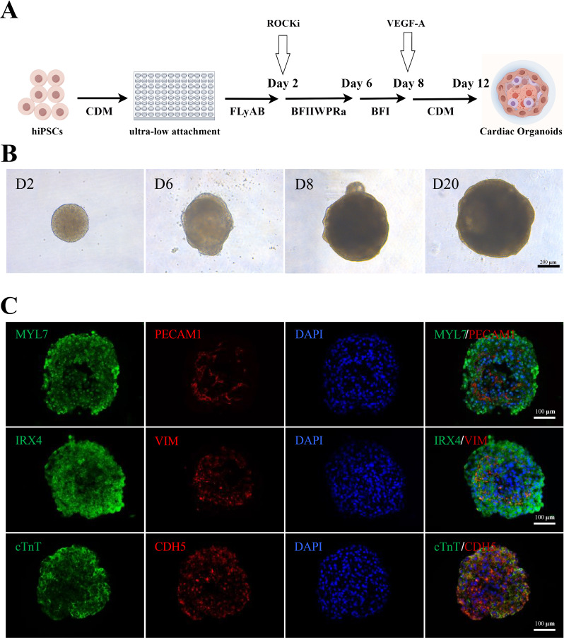
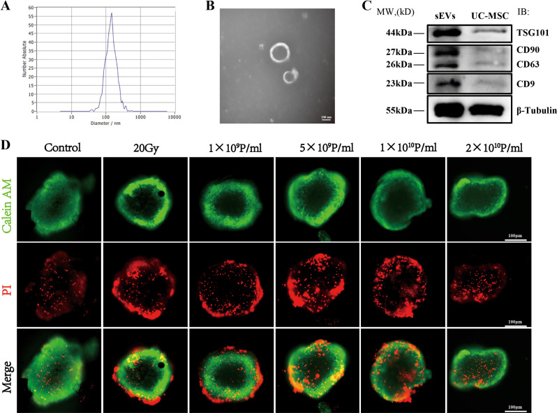
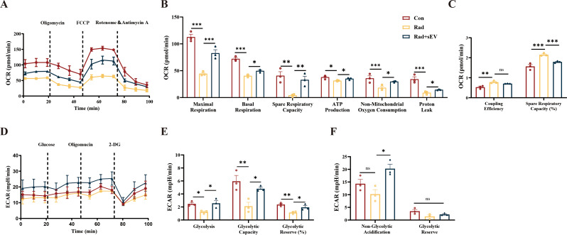
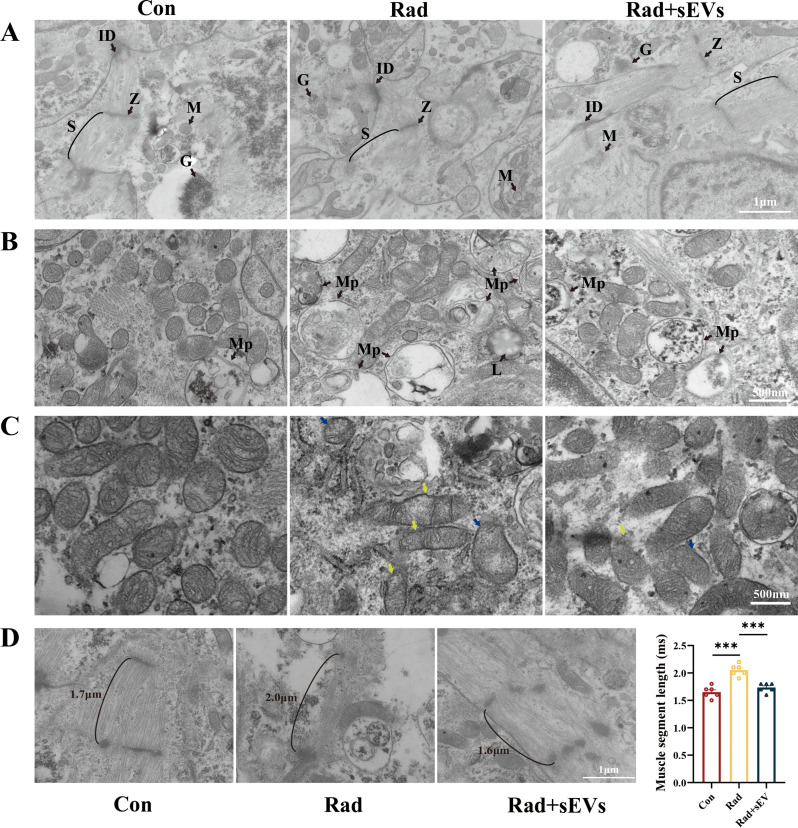
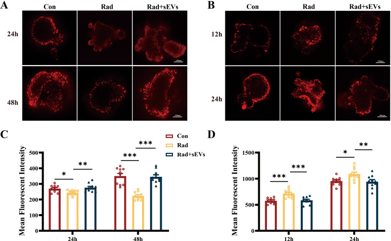
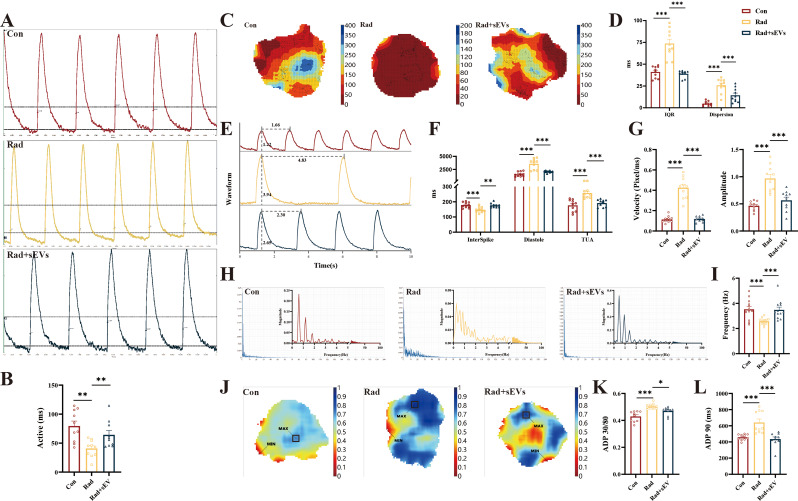
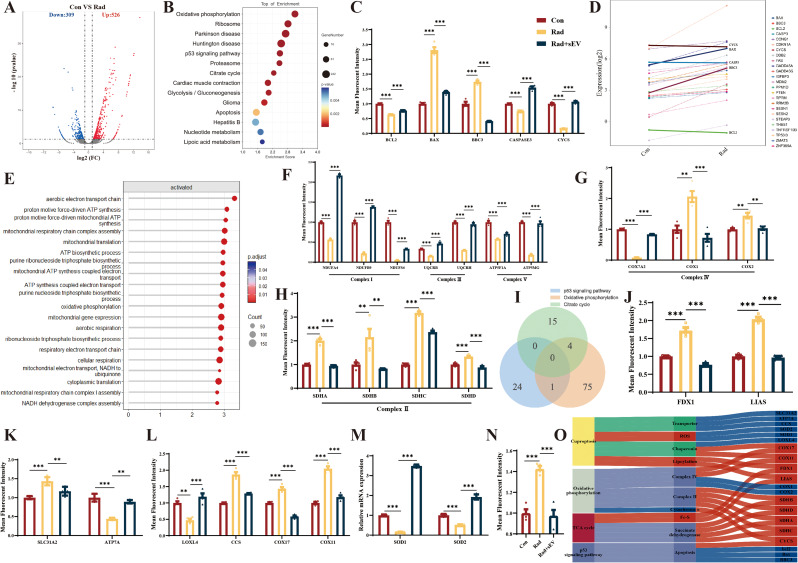
Methods: A radiation-induced cardiac organoid injury model was established by using X-ray radiation, and the optimal radiation dose of 20 Gy was determined by live/dead staining. After radiation, the cardiac organoids were treated with sEVs derived from UCMSCs, and energy metabolism, calcium transient changes and the ultrastructure of the organoids were assessed through Seahorse analysis, optical mapping and transmission electron microscopy, respectively. Confocal microscopy was used to observe the changes in mitochondrial ROS and mitochondrial membrane potential (ΔΨm). Furthermore, real-time quantitative PCR was used to verify the RNA-seq results.

Results: After X-ray radiation, the mortality of cardiac organoids significantly increased, energy metabolism decreased, and calcium transients changed. We also observed that the mitochondrial structure of cardiac organoids was disrupted and that ΔΨm was decreased. These effects could be inhibited by sEVs treatment. sEVs may protect against radiation-induced cardiac organoid injury by regulating oxidative phosphorylation and the p53 signaling pathway.

Conclusion: sEVs derived from UCMSCs can be used as a potential therapeutic strategy for radiation-induced heart disease.

Keywords: Cardiac organoids; Mesenchymal stem cells; Mitochondrial function; Radiation-induced heart disease; Small extracellular vesicles.


Conflict of interest statement

Declarations. Ethics approval and consent to participate: The iPSCs used in our research was obtained from CORIELL Institute (GM25256, USA), which appropriate consent was obtained at the time of biospecimen collection. Clinical-grade UCMSCs was obtained from China Medical Management Consulting (Beijing) LTD. CO. Human umbilical cords were provided by women who gave birth at Yantai Laiyang Central Hospital (Yantai, China). The collection of umbilical cords and the development of mesenchymal stem cells were reviewed and approved by Laiyang Central Hospital of Yantai City on November 14, 2023, with approval number of 2023-002-001. Title of the approved project was “Umbilical cord collection and development of mesenchymal stem cells”. Written informed consent was obtained from the donors participating in the study. All procedures that involved human samples were approved by the ethics committee of Academy of Military Medical Sciences (AF/SC-08/02.425). Title of the approved project was “Experimental study on intervention effect of mesenchymal stem cells derived small extracellular vesicles on radiation-induced cardiac organoid injury”. All procedures that involved animals were approved by the Institutional Animal Care and Use Committee of the Laboratory Animal Center of Academy of Military Medical Sciences (IACUC-DWZX-2023-532). Title of the approved project was “Study on protective effect of stem cells derived small extracellular vesicles on radiation-induced heart damage in mice”. Consent for publication: Not applicable. Competing interests: The authors declare no competing financial interest.

Figures

Fig. 1
Fig. 1
Preparation of cardiac organoids. (A) Schematic of the preparation of cardiac organoids, which started beating on Day 12. By Figdraw. (B) Time course of cardioid formation. Bright-field images of spheroids showing the increase in size and degree of cardioid formation over time. Scale bars: 200 μm. (C) Immunostaining for the CM markers cTnT and MYL7, the endocardium markers PECAM1 and CDH5, the ventricular-specific marker IRX4, and the mesenchymal marker vimentin (VIM) in cardioids on Day 30. Scale bars: 100 μm
Fig. 2
Fig. 2
Identification of UCMSCs-sEVs and optimization of the therapeutic concentration. (A) NTA revealed the particle size distribution of UCMSCs-sEVs. (B) TEM image demonstrating the bilayer membrane structure of sEVs with a diameter of approximately 100 nm. Scale bar: 100 nm. (C) WB image showing the expression of UCMSCs-sEV markers, including TSG101, CD63, CD9, and CD90. (D) Optimization of the therapeutic concentration of UCMSCs-sEVs for radiation-induced cardiac organoid injury. A concentration of 2 × 1010 particles/ml resulted in a notable decrease in red fluorescence intensity. Scale bar: 100 μm
Fig. 3
Fig. 3
UCMSCs-sEVs improved the energy metabolic capacity of radiation-injured cardiac organoids. (A) Changes in the OCR in cardiac organoids after radiation and UCMSCs-sEVs treatment. (B) Statistical analysis of maximal respiration, basal respiration, spare respiratory capacity, ATP production, non-mitochondrial oxygen consumption and proton leak. (C) Changes in coupling efficiency and percentage in spare respiratory capacity. (D) ECAR levels in cardiac organoids after radiation and UCMSCs-sEVs treatment. (E) Glycolysis levels, maximum glycolytic capacity and total glycolytic capacity reserve in cardiac organoids after radiation injury and UCMSCs-sEVs treatment. (F) Nonglycolytic acidification value and glycolytic capacity reserve after radiation and UCMSCs-sEVs treatment. n = 3
Fig. 4
Fig. 4
Ultrastructural features of cardiac organoids after radiation and UCMSCs-sEVs treatment. (A) TEM was used to observe the ultrastructure of cardiac organoids, including myofiber alignment and mitochondria. Z = Z line, ID = intercalated disc, S = sarcomere, M = mitochondria, G = glycogen. Scale bar: 1 μm. (B) Representative images of mitochondrial autophagy and lysosomal activity in cardiac organoids post-irradiation. Mp = Mitophagy, L = lysosomal. Scale bar: 500 nm. (C) Mitochondrial changes observed via TEM. Reductions in mitochondrial cristae (yellow arrow), mitochondrial vacuolation (blue arrow), and enhanced lysosomal activity were shown in radiation-injured cardiac organoids. Scale bar: 500 nm. (D) Myofibril length of cardiac organoids after radiation and UCMSCs-sEVs treatment. n = 6. Scale bar: 1 μm
Fig. 5
Fig. 5
UCMSCs-sEVs treatment improved the mitochondrial membrane potential and decreased oxidative stress in radiation-injured cardiac organoids. At 24 h and 48 h after radiation and UCMSCs-sEVs treatment, the cardiac organoids were stained with TMRM. (A) Representative confocal microscopy images showing the ΔΨm, and (B) the histogram shows the quantitative results. At 12 h and 24 h after radiation and UCMSCs-sEVs treatment, (C) confocal microscopy was used to detect mitochondrial ROS production (red fluorescence), and (D) the histogram shows the quantitative results. n = 10. Scale bar: 100 μm
Fig. 6
Fig. 6
UCMSCs-sEVs restored the normal calcium transients of radiation-injured cardiac organoids. The calcium signal was detected by using optical mapping. (A) Representative images showing the calcium signaling activation time and (B) the histogram showing the quantitative results. (C) Representative images showing the direction of calcium signal conduction and the degree of dispersion with the vertical coordinate of conduction time. (D) Quantitative results of the discrete degree of calcium transients and the calcium signaling dispersion index. (E) Representative images showing calcium signal conduction waveforms and (F-I) the histogram shows the quantitative results of physiological parameters of the calcium signal. (J) Schematic of the change in ADP 30/80. The vertical coordinate is time in seconds. (K) Quantitative results for ADP 30/80. (L) Changes in ADP 90. n = 10
Fig. 7
Fig. 7
The therapeutic effects of UCMSCs-sEVs on radiation-induced cardiac damage in mice. Changes in myocardial enzyme profile on day 14 (A) and day 28 (B) after radiation. n = 5. (C) Representative images of echocardiography at day 42 after radiation. Statistical results of LVEF (D) and LVFS (E). n = 5. (F) Electron microscopy images of mouse apical heart tissue. Yellow arrow: reduced mitochondrial cristae. Scale bar: 1 μm. Representative images of mouse heart HE staining (G) and Masson staining (H). Expression of inflammation related genes (I) and fibrosis related genes (J) in mouse heart. n = 4
Fig. 8
Fig. 8
The potential mechanism of action of UCMSCs-sEVs in radiation-induced cardiac organoid injury. (A) Volcano map of differentially expressed genes. There were a total of 835 DEGs, 526 of which were upregulated and 309 of which were downregulated. (B) KEGG enrichment analysis of RNA-seq data 48 h post-radiation. (C) Caspase 3-related apoptotic genes in the p53 signaling pathway. (D) Differential gene expression in the p53 signaling pathway. (E) GO enrichment was used to analyze gene function. (F) Complex I, complex III and complex V involved in oxidative phosphorylation. (G) Complex IV involved in oxidative phosphorylation. (H) Succinate dehydrogenase and complex II involved in the TCA cycle and oxidative phosphorylation. (I) Venn diagram showing the p53 signaling pathway with overlapping oxidative phosphorylation genes, such as CYCS; the TCA cycle with overlapping oxidative phosphorylation genes, such as SDHA, SDHB, SDHC and SDHD. (J) Lipoylated genes in the TCA cycle. (K) Expression of copper ion transport proteins (SLC31A and ATP7A). (L) The expression of copper-dependent monoamine oxidase (LOXL4), a copper chaperone for superoxide dismutase (CCS), the cytochrome c oxidase copper chaperone COX17 and the cytochrome c oxidase copper chaperone COX11. (M) The expression of superoxide dismutase. (N) The expression of the cytochrome C oxidase 1. (O) Sankey diagram describing the potential mechanisms of X-ray-induced radiation damage in cardiac organoids. n = 4

References

    1. Wang H, Wei J, Zheng Q, et al. Radiation-induced heart disease: a review of classification, mechanism and prevention. Int J Biol Sci. 2019;15(10):2128–38. 10.7150/ijbs.35460. - PMC - PubMed
    1. Lee PJ, Mallik R. Cardiovascular effects of radiation therapy: practical approach to radiation therapy-induced heart disease. Cardiol Rev. 2005;13(2):80–6. 10.1097/01.crd.0000131188.41589.c5. - PubMed
    1. Donnellan E, Phelan D, McCarthy CP, et al. Radiation-induced heart disease: a practical guide to diagnosis and management. Cleve Clin J Med. 2016;83(12):914–22. 10.3949/ccjm.83a.15104. - PubMed
    1. Boero IJ, Paravati AJ, Triplett DP, et al. Modern Radiation Therapy and Cardiac outcomes in breast Cancer. Int J Radiat Oncol Biol Phys. 2016;94(4):700–8. 10.1016/j.ijrobp.2015.12.018. Epub 2015 Dec 17. - PubMed
    1. Boekel NB, Schaapveld M, Gietema JA, et al. Cardiovascular Disease Risk in a large, Population-based cohort of breast Cancer survivors. Int J Radiat Oncol Biol Phys. 2016;94(5):1061–72. Epub 2015 Dec 14. - PubMed

MeSH terms