Review
. 2025 Jan;117(1):e2400096.
doi: 10.1111/boc.202400096. Epub 2024 Dec 20.

Phosphoinositide signaling in the nucleus: Impacts on chromatin and transcription regulation

Affiliations
Review

Phosphoinositide signaling in the nucleus: Impacts on chromatin and transcription regulation

Nesrine Hifdi et al. Biol Cell. 2025 Jan.

Abstract

Phosphoinositides also called Polyphosphoinositides (PPIns) are small lipid messengers with established key roles in organelle trafficking and cell signaling in response to physiological and environmental inputs. Besides their well-described functions in the cytoplasm, accumulating evidences pointed to PPIns involvement in transcription and chromatin regulation. Through the description of previous and recent advances of PPIns implication in transcription, this review highlights key discoveries on how PPIns modulate nuclear factors activity and might impact chromatin to modify gene expression. Finally, we discuss how PPIns nuclear and cytosolic metabolisms work jointly in orchestrating key transduction cascades that end in the nucleus to modulate gene expression.


Conflict of interest statement

The authors declare no conflicts of interest.

Figures
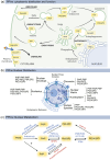
FIGURE 1
FIGURE 1
Cytoplasmic and nuclear PPIns metabolism. (a) PPIns act as second messengers of transduction cascades and organelle trafficking at the plasma membrane and endomembranes. PPIns mark organelles identity allowing endomembrane dynamics to ensure key process such as endocytosis, exocytosis, and autophagy. The major PPIns organelle signature are indicated in yellow. At the plasma membrane, PI(4,5)P2 and PI(3,4)P2 are the most abundant PPIns. The PI(4,5)P2 and PI(3,4,5)P3 interconversion cycle is controlled by the Class‐I PI3Ks and PTEN, a major signaling cascades involved in cell growth. Generation of PI(3,4)P2 mainly by the Class‐II PI3K occurs on nascent endocytic carriers such as clathrin‐coated pits before conversion to PI3P in early endosomal compartments. PI3P conversion to PI(3,5)P2 marks the late endosomes and the lysosomes. PI(3,5)P2 and PI3P are also involved in the genesis of autophagosomes, a degradative organelle that emerges from the endoplasmic reticulum, the major reservoir of PtdIns. The PI4P is highly present at the Golgi apparatus, a compartment where main exocytosis vesicles emerge. The PI4P‐positive vesicles emanating from the Golgi could also fuel additional cell membranes compartment such as the ER through anterograde and retrograde trafficking. (b) Subnuclear distribution of PPIns in different nuclear compartments. Note that PPIns such as PI5P and PI(4,5)P2 are redundant in many compartments suggesting multiple nuclear functions. (c) PPIns (polyphosphoinositide)‐kinases (in blue) and phosphatases (in red) isoforms that are active for PPIns interconversion in the nucleus. Two distinct nomenclatures are indicated.
FIGURE 2
FIGURE 2
PPIns regulate the transcriptional activity of SF‐1. The steroidogenic factor 1(SF‐1) and the liver hormone receptor (LHR‐1) belong to the NR5A family of orphan nuclear receptors. PI(4,5)P2 binds to the ligand binding domain of SF‐1 (LBD) through its acyl chains. PI(4,5)P2 is converted to PI(3,4,5)P3 by the inositol polyphosphate multikinase (IPMK), resulting in SF‐1 transcriptional activation. The phosphatase and tensin homolog deleted on chromosome 10 (PTEN) acts antagonistically to IPMK to shut down SF‐1 transcriptional activity. The inositol headgroup of PI(3,4,5)P3 that faces the cytosol may recruit additional, yet unknown, nuclear transcription factors.
FIGURE 3
FIGURE 3
PI5P links transcription factors to chromatin. The transcription factors ING‐2, TAF‐3, and UHRF1 are regulated by PI5P binding. The PHD domain is present in all proteins, although it was involved in the interaction with PI5P only for ING2 and TAF3. A stretch of polybasic residues (illustrated by a yellow star) is present in the C‐terminal part of both ING2 and TAF3 PHD and is required for PI5P binding. The PHD domain is also involved in the recognition of H3K4me3, associated with active transcription. PI5P enhances the recruitment of ING2 and TAF3 on chromatin via binding of the PHD to H3K4me3. ING2 binds to the Sin3 deacetylase complex that leads to transcriptional repression of p53‐dependent genes by histone deacetylation. TAF3 is a general transcription factor, which forms with TRF3 the pre‐initiation complex required for muscle‐specific gene activation. UHRF1 binds to PI5P via a polybasic region (PBR) independently of the PHD domain. The binding of PI5P to the PBR induces a conformational change that frees the TTD domain from the PBR‐containing region, allowing the TTD to bind to H3K9me3. This configuration triggers the recruitment of the DNA methyltransferase 1 (DNMT1) that interacts with UHRF‐1 to methylate DNA during S‐phase or promotes the repression of transcription of tumor suppressor genes. The hydrophobic diacylglycerol tail of PPIns should be protected from the hydrophilic environment of nucleoplasm, which implies its association with proteins containing hydrophobic pockets such as SF‐1 or PITPs. LZL, leucine zipper‐like domain; PHD, plant homeodomain zinc finger; RING, really interesting new gene domain; SRA, SET and RING‐associated domain; TTD, tandem tudor domain; UBL, ubiquitin‐like domain. Image created with Biorender.
FIGURE 4
FIGURE 4
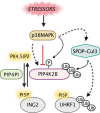
PI5P nuclear levels are modulated by stressors. The antagonizing enzymes PIP4K2B and PIP4PI (PI(4,5)P2‐4‐phosphatase I) regulate nuclear PI5P levels. PIP4K2B is inhibited through direct phosphorylation by p38‐MAPK that is stimulated by genotoxic stress. In addition, in response to genotoxic stress, a fraction of PIP4PI was shown to translocate to the nucleus through an unknown mechanism to dephosphorylate PI(4,5)P2. Alternatively, p38MAPK activates the ubiquitination of PIP4K2B and potentially UHRF‐1 through activation of the SPOP (speckle‐type POZ domain protein)‐Cullin3 (Cul3) E3 ubiquitin ligase complex.
FIGURE 5
FIGURE 5
Cytosolic and nuclear PPIns regulate the Hippo pathway. The Hippo pathway is activated at the plasma membrane by a variety of upstream signals, such as ligands that stimulate tyrosine‐kinase receptors (RTK) or G‐protein‐coupled receptors (GPCR), cell density, or stress signals. The Hippo pathway is activated by phosphorylation cascades that promote YAP/TAZ cytoplasmic sequestration and degradation. When dephosphorylated, YAP/TAZ translocate to the nucleus and associate with TEAD transcription factors to activate target gene expression. PPIns regulate the Hippo pathway at different levels. PI4P, transported from the endoplasmic reticulum (ER) to the plasma membrane by the Phosphatidylinositol transfer proteins (PITPs), restrains the Hippo pathway through an unknown mechanism that may involve NF2, an upstream regulator of the Hippo pathway. Upon osmotic stress, a plasma membrane pool of PI4P is converted to PI(4,5)P2 by the PIP5KI that is stimulated by ARF6. This leads to the recruitment of NF2 to the plasma membrane, its binding to PI(4,5)P2, and the activation of the Hippo pathway. In the cytoplasm, MST‐1/2 phosphorylates and inhibits PI5P4K/PIP4K2, the PI‐kinase that converts PI5P to PI(4,5)P2, which leads to an increase of a cytosolic PI5P pool. PI5P enhances the association of LATS1 to its chaperone MOBP1, triggering the inhibitory phosphorylation of YAP/TAZ. In the nucleus, PI(4,5)P2 and PI(3,4,5)P3 interact with YAP/TAZ, enhance its association with TEAD promoting the transcription of their target genes. This nuclear regulation requires a local production of PI(4,5)P2 and PI(3,4,5)P3 by the PI4P‐5 kinase (PIPKIα) and the PI3‐kinase IPMK. The hydrophobic diacylglycerol tail of PPIns should be protected from the hydrophilic environment of nucleoplasm, which implies its association with proteins containing hydrophobic pockets. To simplify, it was not drawn. LATS1/2, large tumor suppressor kinase 1/2; MOB1A/B, MOB kinase activator 1 A/B; MST1/2, mammalian STE20‐like kinase 1/2; TEAD, transcriptional enhanced associated domain transcription factor; YAP‐1, yes‐associated protein 1; ZAP, WW‐domain‐containing transcription regulator. Image created with Biorender.

References

    1. Andjelkovic, M. , Alessi, D.R. , Meier, R. , Fernandez, A. , Lamb, N.J.C. , Frech, M. , Cron, P. , Cohen, P. , Lucocq, J.M. & Hemmings, B.A. (1997) Role of translocation in the activation and function of protein kinase B. Journal of Biological Chemistry, 272, 31515–31524. Available from: 10.1074/jbc.272.50.31515 - DOI - PubMed
    1. Arrowsmith, C.H. & Schapira, M. (2019) Targeting non‐bromodomain chromatin readers. Nature Structural & Molecular Biology, 26, 863–869. Available from: 10.1038/s41594-019-0290-2 - DOI - PubMed
    1. Ashlin, T.G. , Blunsom, N.J. & Cockcroft, S. (2021) Courier service for phosphatidylinositol: PITPs deliver on demand. Biochimica et Biophysica Acta (BBA)—Molecular and Cell Biology of Lipids, 1866, 158985. Available from: 10.1016/j.bbalip.2021.158985 - DOI - PMC - PubMed
    1. Asmamaw, M.D. , He, A. , Zhang, L.‐R. , Liu, H.‐M. & Gao, Y. (2024) Histone deacetylase complexes: structure, regulation and function. Biochimica et Biophysica Acta (BBA)—Reviews on Cancer, 1879, 189150. Available from: 10.1016/j.bbcan.2024.189150 - DOI - PubMed
    1. Bahmanyar, S. (2015) Spatial regulation of phospholipid synthesis within the nuclear envelope domain of the endoplasmic reticulum. Nucleus, 6, 102–106. Available from: 10.1080/19491034.2015.1010942 - DOI - PMC - PubMed

LinkOut - more resources