. 2025 Mar;66(3):914-928.
doi: 10.1111/epi.18234. Epub 2024 Dec 21.

Rare dysfunctional SCN2A variants are associated with malformation of cortical development

Affiliations

Rare dysfunctional SCN2A variants are associated with malformation of cortical development

Jérôme Clatot et al. Epilepsia. 2025 Mar.

Abstract

Objective: SCN2A encodes the voltage-gated sodium (Na+) channel α subunit NaV1.2, which is important for the generation and forward and back propagation of action potentials in neurons. Genetic variants in SCN2A are associated with a spectrum of neurodevelopmental disorders. However, the mechanisms whereby variation in SCN2A leads to disease remains incompletely understood, and the full spectrum of SCN2A-related disorders may not be fully delineated.

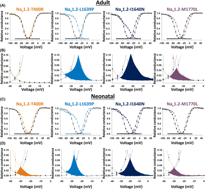
Methods: Here, we identified seven de novo heterozygous variants in SCN2A in eight individuals with developmental and epileptic encephalopathy (DEE) accompanied by prominent malformation of cortical development (MCD). We characterized the electrophysiological properties of Na + currents in human embryonic kidney (HEK) cells transfected with the adult (A) or neonatal (N) isoform of wild-type (WT) and variant NaV1.2 using manual and automated whole-cell voltage clamp recording.

Results: The neonatal isoforms of all SCN2A variants studied exhibit gain of function (GoF) with a large depolarized shift in steady-state inactivation, creating a markedly enhanced window current common across all four variants tested. Computational modeling demonstrated that expression of the NaV1.2-p.Met1770Leu-N variant in a developing neocortical pyramidal neuron results in hyperexcitability.

Significance: These results support expansion of the clinical spectrum of SCN2A-related disorders and the association of genetic variation in SCN2A with MCD, which suggests previously undescribed roles for SCN2A in fetal brain development.

Keywords: SCN2A; Nav1.2; developmental and epileptic encephalopathy; malformation of cortical development; voltage‐gated sodium channel.


Conflict of interest statement

None of the authors has any conflict of interest to disclose.

Figures

FIGURE 1
FIGURE 1
Magnetic resonance imaging (MRI) of the brain of patients in the cohort with SCN2A encephalopathy and malformation of cortical development. (A) Patient 1. T2 axial and coronal MRI images at age 6 weeks show diminutive size of the intraorbital and prechiasmatic optic nerves (at left; right optic nerve indicated by arrowhead); marked cortical and subcortical atrophy with extensive abnormality of gyrification. (B) Patient 2. T2 axial MRI at age 3 weeks shows extensive malformation consistent with polymicrogyria (PMG) of the right frontal and parietal lobes. (C) Patient 3. T2 axial and coronal MRI at age 7 months shows extensive malformation consistent with PMG throughout the neocortex, predominantly affecting both parietal lobes. (D) Patient 4. Axial T2 fluid‐attenuated inversion recovery (FLAIR) and coronal T2 MRI images illustrate diffuse PMG. (E) Patient 5. Coronal and axial T2 and coronal T2 FLAIR MRI images at age 3 years showing marked cortical and subcortical atrophy with extensive dysgyria and areas of extensive malformation with presumptive PMG involving the bilateral frontal and parietal lobes. Note as well the appearance of cerebellar atrophy on the T2 coronal image. (F) Patient 6. Coronal, sagittal, and axial T2 MRI images at age 13 days showing simplified gyral pattern of the left hemisphere consistent with PMG, mostly involving the temporo‐parieto‐insular region (white arrows). (G) Patient 7. Coronal and axial T2 MRI images at age 1 day showing PMG of the left hemisphere, mostly affecting the frontal and temporal lobes (white arrows). (H) Schematic representation of Nav1.2 indicating the locations of the A263V variant in domain I (DI) S4 (red circle), Thr400Arg in the DI S5–6 extracellular linker (orange), I1488V in the DII–DII linker (green), L1614P in the extracellular DIII S3–4 linker (raspberry), Leu1639Pro (light blue) and Ile1640Asn (dark blue) in the DIV voltage‐sensing region, and Met1770Leu variants (purple) in DIV S6. (I) Topology diagram of Nav1.2 showing the predicted location of the affected residues.
FIGURE 2
FIGURE 2
Current–voltage relationship of sodium currents recorded from human embryonic kidney (HEK)‐293 cells expressing wild‐type (WT) SCN2A vs variants associated with early‐onset epilepsy and malformation of cortical development. (A) Representative families of currents recorded from WT and variants SCN2A‐p.Thr400Arg (T400R), Leu1639Pro (L1639P), Ile1640Asn (I1640N), and Met1770Leu (M1770L) adult (A; top) and neonatal (N; bottom) isoforms. (B) Current–voltage (I–V) relationships. Note that only SCN2A‐p.Ile1640Asn displayed significant loss in peak current density compared to WT.
FIGURE 3
FIGURE 3
Voltage dependence of activation and steady‐state inactivation recorded from human embryonic kidney (HEK)‐293 cells expressing wild‐type (WT) SCN2A vs variants associated with early‐onset epilepsy and malformation of cortical development. (A) Shown are the voltage dependence of activation and steady‐state inactivation for the WT SCN2A adult isoform vs variants SCN2A‐p.Thr400Arg (T400R; orange), Ile1640Asn (I1640N; dark blue), and Met1770Leu (M1770L; purple). (B) Insets from (A) showing the window currents. (C) Shown are the voltage dependence of activation and steady‐state inactivation for the WT SCN2A neonatal isoform vs variants SCN2A‐p.Thr400Arg (T400R; orange), Leu1639Pro (L1639P; light blue), Ile1640Asn (I1640N; dark blue), and Met1770Leu (M1770L; purple). (D) Insets from (C) showing the window currents. Note that window currents are significantly augmented for the neonatal isoform of all four variants.
FIGURE 4
FIGURE 4
Automated patch‐clamp recordings from human embryonic kidney (HEK)‐293 cells expressing wild‐type (WT) SCN2A vs variants associated with early‐onset epilepsy and malformation of cortical development. (A) Average normalized whole‐cell current families of SCN2A‐WT (black), T400R (orange), I1640N (dark blue), and M1770L (purple) in the adult (top) and neonatal (bottom) splice isoforms. Summary data showing deviation from WT of each variant for (B) whole‐cell current density, (C) voltage dependence of inactivation, and (D) window current. Circles represent data recorded from the adult splice isoform, whereas squares represent data from the neonatal splice isoform. Open symbols represent data from each recorded cell, and solid symbols show mean ± 95% confidence interval. Asterisks (*) denote p < .05.
FIGURE 5
FIGURE 5
Simulated neuronal excitability in a neocortical pyramidal cell model expressing wild‐type (WT; black) or M1770L variant (purple). (A) Voltage dependence of activation and inactivation for the adult (top) and neonatal (bottom) isoforms of Nav1.2‐WT and Nav1.2‐M1770L. (B) Firing of the model WT (black) and M1770L cell (purple) in response to stimulation using a 500 ms step stimulation (top) or a synaptic stimulation (bottom). F–I curves (left) and example traces (right) of modeled firing in response to 0.8 and 1.6 nA current injections (top) and different multiplications of the synaptic stimulation (bottom).

References

    1. Meisler MH, Hill SF, Yu W. Sodium channelopathies in neurodevelopmental disorders. Nat Rev Neurosci. 2021;22(3):152–166. - PMC - PubMed
    1. Wolff M, Brunklaus A, Zuberi SM. Phenotypic spectrum and genetics of SCN2A‐related disorders, treatment options, and outcomes in epilepsy and beyond. Epilepsia. 2019;60(Suppl 3):S59–S67. - PubMed
    1. Thompson CH, Potet F, Abramova TV, DeKeyser J‐M, Ghabra NF, Vanoye CG, et al. Epilepsy‐associated SCN2A (NaV1.2) variants exhibit diverse and complex functional properties. J Gen Physiol. 2023;155(10):e202313375. - PMC - PubMed
    1. Berg AT, Thompson CH, Myers LS, Anderson E, Evans L, Kaiser AJE, et al. Expanded clinical phenotype spectrum correlates with variant function in SCN2A‐related disorders. Brain. 2024;awae125:2761–2774. - PMC - PubMed
    1. Smith RS, Kenny CJ, Ganesh V, Jang A, Borges‐Monroy R, Partlow JN, et al. Sodium Channel SCN3A (NaV1.3) regulation of human cerebral cortical folding and Oral motor development. Neuron. 2018;99(5):905–913.e7. - PMC - PubMed

Substances