. 2025 Jan 8;147(1):488-497.
doi: 10.1021/jacs.4c11612. Epub 2024 Dec 21.

Parallel Proteomic and Transcriptomic Microenvironment Mapping (μMap) of Nuclear Condensates in Living Cells

Affiliations

Parallel Proteomic and Transcriptomic Microenvironment Mapping (μMap) of Nuclear Condensates in Living Cells

Steve D Knutson et al. J Am Chem Soc. .

Abstract

Cellular activity is spatially organized across different organelles. While several structures are well-characterized, many organelles have unknown roles. Profiling biomolecular composition is key to understanding function but is difficult to achieve in the context of small, dynamic structures. Photoproximity labeling has emerged as a powerful tool for mapping these interaction networks, yet maximizing catalyst localization and reducing toxicity remains challenging in live cell applications. Here, we disclose a new intracellular photocatalyst with minimal cytotoxicity and off-target binding, and we utilize this catalyst for HaloTag-based microenvironment-mapping (μMap) to spatially catalog subnuclear condensates in living cells. We also specifically develop a novel RNA-focused workflow (μMap-seq) to enable parallel transcriptomic and proteomic profiling of these structures. After validating the accuracy of our approach, we generate a spatial map across the nucleolus, nuclear lamina, Cajal bodies, paraspeckles, and PML bodies. These results provide potential new insights into RNA metabolism and gene regulation while significantly expanding the μMap platform for improved live-cell proximity labeling in biological systems.


Conflict of interest statement

The authors declare the following competing financial interest(s): DWCM declares an ownership interest in the company Dexterity Pharma LLC, which has commercialized materi-als used in this work.

Figures
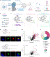
Figure 1.
Figure 1.
Optimizing a live-cell μMap workflow. (a) μMap using a HaloTag-fused POI and an alkyl chloride-functionalized photocatalyst to label proximal protein and/or RNA molecules. (b) μMap proximity labeling workflow with HaloTag-expressing cells. (c) Structures of μMap catalysts. (d) Immunofluorescence and (e) proteomic evaluation of photolabeling in live cells using version 1 (v1) and v2 μMap Ir catalysts in HEK293T cells. Nuclei stained with Hoechst (blue), mitochondria COXIV (green), and biotinylation stained with streptavidin AF555 (red). Scale bar, 10 μm. Volcano plots depict mitochondrial proteins labeled in red and additional top hits are labeled. (f) Human Protein Atlas subcellular annotation of significant interactors (log2FC > 2, p < 0.05) for both catalysts.
Figure 2.
Figure 2.
Nuclear proteomic and transcriptomic labeling. (a) Immunofluorescence validation of photolabeling in HaloTag-NLS cells. Nuclei stained with Hoechst (blue), anti-HaloTag (green), and biotinylation stained with streptavidin AF555 (red). Scale bar, 10 μm. (b) Quantitative proteomics in HaloTag-NLS vs wild-type cells with nuclear-annotated proteins (blue). (c) Human Protein Atlas annotation and (d) GO enrichment of interactors. (e) μMap-seq experimental workflow for RNA photolabeling, enrichment, and sequencing. (f,g) μMap-seq of nuclear transcripts with known nuclear transcripts in purple. Several of the top enriched transcripts are also labeled in the volcano plot, with well-known cytosolic transcripts (MEG3, DANCR, TINCR and 7SL) at the bottom in gray.
Figure 3.
Figure 3.
Nucleolar μMap. (a) Immunofluorescence validation of nucleolar photolabeling. Nuclei stained with Hoechst (blue), anti-HaloTag (green), and biotinylation stained with streptavidin AF555 (red). Scale bar, 10 μm. (b) Quantitative proteomics in HaloTag-NIK vs wild-type cells. Ribosomal protein subunits (orange) and ribosomal biogenesis machinery (green), novel nucleolar interactors (red). (c) Human Protein Atlas annotation and (d) GO enrichment analysis of nucleolar interactors. (e,f) μMap-seq of nucleolar transcripts. Several top hits are annotated and data points colored by transcript type.
Figure 4.
Figure 4.
μMap profiling across nuclear locations. (a) Immunofluorescence validation of photolabeling with each stable cell line. Nuclei stained with Hoechst (blue), anti-HaloTag (green), and biotinylation stained with streptavidin AF555 (red). Scale bars, 10 μm. (b–e) Quantitative μMap proteomics in each HaloTag cell line vs wild-type cells. Highlighted proteins denote hits with RNA or DNA-related functions.
Figure 5.
Figure 5.
Comparative gene ontology (GO) enrichment from μMap proteomic profiling across the nucleolus, nuclear lamina, cajal bodies, paraspeckles, and PML bodies. Red heatmap scores highlight enriched biological processes and molecular functions, with intensity correlating to enrichment significance (−log10 P).
Figure 6.
Figure 6.
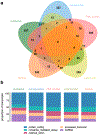
μMap-seq transcriptional profiling across subnuclear locations. (a) Transcript-level comparison of enriched RNAs between locations. (b) Stacked bar graph displaying enriched transcript types.

References

    1. Sabari BR; Dall’Agnese A; Young RA Biomolecular condensates in the nucleus. Trends Biochem. Sci 2020, 45 (11), 961–977. - PMC - PubMed
    1. Boeynaems S; Chong S; Gsponer J; Holt L; Milovanovic D; Mitrea DM; Mueller-Cajar O; Portz B; Reilly JF; Reinkemeier CD; et al. Phase separation in biology and disease; current perspectives and open questions. J. Mol. Biol 2023, 435 (5), 167971. - PMC - PubMed
    1. Mathieu C; Pappu RV; Taylor JP Beyond aggregation: Pathological phase transitions in neurodegenerative disease. Science 2020, 370 (6512), 56–60. - PMC - PubMed
    1. Spannl S; Tereshchenko M; Mastromarco GJ; Ihn SJ; Lee HO Biomolecular condensates in neurodegeneration and cancer. Traffic 2019, 20 (12), 890–911. - PubMed
    1. Geri JB; Oakley JV; Reyes-Robles T; Wang T; McCarver SJ; White CH; Rodriguez-Rivera FP; Parker DL Jr.; Hett EC; Fadeyi OO; et al. Microenvironment mapping via Dexter energy transfer on immune cells. Science 2020, 367 (6482), 1091–1097. - PMC - PubMed

LinkOut - more resources