. 2024 Dec 13;38(24):e70143.
doi: 10.1096/fj.202302453RRR.

Melatonin mitigates intervertebral disc degeneration by suppressing NLRP3 inflammasome activation via the EGR1/DDX3X pathway

Affiliations

Melatonin mitigates intervertebral disc degeneration by suppressing NLRP3 inflammasome activation via the EGR1/DDX3X pathway

Kangcheng Zhao et al. FASEB J. .

Abstract

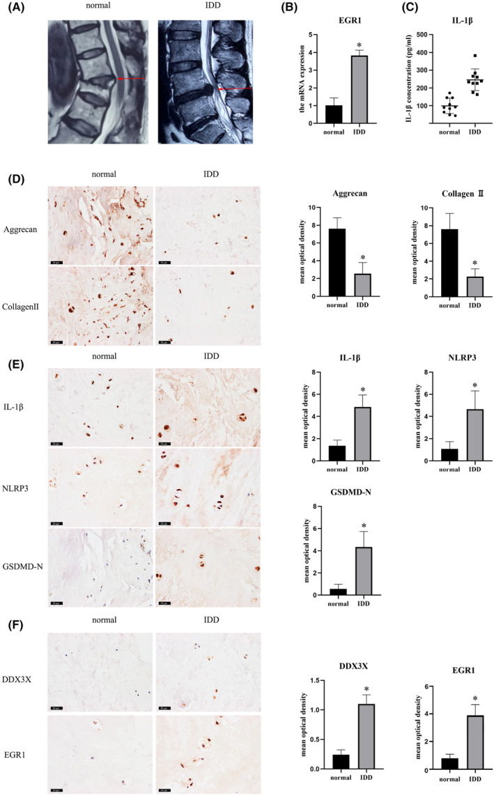
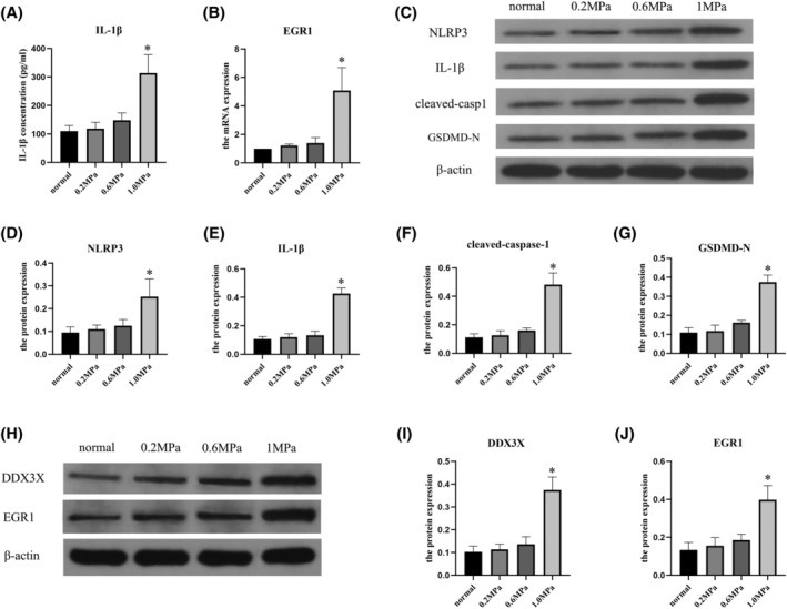
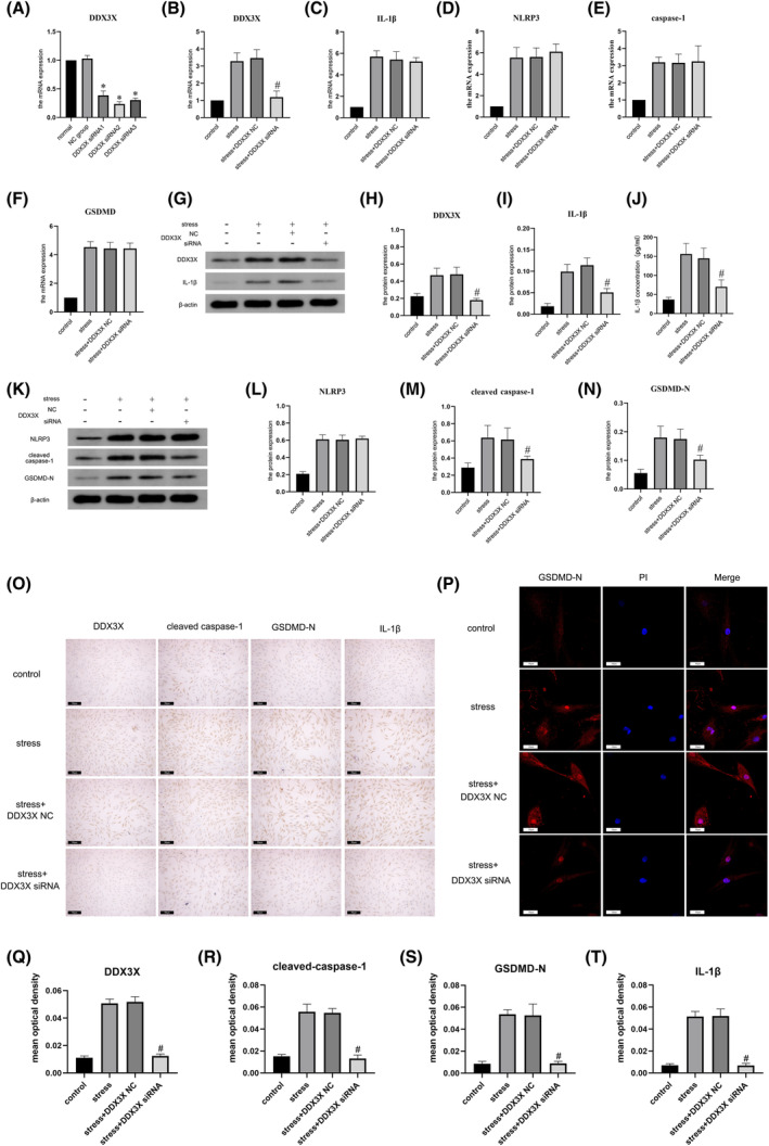
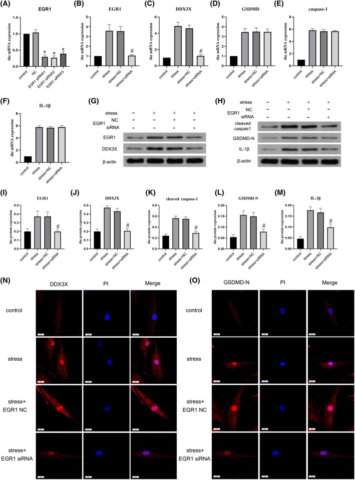
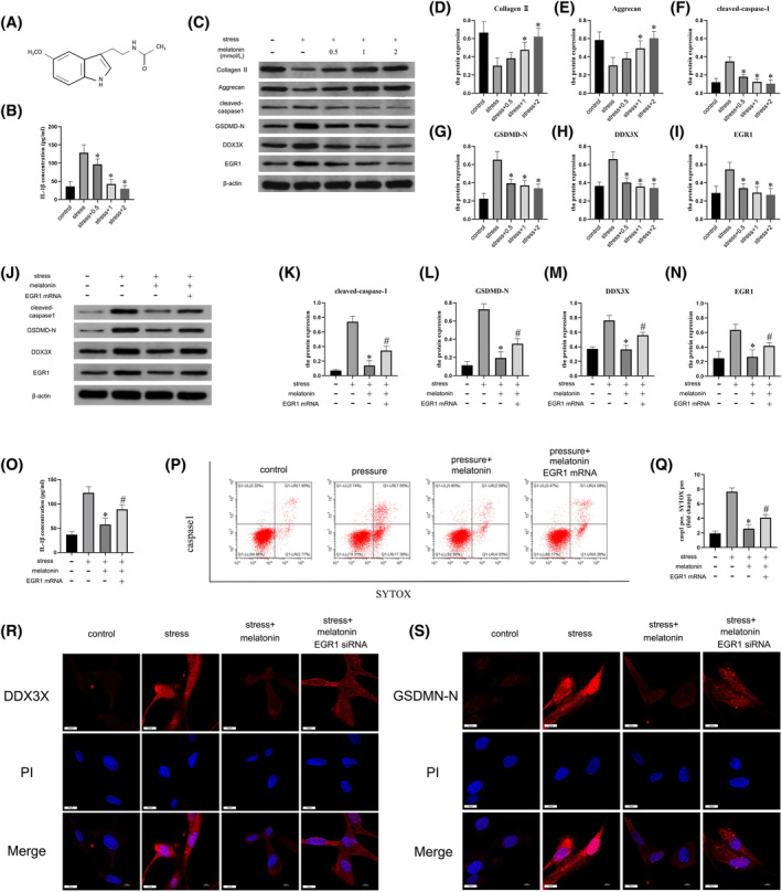
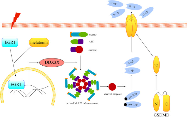
Intervertebral disc degeneration (IVDD), is one of the leading causes of low back pain. Inflammation is considered to be the main pathophysiological process of IVDD. The nucleotide-binding domain and leucine-rich pyrin domain containing 3 (NLRP3) inflammasome-mediated inflammatory responses are critically involved in the progression of IVDD. Melatonin is known for its anti-inflammatory and antioxidant effects. However, little is known about the potential effects of melatonin in the pathological process of IVDD. We found that the expression of EGR1, DDX3X, and NLRP3 inflammasome increased and extracellular matrix (ECM) degraded in IVDD. With the application of EGR1 siRNA, the expression of DDX3X and the activation of NLRP3 inflammasome were inhibited in stress-induced NP cells. DDX3X/NLRP3 was regulated on dependence of EGR1. Besides, the utility of melatonin mitigated the EGR1-induced overproduction of DDX3X and activation of NLRP3 inflammasome, thus protecting cells from pyroptosis and ECM degradation. In vivo, in a rat IVDD model, melatonin was found to be able to delay the development of IVDD by imageological and histological evaluation. In conclusion, our study demonstrated that melatonin prevented IVDD progression by regulating EGR1/DDX3X/NLRP3 axis. Our study provides insight into melatonin as a new target for therapeutic approaches for IVDD.

Keywords: DDX3X; EGR1; NLRP3 inflammasome; intervertebral disc degeneration; melatonin; nucleus pulposus.


Figures

FIGURE 1
FIGURE 1
The expression of EGR1 expression and NLRP3 inflammasome activation rise during IVDD development. (A) Representative MRI images of human normal and IVDD specimens. (B) RT‐PCR analysis of EGR1 in human normal and IVDD specimens. (C) ELISA analysis of IL‐1β in human normal and IVDD specimens. (D–F) Representative images of immunohistochemistry and quantitative analysis of collagen II, aggrecan, IL‐1β, NLRP3, GSDMD‐N, DDX3X, and EGR1 in human normal and IVDD specimens. Scale bar = 20 μm. Significant differences between groups are indicated as *p < .01 compared with the normal group.
FIGURE 2
FIGURE 2
Effect of mechanical stress in human NP cells in vitro. (A) ELISA analysis of IL‐1β in degenerated NP cells. (B) RT‐PCR analysis of EGR1 in degenerated NP cells. (C–G) Western blot and quantitative analysis of NLRP3, IL‐1β, cleaved caspase‐1, and GSDMD‐N in degenerated NP cells under stress stimulation. (H–J) Western blot and quantitative analysis of DDX3X and EGR1 in degenerated NP cells. Data are represented as the mean ± SD (n = 3). Significant differences between groups are indicated as *p < .01, compared with the control group.
FIGURE 3
FIGURE 3
DDX3X emerged as a pivotal factor in NLRP3 inflammasome activation in NP cells. (A) RT‐PCR analysis of knockdown efficiency of three different DDX3X siRNAs in NP cells. (B–F) RT‐PCR analysis of DDX3X, IL‐1β, NLRP3, caspase‐1, and GSDMD in NP cells under stress stimulation. (G–I) Western blotting and quantitative analysis of DDX3X and IL‐1β in NP cells. (J) ELISA analysis of the extracellular IL‐1β in NP cells. (K–N) Western blotting and quantitative analysis of NLRP3, caspase‐1, and GSDMD in NP cells under stress stimulation. (O) Representative images of immunohistochemistry of DDX3X, caspase‐1, GSDMD, and IL‐1β in NP cells under stress stimulation. Scale bar = 50 μm. (P) Representative images of immunofluorescence of GSDMD in NP cells. Scale bar = 10 μm. (Q–T) quantitative analysis of DDX3X, caspase‐1, GSDMD, and IL‐1β immunohistochemistry images in NP cells under stress stimulation. Data are represented as the mean ± SD (n = 3). Significant differences between groups are indicated as *p < .01, compared with the control group; # p < .01, compared with the NC siRNA group.
FIGURE 4
FIGURE 4
EGR1 was involved in the regulation of DDX3X expression in NP cells under stress stimulation. (A) RT‐PCR analysis of knockdown efficiency of three different EGR1 siRNAs in NP cells. (B–F) RT‐PCR analysis of EGR1, DDX3X, GSDMD, cleaved caspase‐1, and IL‐1β in NP cells under stress stimulation. (G, H) Western blotting of DDX3X, EGR1, GSDMD, cleaved caspase‐1, and IL‐1β in NP cells under stress stimulation. (I–M) Quantitative analysis of EGR1, DDX3X, cleaved caspase‐1, GSDMD‐N, and IL‐1β in NP cells detected by Western blot. (N, O) Representative images of immunofluorescence of DDX3X and GSDMD‐N in NP cells. Scale bar = 10 μm. Data are represented as the mean ± SD (n = 3). Significant differences between groups are indicated as *p < .01, compared with the control group; # p < .01, compared with the NC siRNA group.
FIGURE 5
FIGURE 5
Melatonin inhibited NLRP3 inflammasome activation and attenuated degeneration of NP cells through EGR1/DDX3X pathway. (A) Chemical structure formula of EGR1. (B) ELISA analysis of extracellular IL‐1β in NP cells treated with gradient concentrations of melatonin. (C–I) Western blot and quantitative analysis of collagen II, aggrecan, cleaved caspase‐1, GSDMD‐N, DDX3X, and EGR1 in NP cells. (J–N) Western blot and quantitative analysis of cleaved caspase‐1, GSDMD‐N, DDX3X, and EGR1 in NP cells. (O) ELISA analysis of extracellular IL‐1β in NP cells. (P, Q) The pyroptosis of NP cells was detected by flow cytometry assay. (R) Representative images of immunofluorescence of DDX3X in NP cells. Scale bar = 10 μm. (S) Representative images of immunofluorescence of NLRP3 inflammasome in NP cells. Scale bar = 10 μm. Data are represented as the mean ± SD (n = 3). Significant differences between groups are indicated as *p < .01, compared with the control group; # p < .01, compared with the NC siRNA group.
FIGURE 6
FIGURE 6
Melatonin effectively alleviated the progression of IVDD and inflammation in vivo. (A) Representative images of the established model of IVDD. (B) Representative MRI images and HE staining of intervertebral discs in rat tail intervention segments after 6 weeks. (C) Representative images of immunohistochemical staining of collagen II, aggrecan, NLRP3, cleaved caspase‐1, IL‐1β, GSDMD‐N, EGR1, and DDX3X in NP cells in vivo. ×40, Scale bar = 50 μm.
FIGURE 7
FIGURE 7
Schematic illustration of melatonin ameliorating IVDD via inhibiting EGR1/DDX3X/NLRP3 inflammasome axis.

Similar articles

Cited by

References

    1. Lyu FJ, Cui H, Pan H, et al. Painful intervertebral disc degeneration and inflammation: from laboratory evidence to clinical interventions. Bone Res. 2021;9(1):7. - PMC - PubMed
    1. Yang S, Zhang F, Ma J, Ding W. Intervertebral disc ageing and degeneration: the antiapoptotic effect of oestrogen. Ageing Res Rev. 2020;57:100978. - PubMed
    1. Zehra U, Tryfonidou M, Iatridis JC, Illien‐Jünger S, Mwale F, Samartzis D. Mechanisms and clinical implications of intervertebral disc calcification. Nat Rev Rheumatol. 2022;18(6):352‐362. - PMC - PubMed
    1. Wang Z, Hu X, Cui P, et al. Progress in understanding the role of cGAS‐STING pathway associated with programmed cell death in intervertebral disc degeneration. Cell Death Discov. 2023;9(1):377. - PMC - PubMed
    1. Hooftman A, Angiari S, Hester S, et al. The immunomodulatory metabolite Itaconate modifies NLRP3 and inhibits Inflammasome activation. Cell Metab. 2020;32(3):468‐478.e467. - PMC - PubMed

MeSH terms

LinkOut - more resources