. 2024 Dec 21;43(1):327.
doi: 10.1186/s13046-024-03253-y.

Ecteinascidin synthetic analogues: a new class of selective inhibitors of transcription, exerting immunogenic cell death in refractory malignant pleural mesothelioma

Affiliations

Ecteinascidin synthetic analogues: a new class of selective inhibitors of transcription, exerting immunogenic cell death in refractory malignant pleural mesothelioma

I C Salaroglio et al. J Exp Clin Cancer Res. .

Abstract

Background: Malignant pleural mesothelioma (MPM) is a highly chemo-refractory and immune-evasive tumor that presents a median overall survival of 12-14 months when treated with chemotherapy and immunotherapy. New anti-tumor therapies as well as the concomitant reactivation of immune destruction are urgently needed to treat patients with this tumor. The aim of this work is to investigate the potential effect of ecteinascidin derivatives as lurbinectedin as new first-line treatment option in MPM, alone and in combination with immunotherapy.

Methods: The antitumor activity of ecteinascidin synthetic analogues: lurbinectedin, ecubectedin and PM54 was evaluated in an array of patient-derived MPM cells in terms of cell proliferation, cell cycle, apoptosis, DNA damage and repair. Immunoblot was used to assess the cGAS/STING pathway. ELISA and flow cytometry-based assays were used to evaluate immunogenic cell death parameters and the effect on the immunophenotype in autologous peripheral blood monocyte-MPM cells co-cultures. Patient-derived xenografts (PDX) in humanized mice were used to evaluate the efficacy of ecteinascidins in vivo.

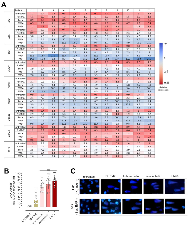
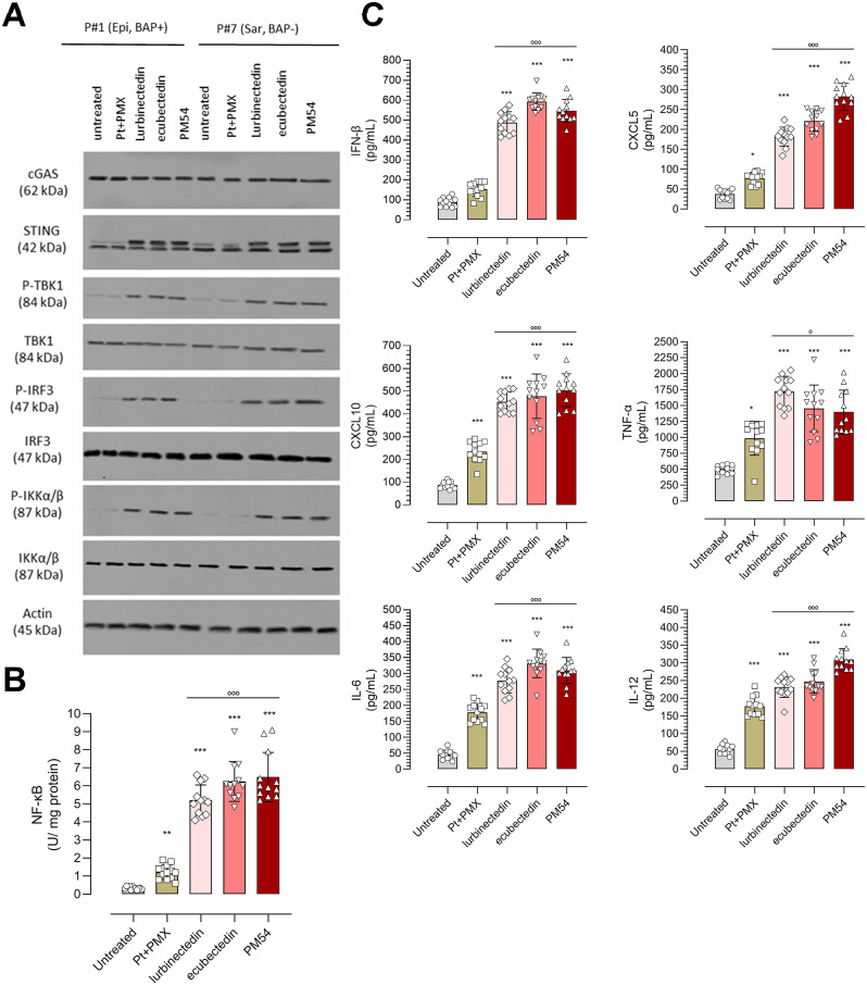
Results: Lurbinectedin, ecubectedin, and PM54 were effective in reducing cell proliferation and migration, as well as inducing S-phase cell cycle arrest and DNA damage in malignant pleural mesothelioma cells. These effects were more pronounced compared to the standard first-line treatment (platinum-based plus pemetrexed). Mechanistically, the drugs downregulated DNA repair genes, activated the cGAS/STING pathway, and promoted the release of pro-inflammatory cytokines. They also induced immunogenic cell death of mesothelioma cells, enhancing the activation of anti-tumor CD8+T-cells and natural killer cells while reducing tumor-tolerant T-regulatory cells and myeloid-derived suppressor cells in ex vivo co-cultures. These promising results were also observed in humanized patient-derived xenograft models, where the drugs were effective in reducing tumor growth and increasing the ratio anti-tumor/pro-tumor infiltrating immune populations, either alone or combined with the anti-PD-1L atezolizumab.

Conclusions: Collectively, these findings reveal a previously unknown mechanism of action of ecteinascidins that merits further investigation for potential clinical applications in the treatment of MPM, as new first line treatment in monotherapy or in association with immunotherapy.

Keywords: Chemo-immunotherapy; DNA damage; Ecteinascidins; Immunogenic cell death; Malignant pleural mesothelioma; cGAS/STING pathway.


Conflict of interest statement

Declarations. Ethics approval and consent to participate: The study was approved by the local Ethical Committee (#128/2016). The patients gave written informed consent to participate in the study. Consent for publication: The patients gave written informed consent to publish the results of the study. Competing interests: PA, HS and CC are PharmaMar S.A. employees or Pharma-Mar S.A. shareholders or both. CR received lurbinectedin and its derivatives, and economical support from PharmaMar S.A. AM received travel expense reimbursement from PharmaMar S.A. outside the present work and is member of the advisory board of Boehringer Ingelheim. LR received grants from AstraZeneca, Roche, Eli Lilly, and Novartis outside the submitted work. SN is speaker’s bureau for: Amgen, AstraZeneca, Boehringer Ingelheim, BeiGene, Eli Lilly, Pfizer, Roche, MSD, Janssen, Novartis, Takeda, Thermo Fisher, member of the advisory board and consultant for Sanofi. GVS has received grants from TESARO/GSK and personal fees as well as supports from Pfizer, AstraZeneca, Eli Lilly and MSD outside the submitted work; personal fees from Roche, Novartis, Clovis, Takeda and BeiGene outside the submitted work.

Figures

Fig. 1
Fig. 1
Antiproliferative and anti-invasive effects of lurbinectedin, ecubectedin and PM54 in patient-derived MPM cells. (A) In vitro IC50 and IC10 median values of cisplatin plus pemetrexed (Pt + PMX), lurbinectedin, ecubectedin and PM54 determined in 12 patient-derived histotypes of MPM (epithelioid: square; sarcomatoid: circle; biphasic: diamond) with BAP1 positive (solid symbol) and BAP1 negative (open symbol). Results are mean of 4 independent experiments. (B) Representative images of crystal violet staining obtained on long-term (10 weeks) assay with APN#7 cells (sarcomatoid, BAP1-). (C). Representative images of the effect induced by the compounds to six different MPM spheroids following 72 h of incubation. Scale bar = 100 μm. (D). Representative phase images of migrated cells (scale bar, 100 μm) and (E) cells in the lower chamber of APN#1 (epithelioid, BAP1+) and APN#7 (sarcomatoid, BAP1-)
Fig. 2
Fig. 2
Effects of lurbinectedin, ecubectedin and PM54 on DNA damage and repair machinery in MPM cells. (A) The expression pattern of DNA damage genes up-regulated (blue) and down-regulated (red) in MPM cell lines after a 24 h treatment with cisplatin + pemetrexed (Pt + PMX), lurbinectedin (L), ecubectedin and PM54 at their IC50 for 24 h, was shown as mRNA abundance versus a pool of housekeeping genes (n = 3 independent experiments, in triplicates). (B) Histograms showing the percentage of total DNA in the tail of Comet assay in MPM cells treated with Pt + PMX, L, ecubectedin and PM54 at IC50 for 24 h. Data are expressed as means ± SD of 12 MPM samples (n = 3 independent experiments, in duplicates). *p < 0.05, ***p < 0.001: vs. untreated cells; °°°p < 0.001: vs. Pt + PMX. (C) Representative Comet assay images of MPM#1 (epithelioid, Bap positive: Epi, BAP+) and MPM#7 (sarcomatoid, BAP1 negative: Sar, BAP-)
Fig. 3
Fig. 3
Effects of lurbinectedin, ecubectedin and PM54 on cGAS/STING pathway activation and downstream cytokines in MPM cells. (A) Representative immunoblot images of the indicated proteins belonging to the cGAS/STING pathway, in MPM#1 (epithelioid, BAP1 positive: Epi, BAP+) and MPM#7 (sarcomatoid, BAP1 negative: Sar, BAP), treated or not (-) with cisplatin + pemetrexed (Pt + PMX), lurbinectedin (L), ecubectedin and PM54 at their IC50 for 24 h, actin was used as a loading control. The figure is representative of 1 out of 3 experiments. (B) Activation of NF-kB after the treatment indicated in (A). Data are expressed as means ± SD of 12 MPM samples (n = 3 independent experiments, in duplicates). **p < 0.01, ***p < 0.001: vs. untreated cells; °°°p < 0.001: vs. Pt + PMX. (C) Pro-inflammatory cytokine/chemokine levels released in the supernatant of MPM cell cultures after the treatments indicated in (A). Data are expressed as means ± SD of 12 MPM samples (n = 3 independent experiments, in duplicates). *p < 0.05, ***p < 0.001: vs. untreated cells; °°°p < 0.001: vs. Pt + PMX
Fig. 4
Fig. 4
Effects of lurbinectedin, ecubectedin and PM54 on immunogenic cell deaths of MPM cells. (A) Workflow of experiment. Dendritic cells (DCs), generated in 4 days from healthy donors PBMC, were co-incubated 18 h with MPM cells, previously grown 24 h in drug-free medium (untreated), cisplatin + pemetrexed (Pt + PMX), lurbinectedin (L), ecubectedin and PM54 at their IC50. An aliquot of DC-MPM co-cultures were collected to measure DC-mediated phagocytosis by flow cytometry. An aliquot of DCs that have been in contact with MPM cells was collected and incubated 10 days with CD8+T-lymphocytes, obtained from PBMC of the same donor. CD8+T-lymphocytes were then collected and co-incubated with the MPM cells of the same patient for 18 h. Finally, CD8+T-lymphocytes were collected and analyzed for the activation markers by flow cytometry, MPM cells were stained with Annexin V-FITC/PI to measure necro-apoptotic cells, as index of immune-killing mediated by CD8+T-lymphocytes. (B) The percentage of cells positive for surface calreticulin (CRT) was measured by flow-cytometry. (C) ATP release was measured by a chemiluminescent-based assay. (D) HMGB1 release was measured by ELISA. (E) Phagocytized MPM cells were counted by flow cytometry. (F) Percentage of CD8+CD107a+INFγ+ cells, as index of cytotoxic T-lymphocyte activation. (G) Percentage of annexin V-FITC+/PI+ MPM cells, as index of tumor cells immune killing by CD8+T-lymphocytes, measured by flow cytometry. In all panels, data are expressed as means ± SD of 12 MPM samples (n = 3 independent experiments, in duplicates). *p < 0.05, ***p < 0.001: vs. untreated cells; °°°p < 0.001: vs. Pt + PMX
Fig. 5
Fig. 5
Effects of chemo-immunotherapy treatments based on lurbinectedin, ecubectedin and PM54 in patient-derived immune-xenografts. MPM#1 (epithelioid, BAP1+) and MPM#7 (sarcomatoid, BAP1) cells were implanted s.c. in 6-week-old female Hu-CD34+ mice and treated as reported under Materials and Methods. (A). Tumor volumes growth and (B) median tumor volume on day 21 (n = 4 mice/group). (C) Percentage of Mo-MDSC, CD8+T-lymphocytes and TAM2 infiltrating the tumors measured by flow cytometry after tumor excision and dissociation. Pt + PMX, cisplatin plus pemetrexed. For both panels: *p < 0.05

References

    1. Janes SM, Alrifai D, Fennell DA. Perspectives on the Treatment of Malignant Pleural Mesothelioma. N Engl J Med. 2021;385:1207–18. - PubMed
    1. Cinausero M, Rihawi K, Sperandi F, Melotti B, Ardizzoni A. Chemotherapy treatment in malignant pleural mesothelioma: a difficult history. J Thorac Dis. 2018;10:S304–10. - PMC - PubMed
    1. Zalcman G, Mazieres J, Margery J, Greillier L, Audigier-Valette C, Moro-Sibilot D, et al. Bevacizumab for newly diagnosed pleural mesothelioma in the Mesothelioma Avastin Cisplatin Pemetrexed Study (MAPS): a randomised, controlled, open-label, phase 3 trial. Lancet. 2016;387:1405–14. - PubMed
    1. Hiltbrunner S, Mannarino L, Kirschner MB, Opitz I, Rigutto A, Laure A, et al. Tumor Immune Microenvironment and Genetic Alterations in Mesothelioma. Front Oncol. 2021;11:660039. - PMC - PubMed
    1. Tagliamento M, Di Maio M, Remon J, Bironzo P, Genova C, Facchinetti F, et al. Meta-Analysis on the Combination of Chemotherapy With Programmed Death-Ligand 1 and Programmed Cell Death Protein 1 Blockade as First-Line Treatment for Unresectable Pleural Mesothelioma. J Thorac Oncol. 2024;19:166–72. - PubMed

LinkOut - more resources