. 2025 Jan 28;44(1):115089.
doi: 10.1016/j.celrep.2024.115089. Epub 2024 Dec 21.

Increased nuclear factor I-mediated chromatin access drives transition to androgen receptor splice variant dependence in prostate cancer

Affiliations

Increased nuclear factor I-mediated chromatin access drives transition to androgen receptor splice variant dependence in prostate cancer

Larysa Poluben et al. Cell Rep. .

Abstract

Androgen receptor (AR) splice variants, of which ARv7 is the most common, are increased in castration-resistant prostate cancer, but the extent to which they drive AR activity is unclear. We generated a subline of VCaP cells (VCaP16) that is resistant to the AR inhibitor enzalutamide (ENZ). AR activity in VCaP16 is driven by ARv7, independently of full-length AR (ARfl), and its cistrome and transcriptome mirror those of ARfl in VCaP cells. ARv7 expression increases rapidly in response to ENZ, but there is a delay in gaining chromatin binding and transcriptional activity, which is associated with increased chromatin accessibility. AR and nuclear factor I (NFI) motifs are most enriched at more accessible sites, and NFIB/X knockdown greatly diminishes ARv7 function. These findings indicate that ARv7 can drive the AR program but that its activity is dependent on adaptations that increase chromatin accessibility to enhance its intrinsically weak chromatin binding.

Keywords: CP: Cancer; CP: Molecular biology; FOXA1; androgen receptor; chromatin; epigenetics; nuclear factor I; prostate cancer; transcription.


Conflict of interest statement

Declaration of interests P.S.N. has received consulting fees from Janssen, Merck, and Bristol Myers Squibb, and research support from Janssen for work unrelated to the present studies.

Figures
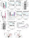
Figure 1.
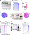
Figure 1.. VCaP16 cells exhibit increased ARv7 expression and resistance to AR blockade
(A) Proliferation for VCaP, VCaP with 16 μM ENZ (VCaP-E), and VCaP16 (maintained in 16 μM ENZ) at 7 days post treatment (*p < 0.05, **p < 0.01, ANOVA). (B) Hallmark androgen response comparing VCaP cells with ENZ for 4 days (VCaP-E) or VCaP16 versus parental VCaP. Significantly differentially expressed genes (adjusted p value <0.05) were input here and below. FDR, false discovery rate; NES, normalized enrichment score. (C) Immunoblot of VCaP16 and parental VCaP. (D) ARfl and ARv7 assessed by RT-qPCR in VCaP (black) and VCaP16 (red) cells (*p < 0.05, **p < 0.01, unpaired t test, two-tailed). (E) (Upper) KLK3 by RT-qPCR and (lower) ARfl and ARv7 in VCaP16 cells after siRNA knockdown with non-targeting control (siNTC), ARv7 (siV7), both ARv7 and ARfl (siEx1), or just ARfl (siEx7) siRNA. Bars represent SEM. (*p < 0.05, **p < 0.01, ns = non-significant, Welch’s t test). (F) Hallmark androgen response in VCaP16 with siRNA knockdown of ARv7 (siV7) or ARfl (siEx7). (G) VCaP16 cells treated with ARCC-32 (500 nM) or DMSO for 36 h. (H) Hallmark androgen response in VCaP16 with ARCC-32 (500 nM) or DMSO for 36 h. Input was all differentially expressed genes. (I) Live-cell DNA fluorescence in VCaP16 treated with non-target control siRNA (siNTC) or ARv7 siRNA (siV7) ± 500 nM ARCC-32. (J) Differential expression of ARv7 gene set from Sharp et al., comparing VCaP16 versus VCaP cells (left) or VCaP16 treated with ARv7 siRNA versus non-target control siRNA (siNTC) (right). Dotted line represents a value of 2 (p = 0.01).
Figure 2.
Figure 2.. ARv7 induced acutely by ENZ is nuclear but lacks chromatin-binding capacity
(A) VCaP with 16 μM ENZ from 2 to 28 days and VCaP16 cells. AR-NT, AR N terminus antibody (long/short exposures). (B) Confocal immunofluorescence of ARv7 and CBP/p300 in DMSO or 16 μM ENZ (4 days) treated VCaP versus VCaP16 (maintained in 16 μM ENZ). (C and D) Quantification of z-stack three-dimensional images showing number of ARv7 puncta per nuclei in four fields (C) and fraction of ARv7 puncta containing CBP/p300 (D). (E) Chromatin fraction from VCaP and VCaP16 following formalin crosslinking for ChIP. (F) ARv7-binding sites based on ChIP-seq in samples from (E) and Venn diagram with overlap between ARv7 sites in VCaP16 (ENZ) in biological replicates. (G) ARfl-binding sites by AR C-terminal antibody ChIP-seq on samples in (E).
Figure 3.
Figure 3.. ARv7 chromatin binding in VCaP16 cells is independent of ARfl
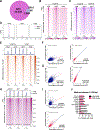
(A) VCaP16 in basal medium (16 μM ENZ) was treated for 24 or 48 h with vehicle (NC) or ARCC-32 (500 nM). Cells were then separated into cytoplasmic, soluble nuclear, or chromatin fractions. (B) ARv7 binding in VCaP16 in basal medium (16 μM ENZ) with addition of DMSO or ARCC-32 (500 nM) for 36 h. ChIP-seq signal is centered at shared ARv7 sites. (C) ARv7-binding sites in VCaP16 in basal medium (with 16 μM ENZ) treated with DMSO or ARCC-32 (500 nM) for 36 h. (D) Intersection of merged ARv7 sites in VCaP16 cells in DMSO or ARCC-32 (500 nM). (E) VCaP16 were double-crosslinked with DSP and formaldehyde. Chromatin was then isolated and solubilized with benzonase, followed by immunoprecipitation. (F) ARv7 and ARfl sites in VCaP16 cells following 4 h of treatment with DHT (10 nM). (G) ARv7 and ARfl binding in VCaP16 following 4-h treatment with DHT (10 nM). ChIP-seq signal is centered at ARv7 sites in DHT-treated VCaP16. (H) Whole-cell lysate (WCL) and chromatin fraction from VCaP16 in basal medium (16-μM ENZ) or treated for 4 h with DHT (10 nM). (I) Enrichment of ARv7 binding in LNCaP95 cells in basal medium treated for 2 h with DMSO, DHT (10 nM), or dexamethasone (DEX, 100 nM). Signal centered at ARv7 sites identified in VCaP16 cells.
Figure 4.
Figure 4.. ARv7- and ARfl-binding sites overlap with proportionate peak intensities
(A) Intersection of ARv7 and ARfl sites in VCaP16 in basal medium (16 μM ENZ). (B) ARfl and ARv7 binding at called ARv7 sites (upper) and at ARfl-unique sites. (C) ARv7- and ARfl-binding intensities across all ARfl sites in VCaP16, and showing ARv7 in VCaP after short-term ENZ (4 days) (VCaP-E). (D) Peak intensities for ARfl and ARv7 in VCaP16 at ARv7 versus ARfl-unique sites. ChIP-seq signal centered at ARv7/ARfl shared (ARv7) and ARfl-unique (ARfl) sites identified in VCaP16. (E) ARfl-binding intensities in (1) VCaP16 in basal medium (16 μM ENZ), (2) VCaP16 with DHT (10 nM) for 4 h, (3) VCaP with ENZ for 72 h followed by removal of ENZ and addition of DHT (10 nM) for 4 h, and (4) VCaP with ENZ for 72 h. ChIP-seq signal is centered at total ARfl sites identified in DHT-stimulated VCaP16. (F and G) Pearson’s correlation of normalized read counts: ARv7 versus ARfl called at shared ARfl/ARv7 and ARfl-unique sites in (F) VCaP16 and (G) VCaP16 treated with DHT. (H) AR signal enrichment with AR N-terminal (GSM2842708), AR C-terminal (GSM2842700), and ARv7 (GSM2842704) antibodies in LNCaP95 cells, centered at shared ARfl/ARv7 (ARv7) sites and ARfl-unique sites identified in VCaP16. (I) Pearson’s correlation of normalized read counts: ARv7 (GSM2842704) versus ARfl (GSM2842700) in LNCaP95 at shared ARfl/ARv7 (ARv7) sites and ARfl-unique sites identified in VCaP16. (J) Top enriched motifs (±100 kb from peak center) at ARfl-unique sites and at shared ARfl/ARv7 sites in VCaP16.
Figure 5.
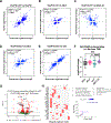
Figure 5.. ARv7 transcriptome in VCaP16 is highly correlated with ARfl in VCaP
(A–E) Log2(fold change) of significantly differentially expressed genes (padj < 0.05). (F) Log2(fold change) of ARv7-regulated genes (ARv7-binding sites ±5 kb from TSS) altered by siV7, siEx1, siEx7, or ARCC-32 in VCaP16. Differentially expressed genes with padj < 0.05 (siV7, siEx1, siEx7) and p < 0.05 (ARCC-32) were included. Mann-Whitney U test, two-tailed: siV7 versus siEx7, p = 0.0308; siV7 versus siEx1, p = 0.8151; siV7 versus ARCC-32, p <0.0001; siEx1 versus siEx7, p = 0.0609; ARCC-32 versus siEx7, p = 0.1148; ARCC-32 versus Ex1, p = 0.0005. (G) Differentially expressed genes linked to ARv7-binding sites in VCaP16 cells (± 100 kb from TSS) in VCaP16 with siRNA knockdown of ARv7 (siV7) versus non-targeting control. (H) Enrichment analysis by Enrichr of downregulated genes from (G).
Figure 6.
Figure 6.. Chromatin accessibility in VCaP16 cells is increased at ARv7-binding sites
(A and B) (A) ATAC-seq signal and (B) numbers of ATAC sites in parental VCaP, VCaP with ENZ for 4 days (VCaP-E) and VCaP16. (C and D) (C) Heatmap and (D) profile plots of ATAC-seq signal in VCaP, VCaP-E and VCaP16 cells. ATAC-seq signal centered at sites with increased ATAC signal (UP sites), sites called as unchanged (Unchanged sites) or sites with decreased ATAC signal (Down sites) in VCaP16 versus parental VCaP cells as identified with CoBRA pipeline. (E) Overlap of differentially more accessible sites (ATAC-UP) with shared ARv7/ARfl (upper panel) and ARfl-unique binding sites (lower panel) in VCaP16. (F and G) (F) ATAC-seq signal enrichment and (G) nucleosome occupancy by MNase-seq at ARv7/ARfl (upper panels) and ARfl-unique binding sites (lower panels) in VCaP16. (H) Motif enrichment analysis by HOMER at ATAC-UP sites in VCaP16. (I) NFI motif enrichment by HOMER at ATAC-seq differentially more accessible sites (UP), unchanged (Unchanged), or differentially closed (Down) sites in VCaP16 versus VCaP with CoBRA pipeline (****p < 0.0001, Fisher’s exact test). (J) V-plot of NFI motif footprint at ATAC-UP sites in VCaP16 (centered on NFI motif).
Figure 7.
Figure 7.. ARv7 activity in VCaP16 is dependent on nuclear factor I transcription factors
(A–D) Enrichment for the (A) ARE, (B) HOXB13, (C) FOXA1, and (D) NFI motifs by HOMER at ARv7/ARfl shared and ARfl-unique sites in VCaP after short-term ENZ treatment (VCaP-E) versus in VCaP16 cells. (E) NFI motif enrichment by HOMER at AR-binding sites with canonical full AREs versus non-consensus AREs (others) in VCaP after short-term ENZ treatment (VCaP-E) versus in VCaP16. (F and G) Pearson’s correlation of log2(fold change) values of significantly differentially expressed genes (padj < 0.1) altered with siRNA knockdown of ARv7 (siV7) versus (F) siRNA knockdown of NFIB and NFIX (siNFIB/X) and versus (G) siRNA knockdown of FOXA1 (siFOXA1) in VCaP16. (H) (Left) ARfl (AR-NT) and ARv7 in VCaP16 and (I) 22RV1 cells after knockdown with both NFIB and NFIX (siNFIB/X), and non-targeting control (siNTC) siRNA. (Right) Quantification of ARfl and ARv7 normalized to siNTC. Numbers indicate levels of ARfl and ARv7 reduction compared to siNTC. (J) ARfl- and ARv7-binding sites based on ChIP-seq in VCaP16 with siRNA knockdown of NFIB and NFIX (siNFIB/X) versus non-targeted siRNA knockdown (siNTC). (K) NFI motif enrichment by HOMER at AR-binding sites in normal prostate tissue (N) versus primary PC in two clinical datasets.

Update of

References

    1. Stanbrough M, Bubley GJ, Ross K, Golub TR, Rubin MA, Penning TM, Febbo PG, and Balk SP (2006). Increased expression of genes converting adrenal androgens to testosterone in androgen-independent prostate cancer. Cancer Res. 66, 2815–2825. - PubMed
    1. Montgomery RB, Mostaghel EA, Vessella R, Hess DL, Kalhorn TF, Higano CS, True LD, and Nelson PS (2008). Maintenance of intratumoral androgens in metastatic prostate cancer: a mechanism for castration-resistant tumor growth. Cancer Res. 68, 4447–4454. - PMC - PubMed
    1. Penning TM, and Detlefsen AJ (2020). Intracrinology-revisited and prostate cancer. J. Steroid Biochem. Mol. Biol. 196, 105499. - PMC - PubMed
    1. Smith MR, Saad F, Chowdhury S, Oudard S, Hadaschik BA, Graff JN, Olmos D, Mainwaring PN, Lee JY, Uemura H, et al. (2018). Apalutamide Treatment and Metastasis-free Survival in Prostate Cancer. N. Engl. J. Med. 378, 1408–1418. - PubMed
    1. Beer TM, Armstrong AJ, Rathkopf DE, Loriot Y, Sternberg CN, Higano CS, Iversen P, Bhattacharya S, Carles J, Chowdhury S, et al. (2014). Enzalutamide in metastatic prostate cancer before chemotherapy. N. Engl. J. Med. 371, 424–433. - PMC - PubMed

Publication types

MeSH terms

Associated data