. 2024 Dec 3;34(12):bhae484.
doi: 10.1093/cercor/bhae484.

Single-pulse transcranial magnetic stimulation of the dorsolateral prefrontal cortex does not directly affect muscle sympathetic nerve activity in humans

Affiliations

Single-pulse transcranial magnetic stimulation of the dorsolateral prefrontal cortex does not directly affect muscle sympathetic nerve activity in humans

Brendan McCarthy et al. Cereb Cortex. .

Abstract

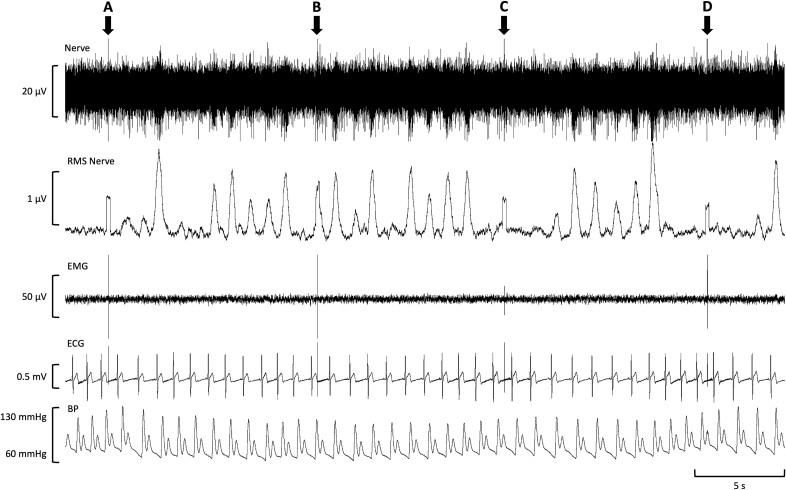
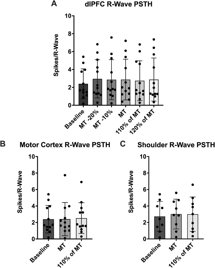
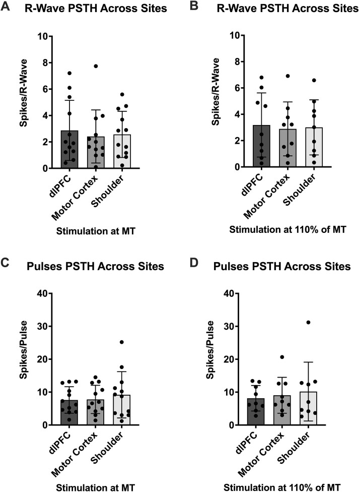
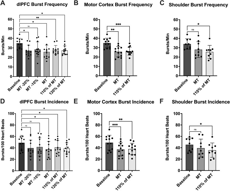
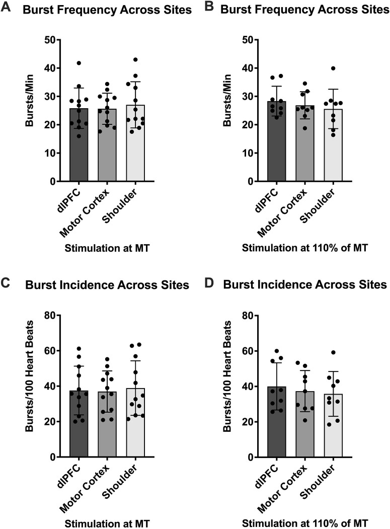
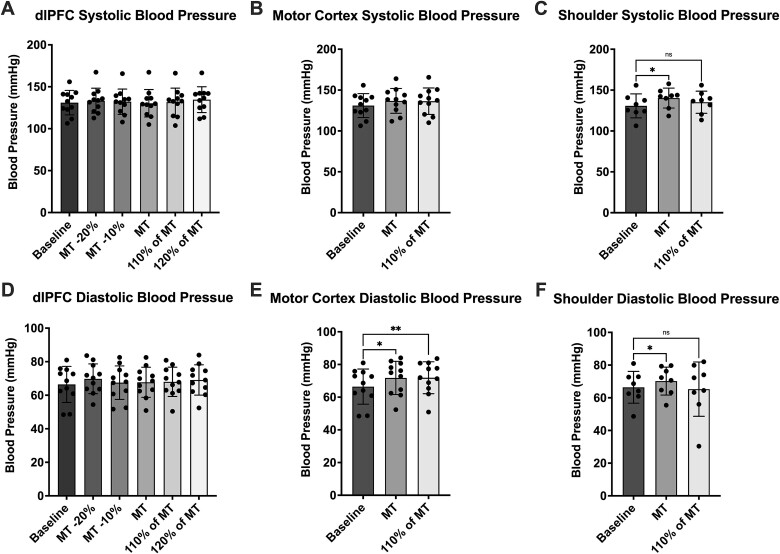
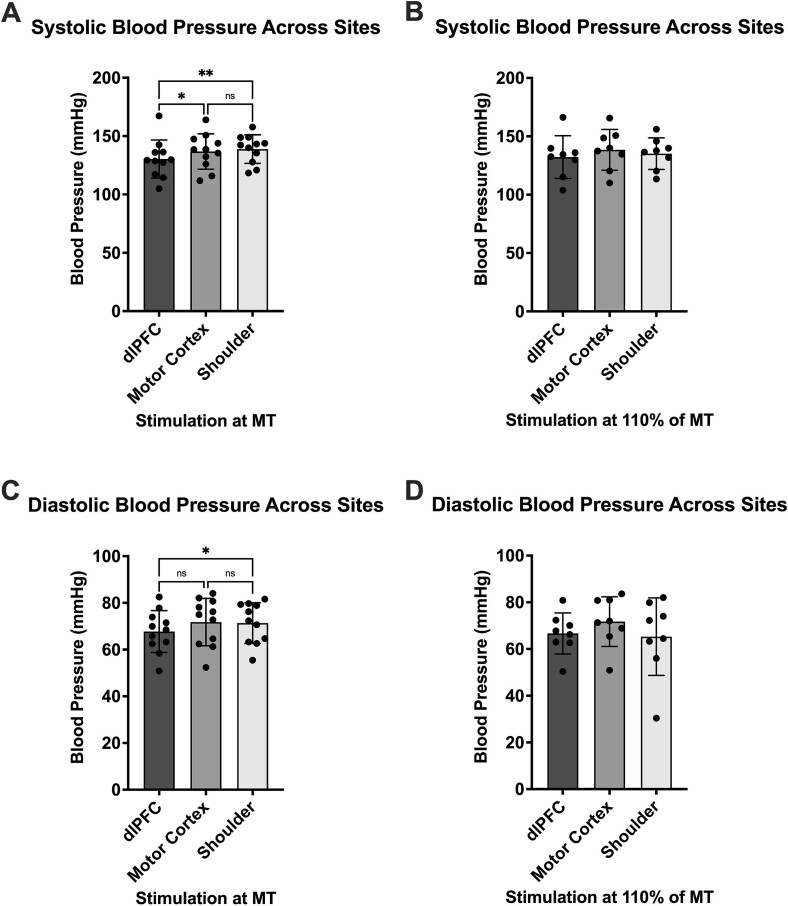
Transcranial magnetic stimulation (TMS) is applied both in research settings and clinically, notably in treating depression through the dorsolateral prefrontal cortex (dlPFC). We have recently shown that transcranial alternating current stimulation of the dlPFC partially entrains muscle sympathetic nerve activity (MSNA) to the stimulus. We, therefore, aimed to further explore the sympathetic properties of the dlPFC, hypothesizing that single-pulse TMS could generate de novo MSNA bursts. Microneurography was performed on the right common peroneal nerve in 12 participants. TMS pulses were then delivered to the ipsilateral dlPFC at resting motor threshold (MT) of the first dorsal interosseous muscle and at powers 20 below, 10 below, 10% above, and 20% above MT. The MT and 10% above MT intensities were also used to stimulate the right motor cortex and shoulder. Comparisons between stimulus intensities at the same site and between sites at the same intensities revealed no differences in MSNA burst frequency, burst incidence, or single MSNA spikes. Most stimulus trains, however, showed reduced burst frequency and incidence from baseline, regardless of site. This suggests that the TMS itself was evoking arousal-based sympathoinhibition, independent of dlPFC influences. It seems the dlPFC is capable of modulating MSNA but cannot directly generate bursts.

Keywords: dorsolateral prefrontal cortex; motor cortex; muscle sympathetic nerve activity; transcranial magnetic stimulation.


Figures

Fig. 1
Fig. 1
Raw physiological data during stimulation of the dorsolateral prefrontal cortex (dlPFC). A single participant’s real-time data during a 40 s time period of right dlPFC stimulation at 110% of motor threshold (MT) is displayed. Muscle sympathetic nerve activity (MSNA) data is shown in the first channel (nerve) and used to calculate the processed signal in the second channel (Root-mean-square (RMS) nerve). Additionally, data from the electromyogram (EMG) and electrocardiogram (ECG), as well as blood pressure (BP), can be seen in the EMG, ECG, and BP channels, respectively. Single transcranial magnetic stimulation (TMS) pulses were delivered at the time points denoted by arrows (A) to (D), each generating a signal artifact that can be seen in all channels except BP. While all pulses were aimed to be delivered out-of-phase with MSNA burst trains, an example of a miss-timed pulse can be seen in (B). The Y-axis on each channel represents signal range.
Fig. 2
Fig. 2
Peristimulus time histogram (PSTH) analysis of MSNA spikes referenced to the R-waves during each stimulus train. (A) Shows spikes of MSNA during stimulation of the dlPFC (n = 12) at five different intensities as well as spikes during baseline. (B) Presents sympathetic spikes during stimulation of the motor cortex (n = 12) and (C) presents spikes during stimulation of the shoulder (n = 9). Baseline values are taken from the same timepoint at the beginning of each experiment and, so, are consistent in (A) and (B). The reduced sample size in (C) has shifted the baseline value slightly. No significant differences (P > 0.05) were found between any stimulation intensities when compared to each other and baseline at each site. Data are presented as mean ± SD.
Fig. 3
Fig. 3
PSTH comparisons of MSNA spikes across different stimulation sites. (A) and (B) depict MSNA spikes referenced to the R-waves of the ECG when stimuli were delivered to the dlPFC, motor cortex and shoulder at MT (n = 12) and at 110% of MT (n = 9), respectively. (C) and (D) depict MSNA spikes referenced to the TMS pulses when stimuli were delivered to the dlPFC, motor cortex and shoulder at MT (n = 12) and at 110% of MT (n = 9), respectively. No significant differences (P > 0.05) were found between sites at either intensity during both PSTH analyses. Data are presented as mean ± SD.
Fig. 4
Fig. 4
RMS-processed MSNA metrics. (A), (B), and (C) present MSNA burst frequency (bursts/min) data during stimulus trains delivered to the dlPFC (n = 12), motor cortex (n = 12) and shoulder (n = 9), respectively. In each graph, stimuli are compared to a baseline value obtained at the beginning of the experiments. (D), (E) and (F) present MSNA burst incidence (bursts/100 heartbeats) data during stimulus trains delivered to the dlPFC (n = 12), motor cortex (n = 12) and shoulder (n = 9), respectively. In each graph, stimuli are compared to a baseline value obtained at the beginning of the experiments. Time points that were found to be significantly different from baseline are denoted with asterisks (*P < 0.05; **P < 0.01; ***P < 0.001). No significant differences (P > 0.05) were found between stimulation intensities at each site. Data are presented as mean ± SD.
Fig. 5
Fig. 5
RMS-processed MSNA metric comparison across different stimulation sites. (A) and (B) show MSNA burst frequency (bursts/min) when stimuli were delivered to the dlPFC, motor cortex and shoulder at MT (n = 12) and at 110% of MT (n = 9), respectively. (C) and (D) show MSNA burst incidence (bursts/100 heartbeats) when stimuli were delivered to the dlPFC, motor cortex and shoulder at MT (n = 12) and at 110% of MT (n = 9), respectively. No significant differences (P > 0.05) were found between sites at either intensity through either metric. Data are presented as mean ± SD.
Fig. 6
Fig. 6
Systolic and diastolic blood pressure during each stimulus train. (A), (B), and (C) present systolic blood pressure data during stimulus trains delivered to the dlPFC (n = 11), motor cortex (n = 11) and shoulder (n = 8), respectively. In each graph, stimuli are compared to a baseline value obtained at the beginning of the experiments. (D), (E), and (F) present diastolic blood pressure data during stimulus trains delivered to the dlPFC (n = 11), motor cortex (n = 11) and shoulder (n = 8), respectively. In each graph, stimuli are compared to a baseline value obtained at the beginning of the experiments. Time points which were found to be significantly different from baseline are denoted with asterisks (*P < 0.05; **P < 0.01). No significant differences (P > 0.05) were found between stimulation intensities at each site. Data are presented as mean ± SD.
Fig. 7
Fig. 7
Blood pressure comparison across different stimulation sites. (A) and (B) show systolic blood pressure when stimuli were delivered to the dlPFC, motor cortex and shoulder at MT (n = 11) and at 110% of MT (n = 8), respectively. (C) and (D) show diastolic blood pressure when stimuli were delivered to the dlPFC, motor cortex and shoulder at MT (n = 11) and at 110% of MT (n = 8), respectively. Significant differences were found between sites only at MT in both metrics and are denoted with asterisks (*P < 0.05; **P < 0.01). Data are presented as mean ± SD.

Similar articles

References

    1. Barker AT, Jalinous R, Freeston IL. Non-invasive magnetic stimulation of human motor cortex. Lancet. 1985:325:1106–1107. - PubMed
    1. Barton DA, Dawood T, Lambert EA, Esler MD, Haikerwal D, Brenchley C, Socratous F, Kaye DM, Schlaich MP, Hickie Iet al. . Sympathetic activity in major depressive disorder: identifying those at increased cardiac risk? J Hypertens. 2007:25:2117–2124. 10.1097/HJH.0b013e32829baae7. - DOI - PubMed
    1. Bent LR, Boulton PS, Macefield VG. Modulation of muscle sympathetic bursts by sinusoidal galvanic vestibular stimulation in human subjects. Exp Brain Res. 2006:174:701–711. 10.1007/s00221-006-0515-6. - DOI - PubMed
    1. Boulton D, Taylor CE, Green S, Macefield VG. The metaboreflex does not contribute to the increase in muscle sympathetic nerve activity to contracting muscle during static exercise in humans. J Physiol. 2018:596:1091–1102. 10.1113/JP275526. - DOI - PMC - PubMed
    1. Bourla A, Mouchabac S, Lorimy L, Crette B, Millet B, Ferreri F. Variability in motor threshold during transcranial magnetic stimulation treatment for depression: neurophysiological implications. Brain Sci. 2023:13:1246. 10.3390/brainsci13091246. - DOI - PMC - PubMed

LinkOut - more resources