

Intrauterine device (IUD) migration completely into the abdominal cavity and half into the bladder to form a stone: a case report and mini-review
- PMID: 39710656
- PMCID: PMC11664840
- DOI: 10.1186/s12894-024-01676-5
Intrauterine device (IUD) migration completely into the abdominal cavity and half into the bladder to form a stone: a case report and mini-review
Abstract
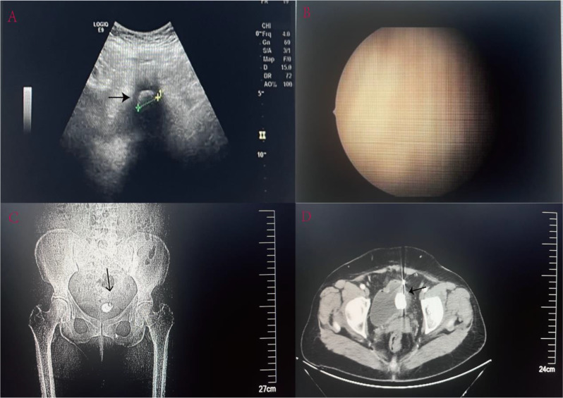
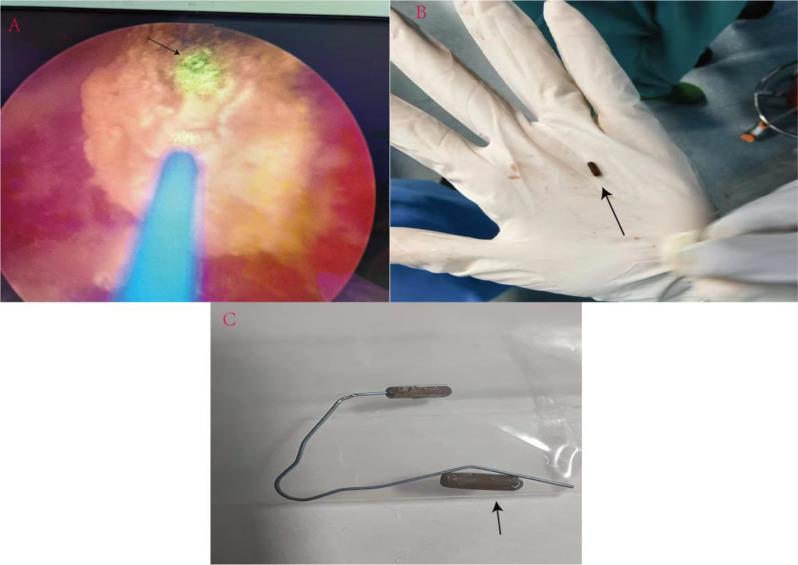
The intrauterine device (IUD) is an important and highly effective means of contraception. Migration of the IUD, post implantation, out of the uterus is an infrequent complication, and its subsequent migration into the urinary bladder with formation of secondary bladder calculi, is even more infrequently reported. The authors report a 51 year old woman who had had her last child delivered via cesarean section 16 years ago. She underwent an IUD insertion in the next few months but was subsequently lost to follow up. Before detection of the ectopic state of the IUD she complained of dysuria and dyspareunia for two weeks. Clinically detected suprapubic discomfort on palpation. After a series of imaging studies and cystoscopy, the presence of an IUD stump with stones attached to it in the patient's bladder were confirmed. Cystoscopy followed by laser lithotripsy of the calculus was performed, as the first step in the treatment. After failure to remove the IUD cystoscopically, further cystotomy was made, however one side end of the cap structure of the IUD could not be located, necessitating abdominal exploration and irrigation till retrieval in totality. The patient made an uneventful recovery.The case report emphasizes the importance of excluding uncommon etiolgies like ectopic IUDs as a cause of immobile adherent bladder calculi in women. Complete removal necessitates good preoperative knowledge of the type of IUD, as structural degradation might lead to separation of its components. An adequate consent for a possible exploration of the abdomen in case of a missing component should also be taken.
Keywords: Bladder calculus; Ectopic; Endoscopy; Intrauterine device migration.
© 2024. The Author(s).
Conflict of interest statement
Declarations. Ethics approval and consent to participate: This institution does not require Institutional Review Board approval for case reports. Considering the ethical principles, the patient’s name was not mentioned in the paper. Consent for publication: Written informed consent was obtained from the patient for publication of this case report and any accompanying images. Clinical trial number: Not applicable. Competing interests: The authors declare no competing interests.
Figures
References
-
- Farmer M, Webb A. Intrauterine device insertion-related complications: can they be predicted? J Fam Plann Reprod Health Care. 2003;29(4):227–31. - PubMed
-
- Kaislasuo J, Suhonen S, Gissler M, Lähteenmäki P, Heikinheimo O. Uterine perforation caused by intrauterine devices: clinical course and treatment. Hum Reprod. 2013;28(6):1546–51. - PubMed
-
- Zong Z, Sun X, Hearst N. Risk factors for delayed intrauterine device removal among menopausal women in rural China. Menopause. 2022;29(9):1077–82. - PubMed
Publication types
MeSH terms
LinkOut - more resources
Full Text Sources
Miscellaneous
