

Experimental Assessment of Traction Force and Associated Fetal Brain Deformation in Vacuum-Assisted Delivery
- PMID: 39710825
- PMCID: PMC11929728
- DOI: 10.1007/s10439-024-03665-z
Experimental Assessment of Traction Force and Associated Fetal Brain Deformation in Vacuum-Assisted Delivery
Abstract
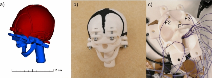
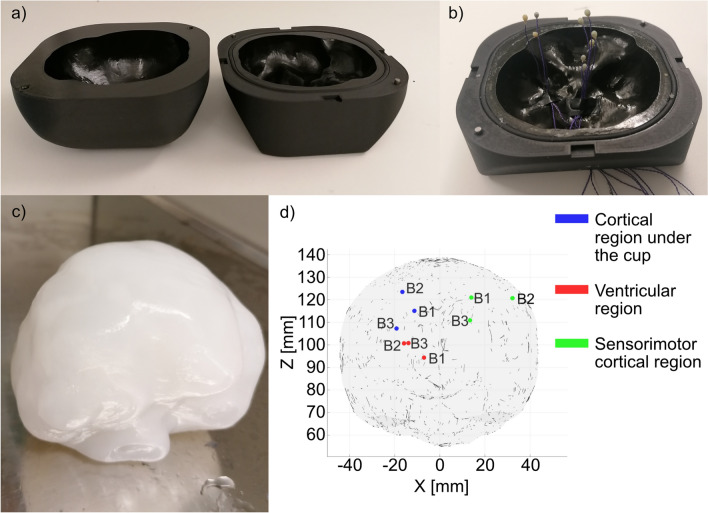
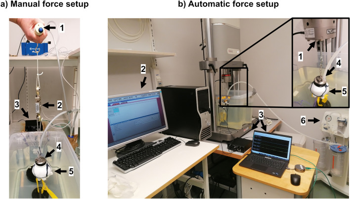
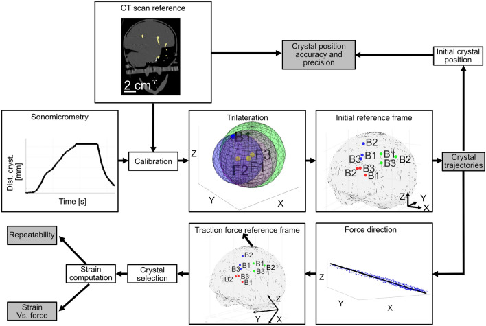
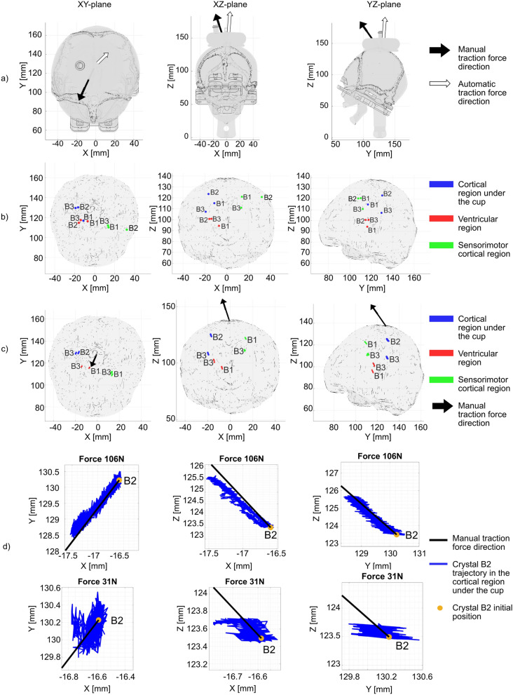
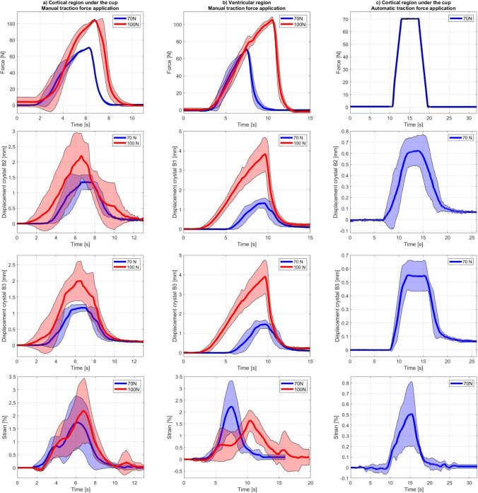
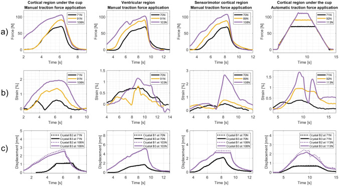
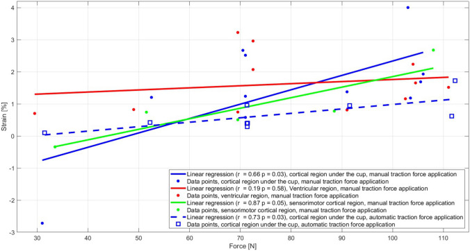
Vacuum-assisted delivery (VAD) uses a vacuum cup on the fetal scalp to apply traction during uterine contractions, assisting complicated vaginal deliveries. Despite its widespread use, VAD presents a higher risk of neonatal morbidity compared to natural vaginal delivery and biomechanical evidence for safe VAD traction forces is still limited. The aim of this study is to develop and assess the feasibility of an experimental VAD testing setup, and investigate the impact of traction forces on fetal brain deformation. A patient-specific fetal head phantom was developed and subjected to experimental VAD in two testing setups: one with manual and one with automatic force application. The skull phantom was 3D printed using multi-material Polyjet technology. The brain phantom was cast in a 3D-printed mold using a composite hydrogel, and sonomicrometry crystals were used to estimate the brain deformation in three brain regions. The experimental VADs on the fetal head phantom allowed for quantifying brain strain with traction forces up to 112 N. Consistent brain crystal movements aligned with the traction force demonstrated the feasibility of the setup. The estimated brain deformations reached up to 4% and correlated significantly with traction force (p < 0.05) in regions close to the suction cup. Despite limitations such as the absence of scalp modeling and a simplified strain computation, this study provides a baseline for numerical studies and supports further research to optimize the safety of VAD procedures and develop VAD training platforms.
Keywords: Brain deformation; Experimental setup; Fetal head phantom; Traction force; Vacuum-assisted delivery.
© 2024. The Author(s).
Conflict of interest statement
Declarations. Conflict of interest: The authors have no relevant financial or non-financial interests to disclose. The authors have no competing interests to declare that are relevant to the content of this article. All authors certify that they have no affiliations with or involvement in any organization or entity with any financial interest or non-financial interest in the subject matter or materials discussed in this manuscript. The authors have no financial or proprietary interests in any material discussed in this article.
Figures
Similar articles
-
Perinatal outcome after vacuum assisted delivery with digital feedback on traction force; a randomised controlled study.BMC Pregnancy Childbirth. 2021 Feb 26;21(1):165. doi: 10.1186/s12884-021-03604-z. BMC Pregnancy Childbirth. 2021. PMID: 33637058 Free PMC article. Clinical Trial.
-
Association of traction force and adverse neonatal outcome in vacuum-assisted vaginal delivery: A prospective cohort study.Acta Obstet Gynecol Scand. 2020 Dec;99(12):1710-1716. doi: 10.1111/aogs.13952. Epub 2020 Sep 9. Acta Obstet Gynecol Scand. 2020. PMID: 32644188
-
Selection of the apposite vacuum extractor during operative delivery: A biomechanical study.J Chin Med Assoc. 2025 Mar 1;88(3):253-260. doi: 10.1097/JCMA.0000000000001204. Epub 2024 Dec 30. J Chin Med Assoc. 2025. PMID: 39787468
-
Vacuum-assisted delivery: a review.J Matern Fetal Neonatal Med. 2004 Sep;16(3):171-80. doi: 10.1080/1476-7050400001706. J Matern Fetal Neonatal Med. 2004. PMID: 15590444 Review.
-
Instrumental delivery: clinical practice guidelines from the French College of Gynaecologists and Obstetricians.Eur J Obstet Gynecol Reprod Biol. 2011 Nov;159(1):43-8. doi: 10.1016/j.ejogrb.2011.06.043. Epub 2011 Jul 28. Eur J Obstet Gynecol Reprod Biol. 2011. PMID: 21802193 Review.
References
-
- Gary Cunningham, F., et al. Williams Obstetrics, 26th ed. New York: McGraw Hill Medical, 2022.
-
- WHO Human Reproduction Programme. WHO Statement on caesarean section rates. Reprod. Health Matters. 2015. 10.1016/j.rhm.2015.07.007. - PubMed
-
- Simms, R., and R. Hayman. Instrumental vaginal delivery. Obstet. Gynaecol. Reprod. Med. 2011. 10.1016/j.ogrm.2010.09.010.
MeSH terms
Grants and funding
LinkOut - more resources
Full Text Sources
Miscellaneous
