. 2025 Jul;77(7):842-853.
doi: 10.1002/art.43095. Epub 2025 Feb 8.

Metabolic Stress Expands Polyfunctional, Proinflammatory Th17 Cells in Patients With Psoriatic Arthritis for Whom There is Interleukin-23-Independent Interleukin-17 Production

Affiliations

Metabolic Stress Expands Polyfunctional, Proinflammatory Th17 Cells in Patients With Psoriatic Arthritis for Whom There is Interleukin-23-Independent Interleukin-17 Production

Carmel B Stober et al. Arthritis Rheumatol. 2025 Jul.

Abstract

Objective: Genetic associations and blockade of the interleukin (IL)-23/IL-17 axis with monoclonal antibodies support a role for this pathway in patients with psoriatic arthritis (PsA). This study examines the requirement of IL-23 for IL-17 production and the role of the metabolic microenvironment in the expansion of Th17-derived cells in patients with PsA.

Methods: Th17 cell frequencies in synovial fluid or peripheral blood from patients with PsA were evaluated by flow cytometry using chemokine receptor 6, CD161, and T-bet as phenotypic markers, and the cytokines interferon γ, granulocyte-macrophage colony-stimulating factor (GM-CSF), and IL-17 were assessed by flow cytometry and enzyme-linked immunosorbent assay. The impact of IL-23 and metabolic stress on T cell differentiation was investigated.

Results: Polyfunctional positive IL-17 (IL-17pos) CD4 (P < 0.0001) and CD8 (P < 0.0001), and GM-CSFpos Th17-derived cells (P < 0.0001) were increased in the inflamed joints of patients with PsA, with a proportional decrease in the peripheral blood of patients. We demonstrate IL-23-independent IL-17 release by CD4 T cells in patients with PsA, in which the absence of IL-23 during Th17 differentiation reduced IL-17 by mean ± SEM 31% ± 5.8%. Exogenous IL-23 increased IL-17, negatively regulated GM-CSF, and cooperated with transforming growth factor β to augment IL-17. Polyfunctional Th17 and Th17-derived cells, but not Th1 cells, were expanded by metabolic stress in patients with PsA.

Conclusion: We confirmed the abundance of polyfunctional type 17 CD4 and CD8 cells in the inflamed joints of patients with PsA. We demonstrate IL-23-independent expansion of Th17 cells, for which IL-23 negatively regulates GM-CSF. This may account for therapeutic differences in IL-17 and IL-23 inhibition in patients with PsA or other spondyloarthritides. Polyfunctional IL-17pos Th17 and Th17-derived but not Th1 cells were expanded by metabolic stress, and metabolic stress may itself represent a unique therapeutic target.


Figures

Figure 1
Figure 1
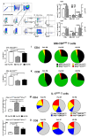
Polyfunctional GM‐CSFpos and IL‐17pos T cells are abundant in inflamed joints of patients with PsA and are decreased in PB of patients with PsA compared with PB from HDs. PB from patients with PsA (n = 60), SF from patients with PsA (n = 10), or PB from HDs (n = 24) were analyzed. PBMCs or SFMCs were stimulated with PMA and ionomycin, before analysis by flow cytometry. (A) PBMCs (or SFMCs) were gated on FSC/SSC to identify lymphocytes, dead cells were excluded, and further gated as (B) CD3+CD4neg (CD8) or (C) CD3+CD4+ (CD4) T cells, expressing either IFNγ, GM‐CSF, or IL‐17. The frequency of (F) CD8 or (G) CD4 T cells that were IFNγpos, GM‐CSFpos, or IL‐17pos were compared in PB and SF from patients with PsA. Polyfunctional cytokine expression was then evaluated where (D, upper) CD8 or (E, lower) CD4 T cells were gated as (D and E, left) IL‐17 negative or (D and E, right) IL‐17 positive, and then gated on IFNγ and GM‐CSF expression. (H) CD4 and (J) CD8 were compared in GM‐CSFpos (IFNγnegIL‐17neg) subsets, and (L) CD4 and (N) CD8 IL‐17posGM‐CSFposIFNγpos subsets were compared in PB and SF from patients with PsA and PB from HDs. The coexpression of GM‐CSF with other cytokines in (I) CD4 and (K) CD8 T cells; and the coexpression of IL‐17 with other cytokines in (M) CD4 and (O) CD8 T cells; in PB and SF from patients with PsA and PB from HDs are demonstrated in pie charts. Data are represented as mean ± SEM and are analyzed by one‐way ANOVA with Tukey's multiple comparison test. *P < 0.05; **P < 0.01; ***P < 0.001; ****P < 0.0001. ANOVA, analysis of variance; FSC, forward scatter; GM‐CSF, granulocyte–macrophage colony‐stimulating factor; HD, healthy donor; IFN, interferon; IL, interleukin; neg, negative; PB, peripheral blood; PBMC, PB mononuclear cells; PMA, phorbol 12‐myristate 13‐acetate; pos, positive; PsA, psoriatic arthritis; SF, synovial fluid; SFMC, SF mononuclear cells; SSC, side scatter.
Figure 2
Figure 2
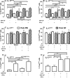
GM‐CSFpos and IL‐17pos polyfunctional CD4 T cells express Th17‐associated phenotypic markers and T‐bet. Cells of PB from patients with PsA (n = 38, open bars) and SF from patients with PsA (n = 9, hatched bars) were incubated with PMA and ionomycin before staining and analysis by flow cytometry. Cells were gated as CD3+CD4+ T cells and evaluated as (A and C) CD161pos or (B and D) CCR6pos cells according to cytokine expression profiles (as in Figure 1E). Alternatively, PB from patients with PsA (n = 10) was stimulated with PMA and ionomycin, gated as CD3+CD4+ T cells, and examined based upon cytokine and T‐bet expression. (E and F) T‐bet expression in (E) IL‐17neg or (F) IL‐17pos CD4 T cells coexpressing GM‐CSF ± IFNγ. Data are represented as mean ± SEM, and analyzed by one‐way ANOVA with Tukey's multiple comparison test. *P < 0.05; **P < 0.01; ***P < 0.001; ****P < 0.0001. ANOVA, analysis of variance; CCR, chemokine receptor; GM‐CSF, granulocyte–macrophage colony‐stimulating factor; HD, healthy donor; IFN, interferon; IL, interleukin; neg, negative; PB, peripheral blood; PMA, phorbol 12‐myristate 13‐acetate; pos, positive; PsA, psoriatic arthritis; SF, synovial fluid.
Figure 3
Figure 3
Exogenous IL‐23 increases IL‐17 but inhibits GM‐CSF release by Teff cells in patients with PsA. PBMCs from patients with PsA (n = 50) and HDs (n = 26) were stimulated with anti‐CD3/28 in the absence or presence of exogenous IL‐23, IL‐12, or IL‐2, and supernatants tested for (A–C) GM‐CSF, (D–F) IL‐17, or (G–I) IFNγ by ELISA. Matched pairs of PBMC from patients with PsA (▲) or HDs (●) without exogenous cytokine were compared with PBMCs from patients with PsA (Δ) or HDs (formula image) with exogenous IL‐23, IL‐12, or IL‐2. Samples from patients with PsA were also compared with PBMCs from HDs within treatment groups. Data are represented as paired samples, with data analyzed by paired or unpaired Student's t‐test. *P < 0.05; **P < 0.01; ***P < 0.001; ****P < 0.0001. ELISA, enzyme‐linked immunosorbent assay; GM‐CSF, granulocyte–macrophage colony‐stimulating factor; HD, healthy donor; IFN, interferon; IL, interleukin; PB, peripheral blood; PBMC, PB mononuclear cell; PsA, psoriatic arthritis.
Figure 4
Figure 4
IL‐17 release by Th17 cells from patients with PsA is only partially IL‐23 dependent, whereas IL‐23 cooperates with TGFβ. Naive CD4 T cells of patients with PsA were differentiated in (A–G) Th17‐polarizing (n = 14) or (C–F) Th1‐polarizing (n = 12) conditions with removal of cytokines as detailed and stimulated with anti‐CD3/28 Dynabeads for five to seven days, and supernatants were tested for (A) IL‐17, (C) GM‐CSF, or (E) IFNγ by ELISA. Cells were harvested and restimulated with PMA and ionomycin before staining and analysis by flow cytometry. Cells were gated as live, CD3+CD4+ T cells, and evaluated as (B) IL‐17pos, (D) GM‐CSFpos, or (F) IFNγpos T cells. Data comparing cytokine removal is normalized relative to Th17‐polarizing conditions with IL‐23 and TGFβ; or Th1 polarizing conditions with IL‐23. (G) Pie charts demonstrating IL‐17pos T cells with the coexpression of GM‐CSF ± IFNγ differentiated under Th17 polarizing conditions in the presence or absence of IL‐23. Data are represented as mean ± SEM, with data analyzed by one‐way ANOVA with Tukey's multiple comparison test. *P < 0.05; **P < 0.01; ***P < 0.001; ****P < 0.0001. ANOVA, analysis of variance; ELISA, enzyme‐linked immunosorbent assay; FACS, fluorescence‐activated cell sorting; GM‐CSF, granulocyte–macrophage colony‐stimulating factor; IFN, interferon; IL, interleukin; PMA, phorbol 12‐myristate 13‐acetate; pos, positive; PsA, psoriatic arthritis; TGF, transforming growth factor. Color figure can be viewed in the online issue, which is available at http://onlinelibrary.wiley.com/doi/10.1002/art.43095/abstract.
Figure 5
Figure 5
Polyfunctional Th17 and Th17‐derived cells are amplified by metabolic stress. Naive CD4 T cells from patients with PsA were differentiated in Th17‐polarizing conditions in the absence (Th17) or presence of the metabolic mediators 2‐DG, NaCl, or TG and stimulated with anti‐CD3/28 Dynabeads for five to seven days, and supernatants were tested for (A) IL‐17, (C) GM‐CSF, or (E) IFNγ by ELISA (normalized to 100% for Th17 cells without metabolic mediators). Cells were then restimulated with PMA and ionomycin, before staining and analysis by flow cytometry. (B, D, and E) Percent change in proportion of single cytokinepos cells (normalized to 100% for Th17 cells without metabolic mediators) and analysis by FACS for (B) IL‐17, (D) GM‐CSF, or (F) IFNγ. (G) GM‐CSFpos cells coexpressing IFNγ ± IL‐17 were displayed in pie charts as Th17 without metabolic mediators, or in the presence of 2‐DG, NaCl, or TG. (H) IL‐17pos cells coexpressing IFNγ ± GM‐CSF were displayed in pie charts as Th17 without metabolic mediators, or in the presence of 2‐DG, NaCl, or TG. Data are represented as mean ± SEM, with data analyzed by paired t‐test using Tukey's multiple comparison test. *P < 0.05; **P < 0.01; ***P < 0.001; ****P < 0.0001. 2‐DG, 2‐deoxy‐d‐glucose; ELISA, enzyme‐linked immunosorbent assay; FACS, fluorescence‐activated cell sorting; GM‐CSF, granulocyte–macrophage colony‐stimulating factor; IFN, interferon; IL, interleukin; pos, positive; PsA, psoriatic arthritis; TG, thapsigargin; TGF, transforming growth factor. Color figure can be viewed in the online issue, which is available at http://onlinelibrary.wiley.com/doi/10.1002/art.43095/abstract.

References

    1. Michalek IM, Loring B, John SM. A systematic review of worldwide epidemiology of psoriasis. J Eur Acad Dermatol Venereol 2017;31(2):205–212. - PubMed
    1. Eder L, Haddad A, Rosen CF, et al. The incidence and risk factors for psoriatic arthritis in patients with psoriasis: a prospective cohort study. Arthritis Rheumatol 2016;68(4):915–923. - PubMed
    1. Bowes J, Budu‐Aggrey A, Huffmeier U, et al. Dense genotyping of immune‐related susceptibility loci reveals new insights into the genetics of psoriatic arthritis. Nat Commun 2015;6:6046. - PMC - PubMed
    1. Stuart PE, Nair RP, Tsoi LC, et al. Genome‐wide association analysis of psoriatic arthritis and cutaneous psoriasis reveals differences in their genetic architecture. Am J Hum Genet 2015;97(6):816–836. - PMC - PubMed
    1. Benham H, Norris P, Goodall J, et al. Th17 and Th22 cells in psoriatic arthritis and psoriasis. Arthritis Res Ther 2013;15(5):R136. - PMC - PubMed

MeSH terms